. 2020 Nov;20(5):161.
doi: 10.3892/ol.2020.12022. Epub 2020 Aug 26.

Nuclear receptor FXR impairs SK-Hep-1 cell migration and invasion by inhibiting the Wnt/β-catenin signaling pathway

Affiliations

Nuclear receptor FXR impairs SK-Hep-1 cell migration and invasion by inhibiting the Wnt/β-catenin signaling pathway

Qianqian Li et al. Oncol Lett. 2020 Nov.

Abstract

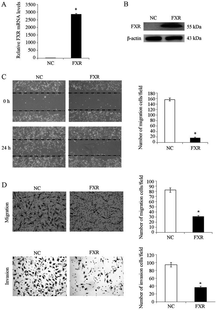
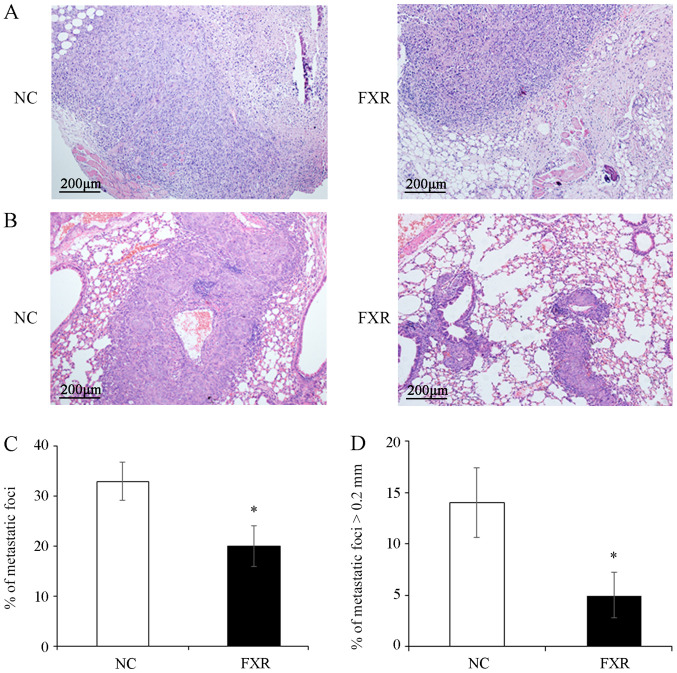
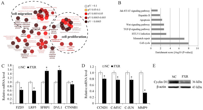
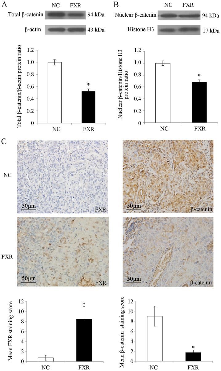
Recently, the nuclear receptor farnesoid X receptor (FXR) has been considered to be a liver tumor suppressor. However, the role of FXR in liver cancer invasion and metastasis remains unclear. The results of the current study demonstrated that FXR suppressed the migratory and invasive capacities of SK-Hep-1 cells in vitro and that FXR overexpression inhibited local invasion and lung metastasis of SK-Hep-1 ×enografts in vivo. Bioinformatics analysis of the gene expression profile of SK-Hep-1 cells with different FXR levels indicated that FXR may regulate the Wnt/β-catenin pathway. Compared with controls, FXR-overexpressing SK-Hep-1 cells exhibited decreased expression of β-catenin target genes and reduced nuclear translocation of β-catenin proteins in vitro and in vivo. In conclusion, these results indicated that FXR may suppress SK-Hep-1 cell invasion and metastasis by suppressing the Wnt/β-catenin signaling pathway. The current study provided novel insight into the diagnosis and treatment of liver cancer.

Keywords: FXR; SK-Hep-1 cell; Wnt/β-catenin; invasion and metastasis.


Figures

Figure 1.
Figure 1.
FXR represses the migration and invasion capacities of SK-Hep-1 cells in vitro. FXR expression in SK-Hep-1-FXR and SK-Hep-1-NC cells were detected by (A) reverse transcription-quantitative PCR and (B) western blotting. SK-Hep-1-FXR and SK-Hep-1-NC cells were pretreated with 2 µM of the FXR agonist GW4064 for 24 h at 37°C, and representative images were captured of the (C) wound healing assay and the (D) Transwell assay for measurement of cell migration and invasion, respectively. The histograms indicate the quantification of migrated or invaded cell numbers determined from three independent experiments. *P<0.05. The ‘FXR’ group represents FXR-overexpressing SK-Hep-1-FXR cells and the ‘NC’ group represents the control SK-Hep-1-NC cells. FXR, farnesoid X receptor; NC, negative control.
Figure 2.
Figure 2.
FXR inhibits the local invasion and metastasis of the SK-Hep-1 ×enograft in vivo. After pretreating with the FXR agonist GW4064 2 µM for 24 h at 37°C, SK-Hep-1-FXR cells and controls were subcutaneously injected into nude mice (n=6 in each group) and tumors and surrounding tissues were harvested three weeks after transplantation. (A) Representative images of hematoxylin and eosin staining. Lung metastasis was induced by tail vein injections of SK-Hep-1-FXR cells and controls pretreated with GW4064 for 24 h (n=4 in each group) into nude mice. (B) Representative images of lung tissue sections. Scale bar, 200 µm (magnification, ×100). (C) Total number of metastatic foci. (D) Number of the metastases with the maximum diameter >0.2 mm. Data are presented as the mean ± standard error of the mean from two experiments with 6 or 4 mice per group for each experiment. *P<0.05. The ‘FXR’ group represents FXR-overexpressing SK-Hep-1-FXR cells and the ‘NC’ group represents the control SK-Hep-1-NC cells. FXR, farnesoid X receptor; NC, negative control.
Figure 3.
Figure 3.
Role of FXR in Wnt/β-catenin pathway. SK-Hep-1-FXR and SK-Hep-1-NC cells were pretreated with GW4064 2 µM for 24 h at 37°C, and harvested for microarray analysis. (A) Gene ontology analysis demonstrated that the differentially expressed genes resulting from FXR upregulation in SK-Hep-1-FXR cells were involved in diverse biological processes. (B) Kyoto Encyclopedia of Genes and Genomes pathway analysis revealed the alteration of diverse cellular signaling pathways due to the FXR overexpression. (C) mRNA expression levels of CTNNB1 and its upstream genes in SK-Hep-1-FXR and SK-Hep-1-NC was detected via RT-qPCR. (D) CTNNB1 target genes were detected using RT-qPCR. (E) Cyclin D1 protein expression was determined via western blotting. Results are presented as mean ± standard error of the mean from three independent experiments. *P<0.05 vs. NC. The ‘FXR’ group represents FXR-overexpressing SK-Hep-1-FXR cells and the ‘NC’ group represents the control SK-Hep-1-NC cells. FXR, farnesoid X receptor; NC, negative control; RT-qPCR, reverse transcription-quantitative PCR; Jak/STAT, Janus kinase/signal transducers and activators of transcription; TGF-β, transforming growth factor β; HTLV-1, human T-cell leukemia virus type 1; FZD5, Frizzled 5; LRP5, low-density lipoprotein receptor-related protein 5; SFRP1, secreted Fzd-related protein; DVL1, Dishevelled 1; CCND1, cyclin D1; CTNNB1, β-catenin.
Figure 4.
Figure 4.
β-catenin expression in SK-Hep-1 cells and lung metastases. After SK-Hep-1-NC and SK-Hep-1-FXR cells were pretreated with GW4064 2 µM for 24 h at 37°C, total and nuclear proteins were isolated. (A) Expression of total β-catenin proteins and (B) nuclear β-catenin proteins were assessed via western blot analysis. The histograms indicate the levels of the protein determined from three independent experiments expressed as the mean ratio relative to that in the SK-Hep-1-NC cells following normalization to β-actin. Data are presented as mean ± standard error of the mean. (C) Representative images of FXR and β-catenin staining in lung metastases of nude mice via immunohistochemistry. Scale bar, 50 µm. Magnification, ×100. The immunoreactivity of tumor cells was quantified using scores based on the staining intensity and the proportion of positively stained tumor cells. Values are presented as mean ± standard error of the mean. *P<0.05. The ‘FXR’ group represents FXR-overexpressing SK-Hep-1-FXR cells and the ‘NC’ group represents the control SK-Hep-1-NC cells. FXR, farnesoid X receptor; NC, negative control.

Similar articles

Cited by

References

    1. Bray F, Ferlay J, Soerjomataram I, Siegel RL, Torre LA, Jemal A. Global cancer statistics 2018: GLOBOCAN estimates of incidence and mortality worldwide for 36 cancers in 185 countries. CA Cancer J Clin. 2018;68:394–424. doi: 10.3322/caac.21492. - DOI - PubMed
    1. Shang N, Wang H, Bank T, Perera A, Joyce C, Kuffel G, Zilliox MJ, Cotler SJ, Ding X, Dhanarajan A, et al. Focal adhesion kinase and β-catenin cooperate to induce hepatocellular carcinoma. Hepatology. 2019;70:1631–1645. doi: 10.1002/hep.30707. - DOI - PMC - PubMed
    1. Massafra V, Pellicciari R, Gioiello A, van Mil SW. Progress and challenges of selective farnesoid X receptor modulation. Pharmacol Ther. 2018;191:162–177. doi: 10.1016/j.pharmthera.2018.06.009. - DOI - PubMed
    1. Tran M, Liu Y, Huang W, Wang L. Nuclear receptors and liver disease: Summary of the 2017 basic research symposium. Hepatol Commun. 2018;2:765–777. doi: 10.1002/hep4.1203. - DOI - PMC - PubMed
    1. Huang XF, Zhao WY, Huang WD. FXR and liver carcinogenesis. Acta Pharmacol Sin. 2015;36:37–43. doi: 10.1038/aps.2014.117. - DOI - PMC - PubMed

LinkOut - more resources