

Periosteal chondroma of pelvis - an unusual location
- PMID: 32934873
- PMCID: PMC7486565
Periosteal chondroma of pelvis - an unusual location
Abstract
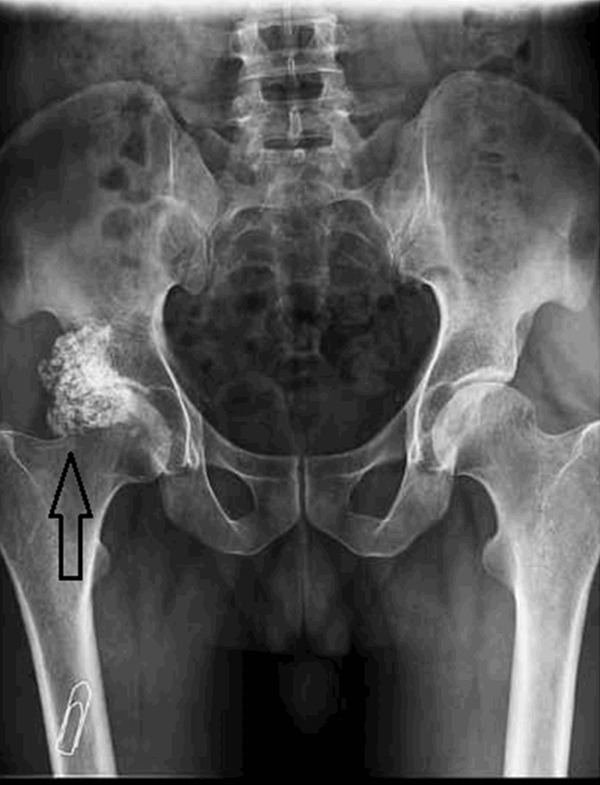
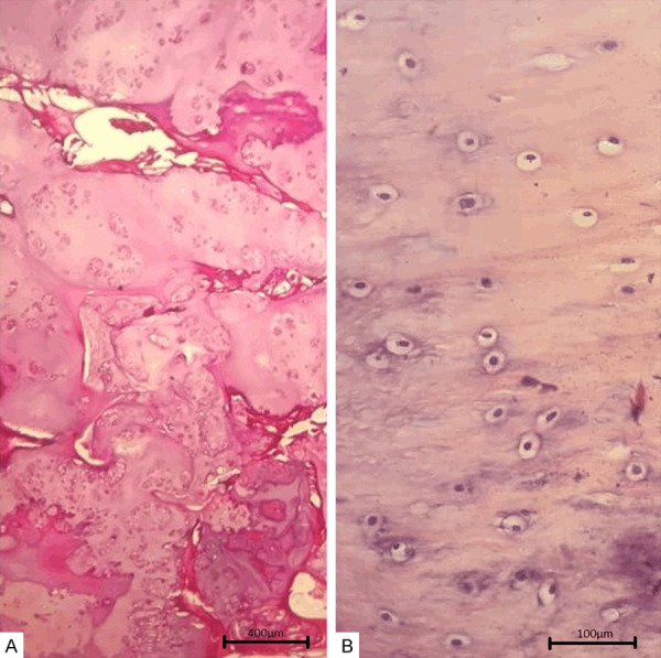
Periosteal chondroma is a slow growing benign tumor with prevalence rate of less than 2% of all chondromas. This tumor is mostly observed in clavicle, ribs and humerus and only one previous case has been reported in pelvis. Here we present an unusual case of periosteal chondroma due to uncommon presentation, location and age range. Our case is a 39 year-old male diagnosed with periosteal chondroma in pelvis. He had unspecific signs and symptoms overlapping with low back pain and disk herniation. By the time of admission he had gluteal muscle atrophy and also claudication. Differentiation of periosteal chondroma from other malignant tumors are pivotal in order to prevent aggressive and inappropriate therapies. He underwent surgical procedures and periosteal chondroma was ascertained by both radiological and Histopathological evidence. 6 months after surgery, he declared no pain, he was able to walk freely. He claimed partial paresthesia but he also declared that his paresthesia has ameliorated.
Keywords: Periosteal chondroma; case report; low back pain; pelvis.
IJBT Copyright © 2020.
Conflict of interest statement
None.
Figures


Similar articles
-
Atypical Presentation of Periosteal Chondroma of the Talus in a 9-Year-Old Boy: A Case Report.J Orthop Case Rep. 2024 Mar;14(3):73-77. doi: 10.13107/jocr.2024.v14.i03.4292. J Orthop Case Rep. 2024. PMID: 38560317 Free PMC article.
-
Periosteal chondroma of the ischium; an unusual location.Turk Patoloji Derg. 2012;28(2):172-4. doi: 10.5146/tjpath.2012.01119. Turk Patoloji Derg. 2012. PMID: 22627638
-
Periosteal chondroma of the clavicle: case report and review of the literature.Int J Surg. 2009 Apr;7(2):140-1. doi: 10.1016/j.ijsu.2008.12.040. Epub 2009 Jan 3. Int J Surg. 2009. PMID: 19185557 Review.
-
Periosteal Chondroma of the Pelvis: An Uncommon Tumor in an Unusual Location.Cureus. 2021 Aug 13;13(8):e17163. doi: 10.7759/cureus.17163. eCollection 2021 Aug. Cureus. 2021. PMID: 34548974 Free PMC article.
-
Periosteal chondroma of the proximal humerus: a case report and review of the literature.J Med Assoc Thai. 2006 Nov;89(11):1970-5. J Med Assoc Thai. 2006. PMID: 17205883 Review.
Cited by
-
Atypical Presentation of Periosteal Chondroma of the Talus in a 9-Year-Old Boy: A Case Report.J Orthop Case Rep. 2024 Mar;14(3):73-77. doi: 10.13107/jocr.2024.v14.i03.4292. J Orthop Case Rep. 2024. PMID: 38560317 Free PMC article.
References
-
- Al-Qudah AS, Abu-Ali HM, Al-Hussaini MA, Massad IM. Periosteal chondroma of the clavicle: case report and review of the literature. Int J Surg. 2009;7:140–1. - PubMed
-
- Lewis MM, Kenan S, Yabut SM, Norman A, Steiner G. Periosteal chondroma. A report of ten cases and review of the literature. Clin Orthop Relat Res. 1990:185–192. - PubMed
-
- Matsushima K, Matsuura K, Kayo M, Gushimiyagi M. Periosteal chondroma of the rib possibly associated with hemothorax: a case report. J Pediatr Surg. 2006;41:E31–3. - PubMed
-
- Goedhart L, Ploegmakers J, Kroon H, Zwartkruis E, Jutte P. The presentation, treatment and outcome of periosteal chondrosarcoma in the Netherlands. Bone Joint J. 2014;96-B:823–8. - PubMed
-
- Chen EM, Masih S, Chow K, Matcuk G, Patel D. Periosteal reaction: review of various patterns associated with specific pathology. Contemporary Diagnostic Radiology. 2012;35:1–5.
Publication types
LinkOut - more resources
Full Text Sources