Review
. 2020 Jul 2;2(1):H29-H43.
doi: 10.1530/VB-20-0007. eCollection 2020.

Endothelial cells on the move: dynamics in vascular morphogenesis and disease

Affiliations
Review

Endothelial cells on the move: dynamics in vascular morphogenesis and disease

Catarina G Fonseca et al. Vasc Biol. .

Abstract

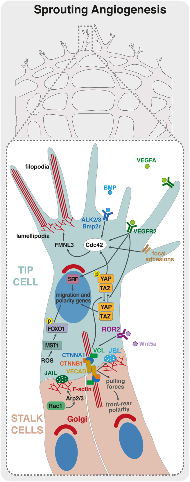
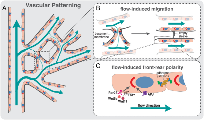
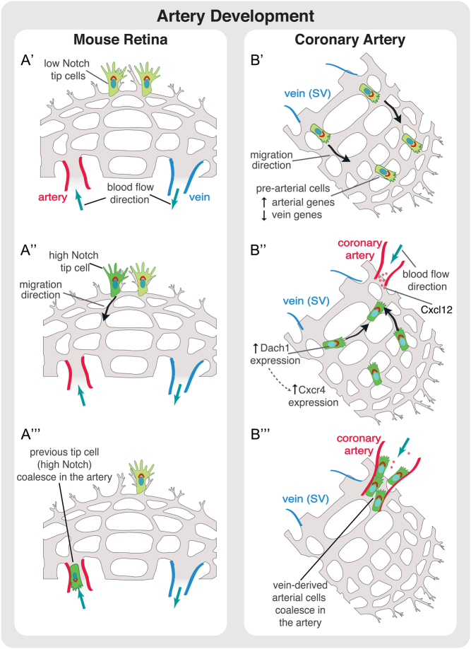
The vascular system is a hierarchically organized network of blood vessels that play crucial roles in embryogenesis, homeostasis and disease. Blood vessels are built by endothelial cells - the cells lining the interior of blood vessels - through a process named vascular morphogenesis. Endothelial cells react to different biomechanical signals in their environment by adjusting their behavior to: (1) invade, proliferate and fuse to form new vessels (angiogenesis); (2) remodel, regress and establish a hierarchy in the network (patterning); and (3) maintain network stability (quiescence). Each step involves the coordination of endothelial cell differentiation, proliferation, polarity, migration, rearrangements and shape changes to ensure network integrity and an efficient barrier between blood and tissues. In this review, we highlighted the relevance and the mechanisms involving endothelial cell migration during different steps of vascular morphogenesis. We further present evidence on how impaired endothelial cell dynamics can contribute to pathology.

Keywords: angiogenesis; cell migration; cell polarity; vascular disease; vascular remodelling.


Figures

Figure 1
Figure 1
Endothelial cell migration during sprouting angiogenesis. Schematic representation of a sprouting front of a vascular plexus with details of a tip cell (in blue) and stalk cells (in red). VEGF/VEGFR2 and BMP/ALK2/3/BMP2R are critical for activation of RHO GTPases, such as CDC42, that controls actin polymerization through formins, such as FMNL3, regulating filopodia and lamellipodia formation. Moreover, VEGF/VEGFR2 activates serum response factor (SRF)-downstream transcription of genes regulating actin cytoskeleton and migration. ROS levels, inside the tip cell, are also important to activate MST1 that phosphorylate FOXO1. Phosphorylated FOXO1 will enter the EC tip nuclei and promote transcription of polarity and migration-related genes. YAP/TAZ transcription factors are crucial for the activation of CDC42 and for adherens junction’s integrity and stabilization. WNT5A/ROR2 signaling pathway is important for the recruitment of vinculin (VCL) to adherens junctions to enhance intercellular force transmission, fundamental to coordinate collective cell polarity and migration. JBLs and JAILs are important for the migration of ECs and junctional stability. Both these structures use RAC1-mediated activation of ARP2/3 in order to promote actin polymerization and lamellipodia extension. These intricate signaling pathways are crucial for the establishment of EC polarity and migration and the collective cell behavior during sprouting angiogenesis.
Figure 2
Figure 2
Vascular patterning depends on blood flow and directional migration. (A) Schematic representation of a vessel network in a developing retina; flow direction and intensity in the plexus are indicated by blue arrows; EC polarity is indicated by the axis of polarity between the nucleus and Golgi complex. (B) Schematic representation of vessel pruning during vascular patterning. Increased levels of shear stress in vessels near the artery induce strong polarization (front-rear polarity) of ECs against the flow direction. The presence of asymmetries in shear stress between juxtaposed vessel segments leads to the migration of ECs away from low-/no-flow vessel segments toward high-flow vessels (black arrows). The directed migration of ECs toward high-flow segments leads to the stabilization of the newly formed vessels and pruning of low-flow vessel segments, detected by the presence of a basement membrane empty sleeve. (C) Molecules involved in flow-induced front-rear polarity. Proper polarization of ECs against flow direction is regulated by non-canonical WNT ligands, WNT5A and WNT11 and by the expression of APLNR (APJ) at EC membrane. Downregulation of these molecules leads to polarity and vascular remodeling defects.
Figure 3
Figure 3
Cell migration during artery development. (A) Proposed model of artery development during angiogenesis in the mouse retina. (A’) Tip cells are activated at the sprouting front by VEGF signaling. (A‘‘) Some tip cells are activated by Notch signaling that promotes their migration away from the sprouting front in direction of the artery. (A‘‘‘) These previous tip cells coalesce in the artery leading to artery growth. (B) Proposed model for coronary artery development. (B’) Vein ECs derived from sinus venosus (SV) sprout by angiogenesis. (B‘‘ During this stage, and prior to the onset of blood flow, some SV-derived ECs start to increase the expression of arterial markers reducing the expression of venous ones. This genetic profile modification, accompanied to the onset of blood flow, induces migration of these pre-arterial cells. The migration of EC against flow direction is regulated by the transcription factor DACH1. DACH1 stimulates expression of CXCR4 in pre-arterial cells enabling these cells to respond to CXCL12, which is expressed by arterial cells. (B‘‘‘) ECs connect to the coronary artery leading to its growth and development.

References

    1. Potente M, Mäkinen T. Vascular heterogeneity and specialization in development and disease. Nature Reviews: Molecular Cell Biology 2017. 18 477–494. (10.1038/nrm.2017.36) - DOI - PubMed
    1. Vaahtomeri K, Karaman S, Mäkinen T, Alitalo K. Lymphangiogenesis guidance by paracrine and pericellular factors. Genes and Development 2017. 31 1615–1634. (10.1101/gad.303776.117) - DOI - PMC - PubMed
    1. Herbert SP, Stainier DYR. Molecular control of endothelial cell behaviour during blood vessel morphogenesis. Nature Reviews: Molecular Cell Biology 2011. 12 551–564. (10.1038/nrm3176) - DOI - PMC - PubMed
    1. Marcelo KL, Goldie LC, Hirschi KK. Regulation of endothelial cell differentiation and specification. Circulation Research 2013. 112 1272–1287. (10.1161/CIRCRESAHA.113.300506) - DOI - PMC - PubMed
    1. Paffett-Lugassy N, Singh R, Nevis KR, Guner-Ataman B, O’Loughlin E, Jahangiri L, Harvey RP, Burns CG, Burns CE. Heart field origin of great vessel precursors relies on nkx2.5-mediated vasculogenesis. Nature Cell Biology 2013. 15 1362–1369. (10.1038/ncb2862) - DOI - PMC - PubMed

LinkOut - more resources