[Preprint]. 2020 Sep 9:rs.3.rs-67888.
doi: 10.21203/rs.3.rs-67888/v1.

How mu-Opioid Receptor Recognizes Fentanyl

Affiliations

How mu-Opioid Receptor Recognizes Fentanyl

Quynh N Vo et al. Res Sq. .

Update in

Abstract

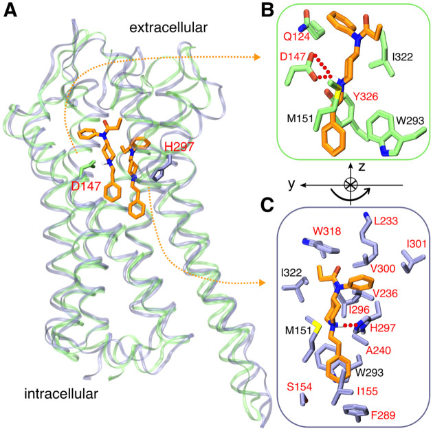
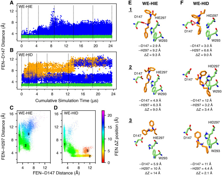
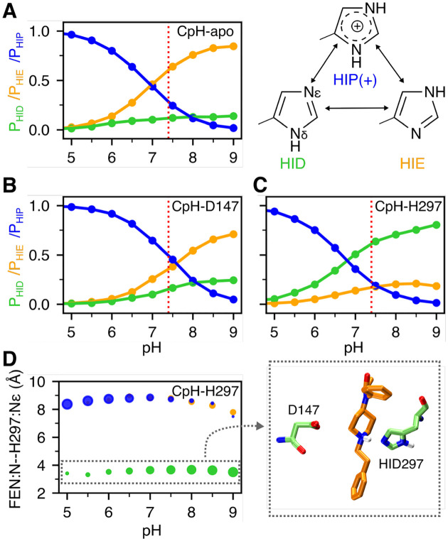
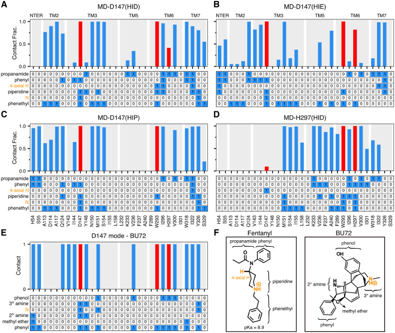
The opioid crisis has escalated during the COVID-19 pandemic. More than half of the overdose-related deaths are related to synthetic opioids represented by fentanyl which is a potent agonist of mu-opioid receptor (mOR). In recent years, crystal structures of mOR complexed with morphine derivatives have been determined; however, structural basis of mOR activation by fentanyl-like synthetic opioids remains lacking. Exploiting the X-ray structure of mOR bound to a morphinan ligand and several state-of-the-art simulation techniques, including weighted ensemble and continuous constant pH molecular dynamics, we elucidated the detailed binding mechanism of fentanyl with mOR. Surprisingly, in addition to the orthosteric site common to morphinan opiates, fentanyl can move deeper and bind mOR through hydrogen bonding with a conserved histidine H297, which has been shown to modulate mOR's ligand affinity and pH dependence in mutagenesis experiments, but its precise role remains unclear. Intriguingly, the secondary binding mode is only accessible when H297 adopts a neutral HID tautomer. Alternative binding modes and involvement of tautomer states may represent general mechanisms in G protein-coupled receptor (GPCR)-ligand recognition. Our work provides a starting point for understanding mOR activation by fentanyl analogs that are emerging at a rapid pace and assisting the design of safer analgesics to combat the opioid crisis. Current protein simulation studies employ standard protonation and tautomer states; our work demonstrates the need to move beyond the practice to advance our understanding of protein-ligand recognition.


Figures

Figure 1.
Figure 1.. Fentanyl binding with mOR.
A. Overlay of the representative simulation snapshots showing mOR is bound to fentanyl in the D147- (green) and H297- (purple) binding modes. B. Zoomed-in view of the D147-binding mode, in which the charged piperidine amine of fentanyl forms a salt bridge with Asp147. C. Zoomed-in view of the H297-binding mode, where the piperidine amine donates a proton to Nϵ of HID297. The curved arrow illustrates the change in the orientation of fentanyl in going from D147- to H297-binding mode. mOR residues making significant contacts with fentanyl (fraction greater than 0.5) are shown (see Fig. 4). Those unique to the two binding modes are labeled in red and otherwise in black.
Figure 2.
Figure 2.. Fentanyl visits the D147-binding mode in the presence of HIE297 but both D147- and H297-binding modes in the presence of HID297.
A, B. The FEN–D147 distance, referred to as the minimum distance between the piperidine nitrogen and the carboxylate oxygen of Asp147, as a function of the cumulative WE simulation time in the presence of HIE297 (A) or HID297 (B). Data with the FEN–D147 and FEN–H297 distances below 3.5 Å are colored green and orange, respectively, and otherwise blue. The unweighted data from all bins were taken and the time refers to the cumulative time. The dotted vertical lines are drawn at every 50 WE iterations. C, D. FEN–H297 vs. FEN–D147 distance from the WE-HIE (C) and WE-HID (D) simulations. The data points are color coded by the fentanyl ΔZ position, defined as the distance between the centers of mass (COM) of fentanyl and mOR in the z direction. N- (52–65) and C-terminal (336–347) residues of mOR were excluded from the COM calculation. The FEN–H297 distance is measured between the piperidine nitrogen and the unprotonated imidazole nitrogen of His297. Three groups of data (labeled in the plots) taken from the last 50 iterations of each simulation were subjected to the hierarchical clustering analysis. For WE-HIE, the three groups were defined as FEN–D147 distance ≤ 3.5 Å; FEN–D147 distance ≥ 4 Å and FEN–H297 distance ≤ 8 Å; and FEN–D147 distance ≥ 4 Å and FEN–H297 distance ≥ 8 Å. For WE-HID, the three groups were defined as FEN–D147 distance ≤ 3.5 Å; FEN–H297 distance ≤ 3.5 Å; and FEN–D147 distance ≥ 8.5 Å and FEN–H297 distance ≥ 4 Å. E, F. Representative structures of the most populated clusters from the WE-HIE (E) and WE-HID (F) data defined in C and D. The FEN–D147 and FEN–H297 distances and the fentanyl ΔZ position are given.
Figure 3.
Figure 3.. Protonation state of His297 is influenced by fentanyl binding.
A, B, C. Occupancies of the HID (green), HIE (orange), and HIP (blue) states of His297 as a function of pH from the replica-exchange CpHMD simulations of the apo mOR (A) and fentanyl-bound complex in the D147- (B) and H297-binding mode (C). The three states are in equilibrium through protonation/deprotonation and tautomerization. D. Average distance between the piperidine nitrogen and His297’s Nϵ at different pH conditions when His297 is in the HID (green), HIE (orange), or HIP (blue) state. The area of the data point is proportional to the occupancy of the protonation state. A zoomed-in snapshot corresponding to the HID state is shown.
Figure 4.
Figure 4.. Fentanyl-mOR interaction profiles in the presence of different protonation state of His297 and comparison to the BU72-mOR contacts in the crystal structure.
A-D Top. Fraction of time that mOR residues form contacts with fentanyl in the equilibrium MD starting from the D147- (A, B, C) and H297-binding modes (D). A contact is considered formed if any sidechain heavy atom is within 4.5 Å of any fentanyl heavy atom. Only residues that form contacts for least 25% of the time in at least one of the six equilibrium simulations are shown. Contacts with Asp147, Trp293, and His297 are highlighted in red. A-D Bottom. Ligand-mOR fingerprint matrix showing the fentanyl groups as rows and mOR residue as columns. 1 represents in contact and 0 represents no contact. E. mOR residues forming contacts with BU72 in the crystal structure (PDB: 5C1M). F. Chemical structure of fentanyl and BU72. Different substituent groups are labeled. The 4-axial hydrogen and amine nitrogen of the piperidine group are indicated.
Figure 5.
Figure 5.. Contact similarity and spatial relationship between the D147- and H297-binding modes.
A. Tanimoto coefficients (Tc) calculated using the binary contacts (details see Methods and Protocols). Tc ranges from 0 to 1, where 1 indicates identical mOR residues are involved in fentanyl binding in both simulations. B, C. Locations of fentanyl (stars) and critical amino acids (circles) plotted on the (Y,Z) and (X,Y) planes. A data points are sampled every 10 ns. The center of mass of mOR is set to origin. The data from MD-D147(HID), MD-D147(HIE), and MD-D147(HIP) are colored light green, orange, and blue, respectively, while the data from MD-H297(HID) are colored dark green. The z axis is the membrane normal, while the x axis is defined by the N2–H vector.

References

    1. Synthetic Opioid Overdose Data | Drug Overdose | CDC Injury Center. https://www.cdc.gov/drugoverdose/data/fentanyl.html, 2020.
    1. Van Bever W. F.; Niemegeers C. J.; Schellekens K. H.; Janssen P. A. N-4-Substituted 1-(2-Arylethyl)-4-Piperidinyl-N-Phenylpropanamides, a Novel Series of Extremely Potent Analgesics with Unusually High Safety Margin. Arzneimittelforschung. 1976, 26, 1548–1551. - PubMed
    1. Volpe D. A.; Tobin G. A. M.; Mellon R. D.; Katki A. G.; Parker R. J.; Colatsky T.; Kropp T. J.; Verbois S. L. Uniform Assessment and Ranking of Opioid Mu Receptor Binding Constants for Selected Opioid Drugs. Regul. Toxicol. Pharmacol. 2011, 59, 385–390. - PubMed
    1. Burns S. M.; Cunningham C. W.; Mercer S. L. DARK Classics in Chemical Neuroscience: Fentanyl. ACS Chem. Neurosci. 2018, 9, 2428–2437. - PubMed
    1. Comer S. D.; Cahill C. M. Fentanyl: Receptor Pharmacology, Abuse Potential, and Implications for Treatment. Neurosci. Biobehav. Rev. 2019, 106, 49–57. - PMC - PubMed

Publication types