

Mast Cells in Diabetes and Diabetic Wound Healing
- PMID: 32935286
- PMCID: PMC7547971
- DOI: 10.1007/s12325-020-01499-4
Mast Cells in Diabetes and Diabetic Wound Healing
Abstract
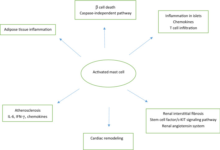
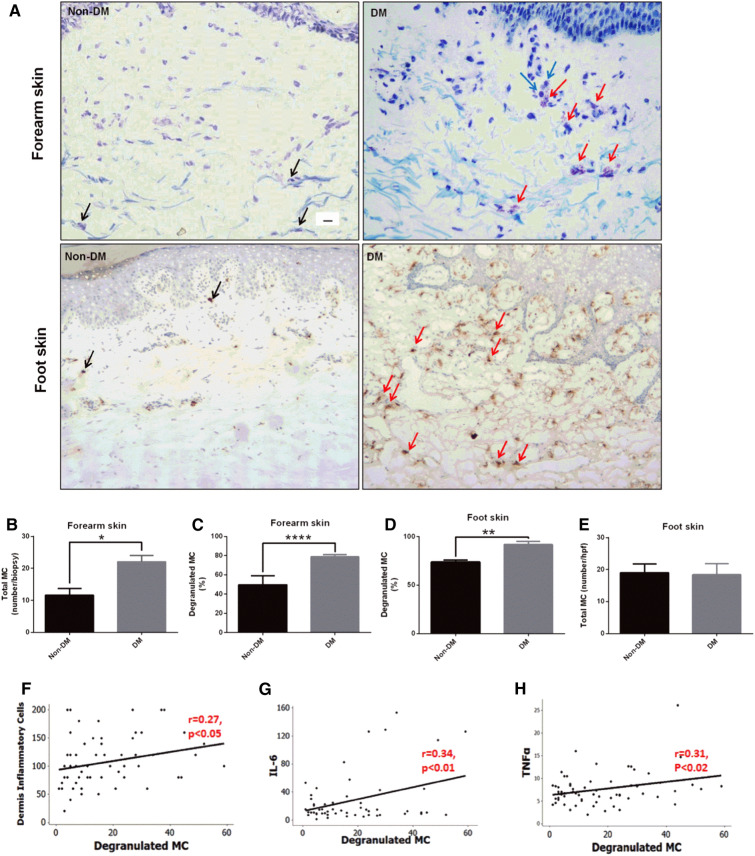
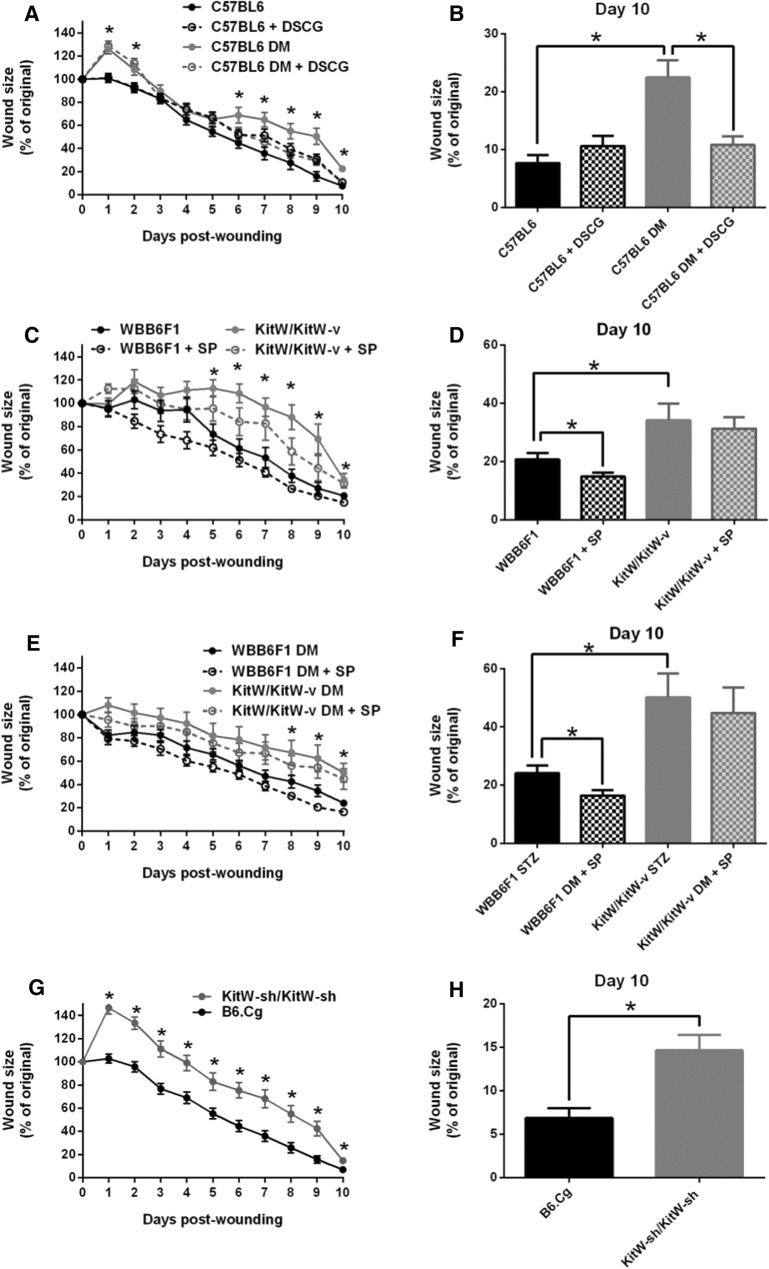
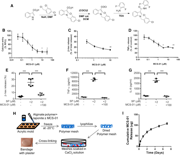
Mast cells (MCs) are granulated, immune cells of the myeloid lineage that are present in connective tissues. Apart from their classical role in allergies, MCs also mediate various inflammatory responses due to the nature of their secretory products. They are involved in important physiological and pathophysiological responses related to inflammation, chronic wounds, and autoimmune diseases. There are also indications that MCs are associated with diabetes and its complications. MCs and MC-derived mediators participate in all wound healing stages and are involved in the pathogenesis of non-healing, chronic diabetic foot ulcers (DFUs). More specifically, recent work has shown increased degranulation of skin MCs in human diabetes and diabetic mice, which is associated with impaired wound healing. Furthermore, MC stabilization, either systemic or local at the skin level, improves wound healing in diabetic mice. Understanding the precise role of MCs in wound progression and healing processes can be of critical importance as it can lead to the development of new targeted therapies for diabetic foot ulceration, one of the most devastating complications of diabetes.
Keywords: Diabetes mellitus; Diabetic foot ulcer; Mast cells; Wound healing.
Figures
Similar articles
-
Topical Application of a Mast Cell Stabilizer Improves Impaired Diabetic Wound Healing.J Invest Dermatol. 2020 Apr;140(4):901-911.e11. doi: 10.1016/j.jid.2019.08.449. Epub 2019 Sep 27. J Invest Dermatol. 2020. PMID: 31568772
-
Mast Cells Regulate Wound Healing in Diabetes.Diabetes. 2016 Jul;65(7):2006-19. doi: 10.2337/db15-0340. Epub 2016 Apr 8. Diabetes. 2016. PMID: 27207516 Free PMC article.
-
Impact of Photobiomodulation and Condition Medium on Mast Cell Counts, Degranulation, and Wound Strength in Infected Skin Wound Healing of Diabetic Rats.Photobiomodul Photomed Laser Surg. 2019 Nov;37(11):706-714. doi: 10.1089/photob.2019.4691. Epub 2019 Oct 7. Photobiomodul Photomed Laser Surg. 2019. PMID: 31589095
-
A Review of the Contribution of Mast Cells in Wound Healing: Involved Molecular and Cellular Mechanisms.Clin Rev Allergy Immunol. 2020 Jun;58(3):298-312. doi: 10.1007/s12016-019-08729-w. Clin Rev Allergy Immunol. 2020. PMID: 30729428 Review.
-
Systematic reviews of wound care management: (3) antimicrobial agents for chronic wounds; (4) diabetic foot ulceration.Health Technol Assess. 2000;4(21):1-237. Health Technol Assess. 2000. PMID: 11074391 Review.
Cited by
-
Immunology of Acute and Chronic Wound Healing.Biomolecules. 2021 May 8;11(5):700. doi: 10.3390/biom11050700. Biomolecules. 2021. PMID: 34066746 Free PMC article. Review.
-
Identifying potential pathogenesis and immune infiltration in diabetic foot ulcers using bioinformatics and in vitro analyses.BMC Med Genomics. 2023 Dec 1;16(1):313. doi: 10.1186/s12920-023-01741-2. BMC Med Genomics. 2023. PMID: 38041124 Free PMC article.
-
Neutrophil heterogeneity and aging: implications for COVID-19 and wound healing.Front Immunol. 2023 Nov 28;14:1201651. doi: 10.3389/fimmu.2023.1201651. eCollection 2023. Front Immunol. 2023. PMID: 38090596 Free PMC article. Review.
-
Deciphering the crosstalk between inflammation and biofilm in chronic wound healing: Phytocompounds loaded bionanomaterials as therapeutics.Saudi J Biol Sci. 2024 Apr;31(4):103963. doi: 10.1016/j.sjbs.2024.103963. Epub 2024 Feb 23. Saudi J Biol Sci. 2024. PMID: 38425782 Free PMC article. Review.
-
Mast Cell Deficiency in Mice Attenuates Insulin Phenolic Preservative-Induced Inflammation.Biomedicines. 2023 Aug 12;11(8):2258. doi: 10.3390/biomedicines11082258. Biomedicines. 2023. PMID: 37626754 Free PMC article.
References
-
- Crivellato E, Beltrami CA, Mallardi F, Ribatti D. Paul Ehrlich’s doctoral thesis: a milestone in the study of mast cells. Br J Haematol. 2003;123:19–21. - PubMed
-
- Maurer M, Taube C, et al. Mast cells drive IgE-mediated disease but might be bystanders in many other inflammatory and neoplastic conditions. J Allergy Clin Immunol. 2019;144:S19–S30. - PubMed
Publication types
MeSH terms
Grants and funding
LinkOut - more resources
Full Text Sources
Medical
