

Maximal Axial Vertebral Rotation in Adolescent Idiopathic Scoliosis: Is the Apical Vertebra the Most Rotated?
- PMID: 32935571
- PMCID: PMC8907634
- DOI: 10.1177/2192568220948830
Maximal Axial Vertebral Rotation in Adolescent Idiopathic Scoliosis: Is the Apical Vertebra the Most Rotated?
Abstract
Study design: Retrospective cross-sectional study.
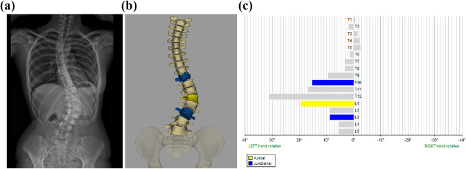
Objectives: It is generally believed that the apical vertebra has the largest axial rotation in adolescent idiopathic scoliosis. We investigated the relationship between apical axial vertebral rotation (apicalAVR) and maximal axial vertebral rotation (maxAVR) in both major and minor curves using biplanar stereo-imaging.
Methods: EOS 2D/3D biplanar radiograph images were collected from 332 patients with adolescent idiopathic scoliosis (Cobb angle range 10°-122°, mean age 14.7 years). Based on the X-ray images, with the help of 3D full spine reconstructions Cobb angle, curvature level, apicalAVR and maxAVR were determined. These parameters were also determined for minor curves in Lenke 2, 3, 4, 6 type patients. Maximal thoracic rotation and maximal thoracolumbar/lumbar rotation were calculated. Statistical analysis was performed with descriptive statistics, Shapiro-Wilk test, and Wilcoxon signed-rank test.
Results: The apical vertebrae were the most rotated vertebra in only 40.4% of the major curves, and 31.7% in minor curves. MaxAVR significantly exceeded apicalAVR values in the major curves (P < .001) as well as in minor curves (P < .001). The 2 parameters differed significantly in each severity group and Lenke type.
Conclusions: The apical vertebrae were not the most rotated vertebra in more than half of cases investigated indicating that apicalAVR and maxAVR should be considered as 2 distinct parameters, of which maxAVR fully describes the axial dimension of scoliosis. Furthermore, the substitution of maxAVR for the apicalAVR should be especially avoided in double and triple curves, as the apical vertebra was even less commonly the most rotated in minor curves.
Keywords: EOS 2D/3D; adolescent idiopathic scoliosis; apical vertebral rotation; biplanar imaging; maximal vertebral rotation.
Conflict of interest statement
Figures
References
-
- Mehta MH. Radiographic estimation of vertebral rotation in scoliosis. J Bone Joint Surg Br. 1973;55:513–520. - PubMed
-
- Perdriolle R, Vidal J. Morphology of scoliosis: three-dimensional evolution. Orthopedics. 1987;10:909–915. - PubMed
-
- Stokes IA. Axial rotation component of thoracic scoliosis. J Orthop Res. 1989;7:702–708. - PubMed
-
- Marchesi DG, Transfeldt EE, Bradford DS, Heithoff KB. Changes in vertebral rotation after Harrington and Luque instrumentation for idiopathic scoliosis. Spine (Phila Pa 1976). 1992;17:775–780. - PubMed
-
- Willers U, Transfeldt EE, Hedlund R. The segmental effect of Cotrel-Dubousset instrumentation on vertebral rotation, rib hump and the thoracic cage in idiopathic scoliosis. Eur Spine J. 1996;5:387–393. - PubMed
LinkOut - more resources
Full Text Sources
