Review
. 2021 Oct;27(5):506-528.
doi: 10.1177/1073858420954826. Epub 2020 Sep 16.

Meninges: A Widespread Niche of Neural Progenitors for the Brain

Affiliations
Review

Meninges: A Widespread Niche of Neural Progenitors for the Brain

Ilaria Decimo et al. Neuroscientist. 2021 Oct.

Abstract

Emerging evidence highlights the several roles that meninges play in relevant brain functions as they are a protective membrane for the brain, produce and release several trophic factors important for neural cell migration and survival, control cerebrospinal fluid dynamics, and embrace numerous immune interactions affecting neural parenchymal functions. Furthermore, different groups have identified subsets of neural progenitors residing in the meninges during development and in the adulthood in different mammalian species, including humans. Interestingly, these immature neural cells are able to migrate from the meninges to the neural parenchyma and differentiate into functional cortical neurons or oligodendrocytes. Immature neural cells residing in the meninges promptly react to brain disease. Injury-induced expansion and migration of meningeal neural progenitors have been observed following experimental demyelination, traumatic spinal cord and brain injury, amygdala lesion, stroke, and progressive ataxia. In this review, we summarize data on the function of meninges as stem cell niche and on the presence of immature neural cells in the meninges, and discuss their roles in brain health and disease. Furthermore, we consider the potential exploitation of meningeal neural progenitors for the regenerative medicine to treat neurological disorders.

Keywords: meninges; neural progenitors; neurogenesis; oligodendrocyte precursor cells; regenerative medicine; stem cell niche.


Conflict of interest statement

Declaration of Conflicting Interests: The author(s) declared no potential conflicts of interest with respect to the research, authorship, and/or publication of this article.

Figures

Figure 1.
Figure 1.
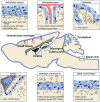
Meninges are widespread in human and rodent central nervous system (CNS). Meningeal distribution of human (A, B, C) and rodent (D, D′, D′) brain are shown. (A) Sagittal depiction of the human encephalon and (C) the corresponding paramedian T2-weighted magnetic resonance (MR) scan are reported, highlighting the wide distribution of the meningeal layers, excluding the dura mater, as a tissue covering and penetrating inside the cerebral and cerebellar parenchyma, following vessel branches, sulci, and stroma gyration. (B) Coronal section of the human brain stained by hematoxylin and eosin shows meninges penetrating trough the gyri into the sulci. (D) Sagittal graphic view of the rodent brain is reported with enlarged view of the superficial meningeal layer covering the parenchyma at the convexity (D′) and the meningeal substructure penetrating the choroid plexus (D′). (D′) The meningeal arachnoid layer defines the subarachnoid space that is hosting blood vessels, as they deeply penetrate into the sulci and parenchyma in the perivascular spaces projecting through the main brain substructures. The pia mater adheres to the parenchyma and its basal membrane and divides the arteriolar endothelium from the parenchyma. (D′) The pia mater also wraps the choroid plexus (tela choroidea, D′).
Figure 2.
Figure 2.
Meningeal cell heterogeneity in healthy and pathological conditions. Schematic representation showing meningeal cell heterogeneity in healthy and pathological conditions. Meninges are formed by three tissue membranes: dura mater, arachnoid, and pia mater. The dura mater contains lymphatic vessels (green) which spread in the arachnoid space with their capillaries. The arachnoid is an epithelial layer filled by cerebrospinal fluid (CSF) (light blue) in which fibroblasts create the typical arachnoid trabeculae. Pia mater (blue) made of a single layer of pial cells, adheres to the surface of the brain parenchyma (astrocytic basal membrane, bordeaux). In physiological conditions, the leptomeninges (arachnoid and pia) host three main different classes of cell populations: neural precursors, resident stromal cells (pericyte, telocyte, smooth muscle cells, fibroblast, and pial cells) and resident immune cells (border-associated macrophages, dendritic cells, and mastocytes). Following disease, meninges increase their thickness and meningeal cell populations react and proliferate increasing their number (stromal cells and neural precursors). Moreover, the pathological condition causes the activation of the resident immune cells and the recruitment of blood-borne immune cells (circulating monocytes, lymphocytes, neutrophils).
Figure 3.
Figure 3.
Meningeal neural progenitors are widespread in the central nervous system (CNS). Schematic representation of a sagittal section of rodent brain and spinal cord showing the distribution of neural stem cells (NSCs, blue), immature neurons (brown), and oligodendrocyte precursor cells (OPCs, green) in CNS meninges (light blue). The specific markers expressed by each neural progenitor subclass are shown for each area accordingly with the reviewed literature. As meninges cover the entire CNS (brain and spinal cord) and are widely distributed, also meningeal neural progenitors are not restricted to a defined meningeal area of the brain. Specifically, they have been found in the external brain meninges (upper left panel), in the meninges of perivascular space (upper middle panel), in the cerebellar meninges (upper right panel), along the meninges of hippocampal fimbria (lower left panel), in the meningeal substructures (lower middle panel), and in spinal cord meninges (lower right panel).
Figure 4.
Figure 4.
Meningeal neural progenitors generate parenchymal neurons and oligodendrocytes in physiological condition. (A) Schematic representation showing that immature neural progenitor cells and neuroblasts in meninges generate meningeal-derived neurons or rare oligodendrocytes in the brain parenchyma. In (B) meningeal-derived neurons (YFP+/CFP+, green and tdTomato, red) in the brain cortex of a postnatal day 30 (P30) PDGFRβ-Cre mouse (upper panel) expressing the neuronal markers NeuN and Satb2 (lower panel, arrowhead) are shown. Meningeal cells were labelled by injecting PDGFRβ-Cre P0 mice with a lentiviral vector expressing the Brainbow 1.0(L) reporter in the meninges allowing to trace the Cre expressing PDGFRβ meningeal cells (YFP+/CFP+ cells, green). tdTomato cells (red) are meningeal derived cells that do not express PDGFRβ. The upper panel shows that the meningeal cells migrated into cortical layers II to IV were mostly PDGFRβ-Cre-derived YFP+/CFP+ cells (green). In the lower panel, YFP/CFP meningeal-derived cells (green), NeuN (red), and Satb2 (blue), showing that the PDGFRβ-Cre-derived YFP+/CFP+ cells were NeuN+/Satb2+ neurons (arrows). Modified from Bifari and others (2017).
Figure 5.
Figure 5.
Functional features of meningeal derived neurons. Electrical phenotype of resident and meningeal-derived neurons in somatosensory cortex (A, left panel). Patch current-clamp recordings of action potentials evoked by direct positive current injection in a resident principal cell and interneuron (black) and in cells of glutamatergic and GABAergic phenotype of meningeal origin (red). In B (left panel) the traces are the frequency-current curves obtained by direct positive current injection above rheobase. Meningeal-derived glutamatergic cells exhibit a higher action potential rate and a higher frequency gain as compared to their resident counterparts. The frequency gain of resident and meningeal-derived interneurons is similar, although the meningeal-derived interneuron generates action potentials at a lower rate as compared to its resident counterpart. Modified with permission from Bifari and others (2017). For comparison, the patch current-clamp recordings of action potentials evoked by direct positive current injection in a mature resident principal cell (black) and a postnatal differentiated mature complex cell (red) of the piriform cortex are shown (A, right panel). In B (right panel) the traces are the frequency-current curves obtained by direct positive current injection above rheobase. Complex cells exhibit different action potential rate and frequency gain as compared to their resident counterparts, similarly to meningeal-derived neuronal cells. Patch-clamp traces modified with permission from Benedetti and others (2020). Frequency-current curves obtained from the original dataset from Benedetti and others (2020) kindly provided by the authors.
Figure 6.
Figure 6.
Meningeal neural progenitors in diseases. Schematic representation showing the meningeal environment in the central nervous system pathological conditions (left panel). Meninges are formed by three tissue membranes: dura mater, arachnoid and pia mater. Following diseases, different signals that include tissue damage, vascular and blood perfusion impairment, cell death, and inflammatory signals activate the meningeal niche. Meningeal progenitor cells, promptly react, proliferate, and migrate from the meninges to the brain parenchyma and differentiate into immature neurons and functional cortical oligodendrocytes. In the right panels, the injury induced meningeal-derived neural cell contribution to three different pathological conditions is shown. In the upper right panel, following transient depletion of oligodendrocyte precursor cells (OPCs), meningeal derived OPCs migrate to the injured parenchyma and differentiate into oligodendrocytes; modified from Dang and others (2019). In the middle right panel, following spinal cord injury, meningeal neural precursors (nestin+, red) migrate to the glial scar site; modified from Decimo and others (2011). In the lower right panel, after brain stroke, meninges increase the expression of nestin- and DCX-positive cells, which migrate to the injured cortex and potentially contribute to cortical regeneration/repair; modified from Nakagomi and others (2012).
Figure 7.
Figure 7.
Schematic representation of the migratory pathway of meningeal neural progenitors to the parenchyma. In the left panel proposed mechanisms of the meningeal neural progenitor cell migration to the underlying parenchyma through the pial basal lamina (dashed line). Meningeal neural progenitors migrate from specific meningeal areas, including brain and spinal cord meninges, meninges of the perivascular space and of the cerebellum. The precise mechanisms of accessing to the brain and the chemotactic guidance signaling regulating this migration have not yet been clearly described. In the right upper panel, the migration path of meningeal progenitor cells to the cortex during perinatal stage (black line). Specifically, meningeal neural progenitors migrate to the cortex via the meningeal substructure (right middle panel) and tela choroidea (lower right panel). Modified from Bifari and others (2017).
Figure 8.
Figure 8.
Regenerative potential of meningeal neural progenitors. In the upper panel image, the human brain meninges exposed during neurosurgery. Meningeal neural progenitors can be isolated from meningeal samples by mechano-enzymatic dissociation and in vitro cultured. Meningeal neural stem cells (NSCs) can be expanded in vitro and subsequently differentiated into oligodendrocytes and neurons (light microscope images in the lower panel). Meningeal NSCs can potentially be used for regenerative medicine in autologous graft setting. Scale bars are 20 µm.

References

    1. Aviezer D, Hecht D, Safran M, Eisinger M, David G, Yayon A.1994. Perlecan, basal lamina proteoglycan, promotes basic fibroblast growth factor-receptor binding, mitogenesis, and angiogenesis. Cell 79(6):1005–13. - PubMed
    1. Barber M, Andrews WD, Memi F, Gardener P, Ciantar D, Tata M, and others. 2018. Vascular-derived vegfa promotes cortical interneuron migration and proximity to the vasculature in the developing forebrain. Cereb Cortex 28(7):2577–93. - PMC - PubMed
    1. Belmadani A, Ren D, Bhattacharyya BJ, Rothwangl KB, Hope TJ, Perlman H, and others. 2015. Identification of a sustained neurogenic zone at the dorsal surface of the adult mouse hippocampus and its regulation by the chemokine SDF-1. Hippocampus 25(11):1224–41. - PMC - PubMed
    1. Benakis C, Llovera G, Liesz A.2018. The meningeal and choroidal infiltration routes for leukocytes in stroke. Ther Adv Neurol Disord 11:1756286418783708. - PMC - PubMed
    1. Benedetti B, Dannehl D, Konig R, Coviello S, Kreutzer C, Zaunmair P, and others. 2020. Functional integration of neuronal precursors in the adult murine piriform cortex. Cereb Cortex 30(3):1499–515. - PMC - PubMed

Publication types