Review
. 2020 Oct 19;49(20):7252-7270.
doi: 10.1039/d0cs00455c.

Design of small molecules targeting RNA structure from sequence

Affiliations
Review

Design of small molecules targeting RNA structure from sequence

Andrei Ursu et al. Chem Soc Rev. .

Abstract

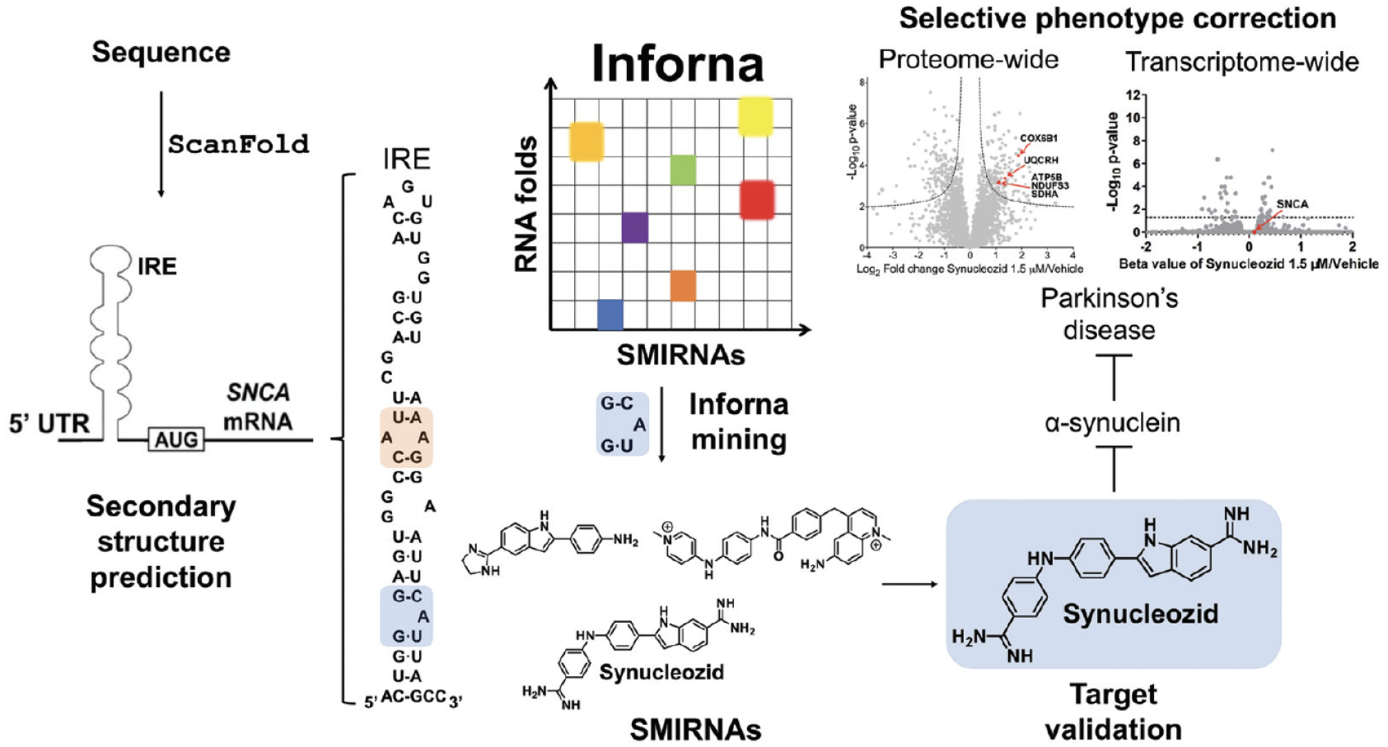
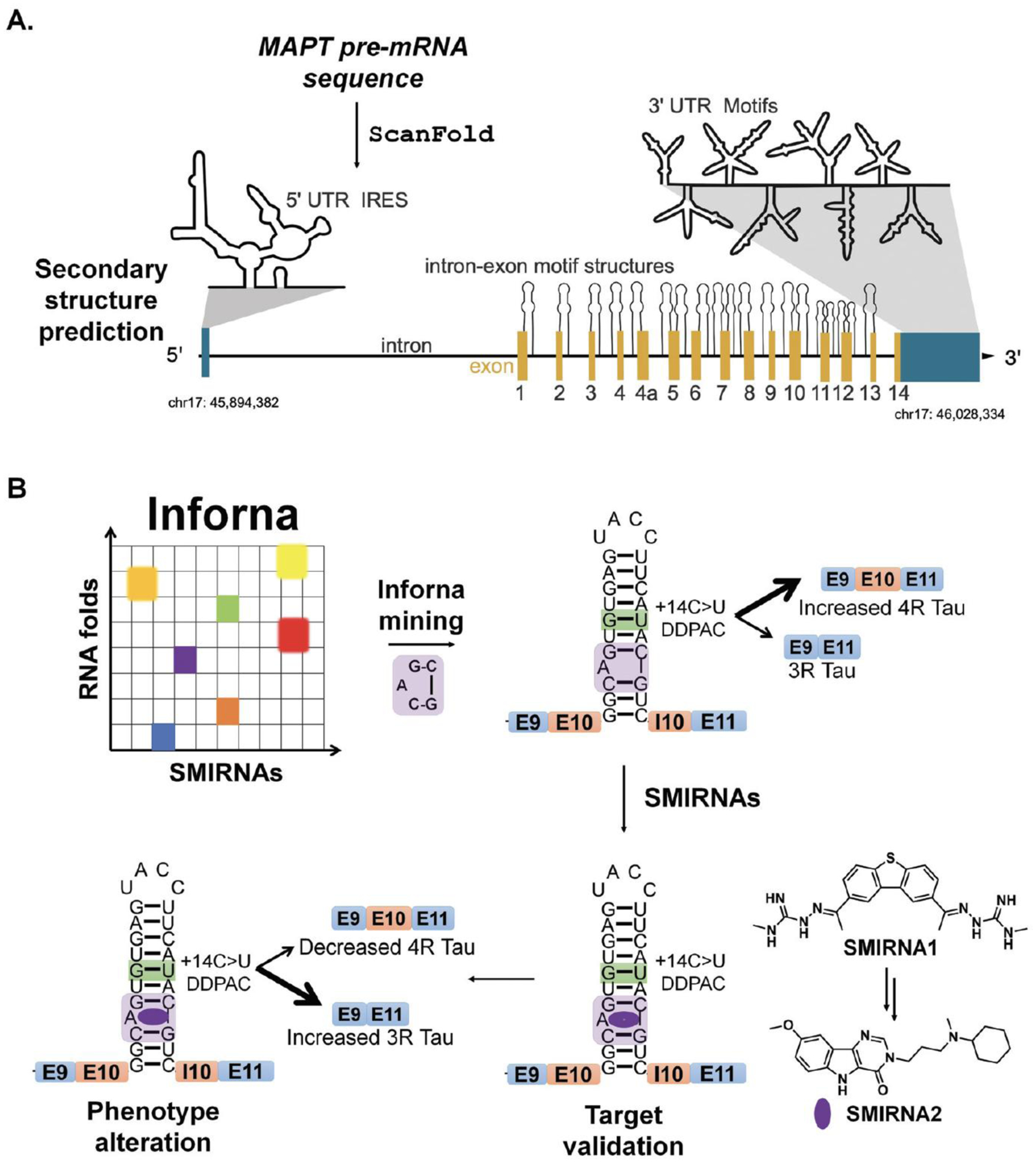
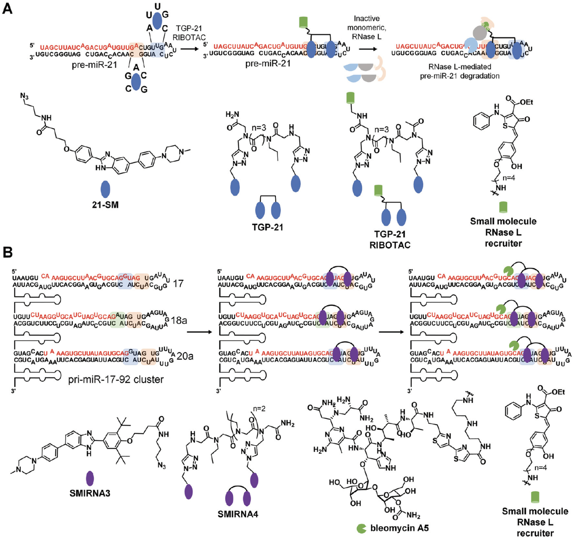
The design and discovery of small molecule medicines has largely been focused on a small number of druggable protein families. A new paradigm is emerging, however, in which small molecules exert a biological effect by interacting with RNA, both to study human disease biology and provide lead therapeutic modalities. Due to this potential for expanding target pipelines and treating a larger number of human diseases, robust platforms for the rational design and optimization of small molecules interacting with RNAs (SMIRNAs) are in high demand. This review highlights three major pillars in this area. First, the transcriptome-wide identification and validation of structured RNA elements, or motifs, within disease-causing RNAs directly from sequence is presented. Second, we provide an overview of high-throughput screening approaches to identify SMIRNAs as well as discuss the lead identification strategy, Inforna, which decodes the three-dimensional (3D) conformation of RNA motifs with small molecule binding partners, directly from sequence. An emphasis is placed on target validation methods to study the causality between modulating the RNA motif in vitro and the phenotypic outcome in cells. Third, emergent modalities that convert occupancy-driven mode of action SMIRNAs into event-driven small molecule chemical probes, such as RNA cleavers and degraders, are presented. Finally, the future of the small molecule RNA therapeutics field is discussed, as well as hurdles to overcome to develop potent and selective RNA-centric chemical probes.


Conflict of interest statement

Conflicts of interest

M. D. D. is a founder of Expansion Therapeutics.

Figures

Fig. 1
Fig. 1
Overview of RNA structure and its prediction directly from sequence using ScanFold. (A) The primary structure of RNA, i.e., sequence, consists of four bases; two purines, adenine (A) and guanine (G), and two pyrimidines, cytosine (C) and uracil (U). (B) The secondary structure of an RNA consists of the non-covalent bonds that form between A and U, G and C, or G and U, bases. These pairings consist of hydrogen bonds and base stacking interactions which form stems (light green) and are often punctuated with internal loops (blue), bulges (pink), and hairpin loops (dark green). (C) The tertiary structure of RNA is largely dictated by the base pairs that form the secondary structure. Stems (light green) will form structured A-form helices and internal loops (blue), bulges (pink), and hairpin loops (dark green) will be less structured, more accessible regions that distort the more rigid helix and offer sites for trans-acting factors to bind in a sequence specific manner. Here, the dotted black line represents the single strand between the two more structured hairpins. (D) Identification of structured RNA motifs within the mRNA sequence of MYC via ScanFoId. Portions of the MYC mRNA coding region and 3′ untranslated region (UTR) are depicted with overlapping ScanFold analysis windows below. In each scanning analysis window, ScanFold calculates numerous folding metrics including the minimum free energy (MFE), ensemble diversity, and z-scores which are depicted as bar graphs. It is important to note that a window will be represented by a single bar, but the downstream nucleotides (nt) (corresponding to the window size) are used to predict the metrics. ScanFoId then determines the most stable and significant base pairs and uses them to generate a consensus structure (displayed as an arc diagram). Regions with highly negative z-scores and low ensemble diversity indicate regions of presumed function, with one (or few) dominating structures and that may merit further, in-depth analyses: e.g., comparative analysis, additional bioinformatics analyses, functional assays, and structure probing assays. These techniques can further characterize and validate the biological function of the structured RNA motif.
Fig. 2
Fig. 2
Overview of two-dimensional combinatorial screening (2DCS) and Inforna. In 2DCS, a small molecule library is spatially arrayed onto a microarray, either through covalent attachment or absorption (AbsorbArray). Compounds are then incubated with a labeled RNA motif library, e.g., 3 × 2 internal loops, containing randomized regions that form structured RNA motifs found in disease-causing RNAs. Unlabeled competitor oligonucleotides that mimic regions common to all library members, r(AU) and r(GC) base pairs, DNA oligonucleotides, and other RNAs are added to eliminate non-specific binding. Small molecules that bind RNAs are excised, amplified by RT-PCR, sequenced by RNA-seq, and analyzed by High Throughput Structure-Activity Relationships Through Sequencing (HiT-StARTS). HiT-StARTS calculates the statistical significance of the enrichment of an RNA in the selection, reported as a Z-score (Zobs). Selective small molecule-RNA motif interactions generally exhibit Zobs > 8. These small molecule-RNA motif interactions and their corresponding Z-scores comprise Inforna. Using Inforna, privileged SMIRNAs can be identified for functionally relevant RNA 3D folds within disease-causing RNAs, such as miRNAs. In addition to mining for SMIRNAs with favorable affinity landscapes for the RNA target of interest, Inforna can also predict potential off-target RNAs.
Fig. 3
Fig. 3
Methods to validate the targets of SMIRNAs, to study cellular selectivity, and to map SMIRNA binding sites within an RNA target. Schematics of target validation techniques for SMIRNAs. In ASO-Bind-Map, unmodified SMIRNAs are used to prevent hybridization of complementary ASOs, thus preventing cleavage. In Chemical Cross-Linking and Isolation by Pull-Down (Chem-CLIP) and related methods (competitive Chem-CLIP (C-Chem-CLIP) and Chem-CLIP-Map-Seq), SMIRNAs are functionalized with cross-linking (chlorambucil or diazirine) modules and a purification module (biotin) at positions that do not affect binding to the intended RNA target. In small molecule nucleic acid profiling by cleavage applied to RNA (RiboSNAP) and its competitive variant, the SMIRNA is appended with the natural product bleomycin A5.
Fig. 4
Fig. 4
Using Inforna to identify SMIRNAs targeting disease-causing miRNAs. (A) Schematic representation of miRNA biogenesis, where SMIRNA binding can inhibit processing by binding to either Drosha or Dicer sites and thereby reduce the levels of the mature miRNA. Reduction of mature miRNA levels results in decreased translational inhibition of target mRNAs by the RNA-induced silencing complex (RISC). Thus, SMIRNA inhibition of miRNA biogenesis derepresses the miRNA’s protein targets, resulting in phenotype modulation. Structure of pri-miR-96 and chemical structures of monomeric compounds 96-SM1 and 96-SM2 that target 1 × 1 GG and UU internal loops (blue and orange, respectively) in the Drosha processing site. Covalent attachment of 96-SM1 to 96-SM2 via a peptoid linker yields dimeric compound TGP-96, a more potent and selective SMIRNA compared to the monomeric units. Indeed, TGP-96 decreases tumor burden in a mouse xenograft model. (B) Secondary structure of pre-miR-210 and chemical structure of TGP-210 which targets a 1 × 1 CC internal loop in the Dicer processing site (highlighted in purple). (C) Secondary structure of pri-miR-885 and pri-miR-515, with the Drosha processing sites highlighted in blue and the adjacent 5′UCA/3′AUU motif present in pri-miR-515 highlighted in orange. The chemical structures of monomeric TCP-515/885 and dimeric compound TGP-515 are also shown. TGP-515 is an example of a potent and selective SMIRNA, generated by simultaneously targeting two 1 × 1 CU internal loops near the Drosha processing site.
Fig. 5
Fig. 5
Mining Inforna to identify Synucleozid, which targets the iron responsive element (IRE) within α-synclein’s (SNCA) mRNA and inhibits translation in cellulis. The 5’ UTR of SNCA mRNA sequence contains ligandable structured RNA motifs within the IRE (highlighted in orange and blue). Mining Inforna for small molecules targeting these RNA motifs yielded potential candidates, the most potent of which named Synucleozid binds to the 5′G_G/3′CAU A-bulge of the IRE. Among the 3300 proteins detectable in the proteome-wide analysis, only ~8% were significantly down- or upregulated (p-value < 0.01) upon treatment with Synucleozid (1.5 μM). Various proteins involved in the oxidative phosphorylation pathway, such as the mitochondrial ATP synthase subunit beta (ATP5B), NADH dehydrogenase [ubiquinone] iron-sulfur protein 3 (NDUFS3), cytochrome c oxidase subunit 6B1 (COX6B1), succinate dehydrogenase [ubiquinone] flavoprotein subunit (SDHA), and cytochrome b-c1 complex subunit 6 (UQCRH), were downregulated upon Synucleozid treatment. RNA-sequencing (RNA-seq) analysis revealed limited off-target effects transcriptome-wide (99.7% of the differentially expressed genes were unchanged) following treatment with Synucleozid. Note: Synucleozid has no effect on SNCA RNA levels as its mode of action is binding the RNA and inhibiting its translation.
Fig. 6
Fig. 6
RNA structure prediction and design of SMIRNAs that target structured RNA motifs within tau’s pre-mRNA. (A) Secondary structure prediction via Scanfold of microtubule associated protein tau’s (MAPT) pre-mRNA sequence. The MAPT pre-mRNA is depicted with 5′ and 3′ UTRs (blue regions), introns (solid, black lines), and exons (yellow regions), along with its chromosomal location (chr17: 45,894,382–46,028,334). The 5′ UTR contains a single, large, structured region that encompasses a known internal ribosome entry site (IRES). ScanFold predicted structured RNA motifs, depicted as hairpins, at exon-intron junctions throughout the MAPT pre-mRNA. These structures are expected to affect which regions are effectively spliced out of the final mRNA product. In the 3′ UTR, eight structured regions were predicted and presumed to confer regulatory effects on mRNA processing. (B) A mutation in MAPT exon 10 (+14C > U, green box around GU pair) destabilizes a splicing regulatory element (SRE) at the exon 10-intron junction, resulting in increased inclusion of exon 10 and increased production of 4R tau. This form of tau is prone to aggregation, triggering neurotoxicity. Chemical similarity searching identified SMIRNA1 that binds the A bulge of the exon 10-intron hairpin (highlighted in purple). Further optimization of SMIRNA1 yielded compound SMIRNA2 with improved properties. Both compounds stabilize the SRE’s RNA structure at the exon 10-intron junction, consequently increasing production of 3R tau and reducing production of the aggregation-prone 4R form.
Fig. 7
Fig. 7
Developing SMIRNAs into chimeric probes that degrade and cleave disease-causing miRNAs. (A) Inforna identified 21-SM that targets the Dicer processing site within pre-miR-21 (highlighted in blue). From monomeric 21-SM, the dimeric compound TGP-21 was generated to target the Dicer processing site and an adjacent bulge (highlighted in orange). A RIBOTAC probe (TGP-21 RIBOTAC) was then synthesized by appending dimeric compound TGP-21 with a small molecule that recruits endogenous RNase L. TGP-21 RIBOTAC more potently and selectively inhibits mature miR-21 levels as a result of the selective RNase L-mediated degradation of pre-miR-21. (B) Inforna identified SMIRNA3 that binds structured RNA motifs (highlighted in green, blue and orange), within the Dicer sites of pre-miR-17, −18a, and −20a in the miR-17–92 cluster. Dimeric compound SMIRNA4 was generated from monomeric SMIRNA3 units connected via a peptoid linker. SMIRNA4 simultaneously targets adjacent bulges present in pre-miR-17, −18a, and −20a, respectively. SMIRNA4 was appended with bleomycin A5 as a cleaving module, yielding SMIRNA4-bleo, and with an RNase L recruiting module, generating SMIRNA4 RIBOTAC. SMIRNA4-bleo selectively ablated the pri-miR-17–92 cluster resulting in a reduction of all mature miRNAs from this cluster. In contrast, SMIRNA4 RIBOTAC only degraded pre-miR-17, pre-miR-18a, and pre-miR-20, as RNase L is cytoplasmic and its interaction is restricted to RNAs present in the cytoplasm.

Similar articles

Cited by

References

    1. Disney MD J. Am. Chem. Soc, 2019, 141, 6776–6790. - PMC - PubMed
    1. Hendrix DK, Brenner SE and Holbrook SR, Q. Rev. Biophys, 2005, 38, 221–243. - PubMed
    1. Costales MG, Aikawa H, Li Y, Childs-Disney JL, Abegg D, Hoch DG, Velagapudi SP, Nakai Y, Khan T, Wang KW, Yildirim I, Adibekian A, Wang ET and Disney MD, Proc. Natl. Acad. Sci. U. S. A, 2020, 117, 2406–2411. - PMC - PubMed
    1. Meyer SM, Williams CC, Akahori Y, Tanaka T, Aikawa H, Tong Y, Childs-Disney JL and Disney MD, Chem. Soc. Rev, 2020, DOI: 10.1039/d0cs00560f. - DOI - PMC - PubMed
    1. Cooper TA, Wan L and Dreyfuss G, Cell, 2009, 136, 777–793. - PMC - PubMed