. 2020 Dec 5;143(11):3343-3351.
doi: 10.1093/brain/awaa266.

Neuropathological correlates of cortical superficial siderosis in cerebral amyloid angiopathy

Affiliations

Neuropathological correlates of cortical superficial siderosis in cerebral amyloid angiopathy

Andreas Charidimou et al. Brain. .

Abstract

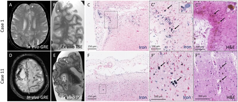
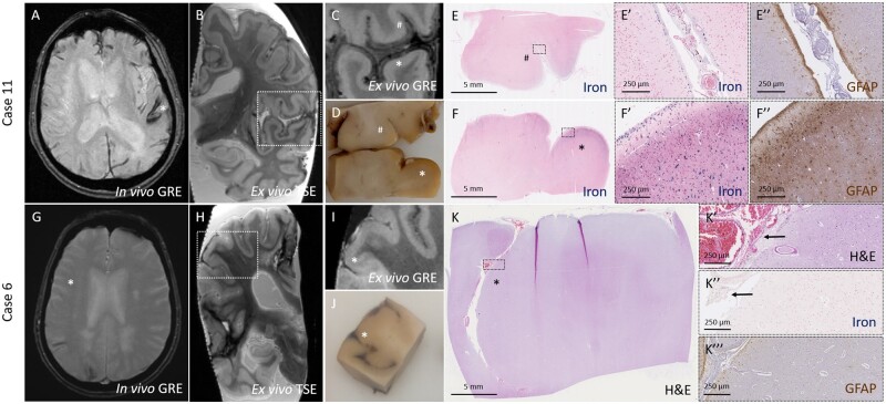
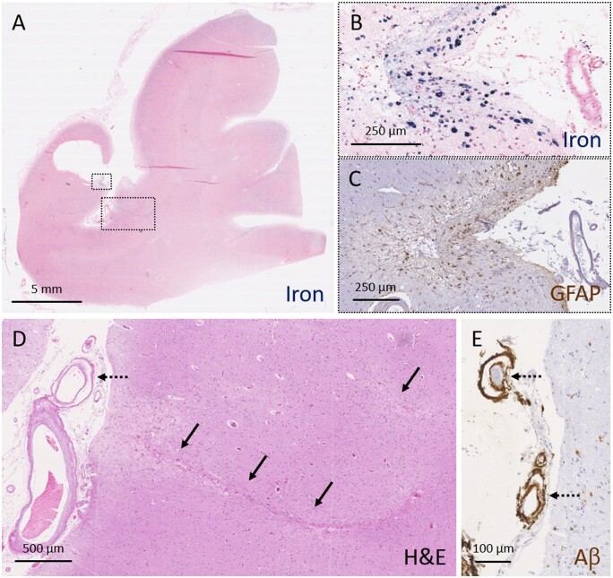
Cortical superficial siderosis is an established haemorrhagic neuroimaging marker of cerebral amyloid angiopathy. In fact, cortical superficial siderosis is emerging as a strong independent risk factor for future lobar intracerebral haemorrhage. However, the underlying neuropathological correlates and pathophysiological mechanisms of cortical superficial siderosis remain elusive. Here we use an in vivo MRI, ex vivo MRI, histopathology approach to assess the neuropathological correlates and vascular pathology underlying cortical superficial siderosis. Fourteen autopsy cases with cerebral amyloid angiopathy (mean age at death 73 years, nine males) and three controls (mean age at death 91 years, one male) were included in the study. Intact formalin-fixed cerebral hemispheres were scanned on a 3 T MRI scanner. Cortical superficial siderosis was assessed on ex vivo gradient echo and turbo spin echo MRI sequences and compared to findings on available in vivo MRI. Subsequently, 11 representative areas in four cases with available in vivo MRI scans were sampled for histopathological verification of MRI-defined cortical superficial siderosis. In addition, samples were taken from predefined standard areas of the brain, blinded to MRI findings. Serial sections were stained for haematoxylin and eosin and Perls' Prussian blue, and immunohistochemistry was performed against amyloid-β and GFAP. Cortical superficial siderosis was present on ex vivo MRI in 8/14 cases (57%) and 0/3 controls (P = 0.072). Histopathologically, cortical superficial siderosis corresponded to iron-positive haemosiderin deposits in the subarachnoid space and superficial cortical layers, indicative of chronic bleeding events originating from the leptomeningeal vessels. Increased severity of cortical superficial siderosis was associated with upregulation of reactive astrocytes. Next, cortical superficial siderosis was assessed on a total of 65 Perls'-stained sections from MRI-targeted and untargeted sampling combined in cerebral amyloid angiopathy cases. Moderate-to-severe cortical superficial siderosis was associated with concentric splitting of the vessel wall (an advanced form of cerebral amyloid angiopathy-related vascular damage) in leptomeningeal vessels (P < 0.0001), but reduced cerebral amyloid angiopathy severity in cortical vessels (P = 0.048). In terms of secondary tissue injury, moderate-to-severe cortical superficial siderosis was associated with the presence of microinfarcts (P = 0.025), though not microbleeds (P = 0.973). Collectively, these data suggest that cortical superficial siderosis on MRI corresponds to iron-positive deposits in the superficial cortical layers, representing the chronic manifestation of bleeding episodes from leptomeningeal vessels. Cortical superficial siderosis appears to be the result of predominantly advanced cerebral amyloid angiopathy of the leptomeningeal vessels and may trigger secondary ischaemic injury in affected areas.

Keywords: cerebral amyloid angiopathy; cortical superficial siderosis; magnetic resonance imaging; microbleeds; microinfarcts.


Figures

Figure 1
Figure 1
Histopathological verification of cSS observed on in vivo MRI in CAA cases.Top row: CSS (asterisk) was observed on an in vivo gradient echo MRI scan performed ∼7 years prior to death in an individual with a clinical diagnosis of probable CAA (Case 1) (A). On the ex vivo turbo spin echo MRI scan the same area was positive for cSS (B, asterisk). On the corresponding histopathological section, iron-positive deposits were observed in the subarachnoid space and superficial layers of the cortex (C). The insets reveal greater detail of the intracellular iron deposits (C′, arrows), corresponding to haemosiderin-containing macrophages on the adjacent haematoxylin and eosin-stained section (C′′, arrows). Bottom row: Disseminated cSS (asterisk) was observed on an in vivo gradient echo MRI scan performed ∼1.5 years prior to death in an individual with a clinical diagnosis of probable CAA (Case 11) (D). On the ex vivo turbo spin echo MRI scan the same area was positive for cSS (E, asterisk) (note the same enlarged perivascular space on the in vivo and ex vivo scan). On the corresponding histopathological section, iron-positive deposits were observed in the subarachnoid space and superficial layers of the cortex (F). The insets reveal greater detail of the intracellular iron deposits (F′, arrows), corresponding to haemosiderin-containing macrophages on the adjacent haematoxylin and eosin-stained section (F′′, arrows). GRE = gradient echo; H&E = haematoxylin and eosin; TSE = turbo spin echo.
Figure 2
Figure 2
Degree and pattern of cSS on MRI closely corresponds to degree and pattern of iron-positive deposits on histopathology.Top row: CSS (asterisk) was observed on an in vivo gradient echo MRI scan performed ∼1.5 years prior to death in an individual with a clinical diagnosis of probable CAA (Case 11) (A). On the ex vivo gradient echo scan varying degree of cSS can be observed (B), including subtle (number sign) and extensive (asterisk) hypointense signal (C). On gross examination of the formalin-fixed tissue, no visible alterations were observed in the area with subtle cSS (number sign), whereas orange discolouration was present in the superficial cortex in the area with extensive cSS (asterisk) (D). The degree of hypointense signal on MRI matched closely to the degree of iron-positive deposits on the corresponding histopathological sections (E and F). The insets reveal greater detail of the degree of iron deposits, which was subtle and restricted to the subarachnoid space and pial surface in E′ and extensive and involving the superficial and deeper layers of the cortex in F′. Many reactive astrocytes were found in the area with extensive iron deposits (F′′), but not in the area with subtle iron deposits (E′′). Bottom row: No evidence of cSS was observed on in vivo gradient echo MRI performed ∼1 year prior to death in an individual with a clinical diagnosis of probable CAA (Case 6) (G) in the area (asterisk) corresponding to an area with cSS on ex vivo turbo spin echo MRI (H). On the ex vivo gradient echo scan cSS is subtle and only observed in the subarachnoid space (asterisk) (I). On gross examination of the formalin-fixed tissue, fresh blood was observed in the subarachnoid space of the sulcus (asterisk) (J), corresponding to intact red blood cells on haematoxylin and eosin (K). The insets reveal greater detail of the red blood cells in K′ (arrows), which were only mildly positive for iron (K′′, arrows). No reactive astrocytes were found in this area (K′′′). GRE = gradient echo; H&E = haematoxylin and eosin; TSE = turbo spin echo.
Figure 3
Figure 3
Moderate-to-severe cSS is associated with advanced leptomeningeal CAA and secondary tissue injury. A representative example of severe cSS as assessed on a Perls’ Prussian blue-stained section (A). Higher magnification of the area outlined with the small box in A, reveals greater detail of severe cSS (B). An adjacent section stained for GFAP revealed many reactive astrocytes in the superficial cortical layers in this area (C). Two cortical microinfarcts (indicated with solid arrows) were observed in the neighbouring cortical area (D, area corresponds to larger box in A). Note the presence of vessel-within-vessel pathology (indicated with broken arrows) in combination with severe CAA of the leptomeningeal vessels (D and E). Aβ = amyloid-β; H&E = haematoxylin and eosin.

References

    1. Arvanitakis Z, Leurgans SE, Barnes LL, Bennett DA, Schneider JA. Microinfarct pathology, dementia, and cognitive systems. Stroke 2011; 42: 722–7. - PMC - PubMed
    1. Beitzke M, Enzinger C, Pichler A, Wünsch G, Fazekas F. Acute diffusion-weighted imaging lesions in cerebral amyloid angiopathy-related convexal subarachnoid hemorrhage. J Cereb Blood Flow Metab 2018; 38: 225–9. - PMC - PubMed
    1. Castellano JM, Kim J, Stewart FR, Jiang H, DeMattos RB, Patterson BW, et al. Human apoE isoforms differentially regulate brain amyloid-β peptide clearance. Sci Transl Med 2011; 3: 89ra57. - PMC - PubMed
    1. Charidimou A, Baron JC, Werring DJ. Cerebral amyloid angiopathy and transient focal neurological episodes. Cerebrovasc Dis 2013. b; 36: 245–6. - PubMed
    1. Charidimou A, Boulouis G, Greenberg SM, Viswanathan A. Cortical superficial siderosis and bleeding risk in cerebral amyloid angiopathy: a meta-analysis. Neurology 2019. a; 93: e2192–e202. - PMC - PubMed

Publication types

MeSH terms

Substances