Observational Study
. 2020 Sep 1;5(9):1000-1005.
doi: 10.1001/jamacardio.2020.1555.

Hybrid Positron Emission Tomography/Magnetic Resonance Imaging in Arrhythmic Mitral Valve Prolapse

Affiliations
Observational Study

Hybrid Positron Emission Tomography/Magnetic Resonance Imaging in Arrhythmic Mitral Valve Prolapse

Marc A Miller et al. JAMA Cardiol. .

Abstract

Importance: Myocardial replacement fibrosis has been reported to occur in one-third of patients with mitral valve prolapse (MVP) and significant mitral regurgitation (MR). However, it remains unknown whether there are detectable changes in myocardial metabolism suggestive of inflammation or ischemia that accompany the development of fibrosis.

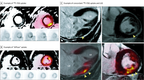
Objectives: To characterize the burden and distribution of fluorine 18-labeled (18F) fluorodeoxyglucose (FDG) uptake and late gadolinium enhancement (LGE) in patients with degenerative MVP and ventricular ectopy.

Design, setting, and participants: Prospective observational study of 20 patients with MVP and significant primary degenerative MR who were referred for mitral valve repair and underwent hybrid positron emission tomography/magnetic resonance imaging (PET/MRI). Ventricular arrhythmias were categorized as either complex (n = 12) or minor (n = 8). Coregistered hybrid 18F FDG-PET and MRI LGE images were assessed and categorized. Recruitment occurred in the new patient clinic of a mitral valve repair reference center. This study was conducted from January 11, 2018, to June 26, 2019.

Exposures: Simultaneous cardiac 18F FDG-PET and MRI with LGE imaging on a hybrid PET/MRI system and ambulatory rhythm monitoring.

Main outcomes and measures: Patients were categorized by the presence and pattern of FDG uptake and LGE, the severity of ventricular arrhythmias, and the indication for mitral valve surgery.

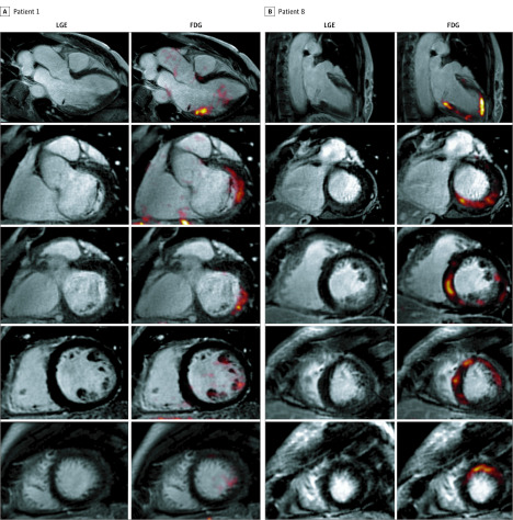
Results: In the cohort of 20 patients, the median age was 59.5 years (interquartile range, 52.5-63.2 years). Focal, or focal-on-diffuse uptake, of 18F-FDG (PET positive) was detected in 17 of 20 patients (85%). The FDG uptake coexisted with areas of LGE (PET/MRI positive) in 14 patients (70%). Of the 5 asymptomatic patients with normal ventricular indices and absence of any surgical indications, all were PET/MRI positive.

Conclusions and relevance: In this pilot study, we demonstrate a novel association between degenerative MVP and FDG uptake, a surrogate for myocardial inflammation and/or ischemia. Such evidence of myocardial injury, even in asymptomatic patients, suggests an ongoing subclinical disease process. These findings warrant further investigation into whether imaging for myocardial inflammation, ischemia, and scar has a role in arrhythmic risk stratification and whether it provides incremental prognostic value in patients with chronic severe mitral regurgitation undergoing active surveillance.


Conflict of interest statement

Conflict of Interest Disclosures: Dr Miller has served as a consultant to Boston Scientific. Dr Dukkipati has received research grant support from Biosense Webster. Dr Reddy has received research grants from and served as a consultant for Biosense Webster, Boston Scientific, Biotronik, and Abbott. Dr Adams declares that The Icahn School of Medicine at Mount Sinai receives royalty payments from Edwards Lifesciences for intellectual property related to the development of 2 mitral valve repair rings, and from Medtronic for intellectual property related to the development of two tricuspid valve repair rings. Dr Adams is the National co-Principal Investigator for the Medtronic CoreValve, NeoChord System, Medtronic Apollo, and Triluminate-II US Pivotal Trials, respectively. Icahn School of Medicine receives royalties for Dr Adams’ intellectual property from Edwards Lifesciences and Medtronic, related to valve repair rings. Dr Pandis reported nonfinancial support from TomTec Imaging Systems GmbH during the conduct of the study. Dr Robson reported grants from the National Institutes of Health during the conduct of the study. Dr Reddy is a consultant for Abbott, Axon, Biosense-Webster, Biotronik, Boston Scientific, Cardiofocus, Cardionomic, CardioNXT/AFTx, EBR, Impulse Dynamics, Medtronic, Philips, Stimda, and Thermedical; is a consultant and holds equity in Ablacon, Acutus Medical, Affera, Apama Medical, Aquaheart, Autonomix, Backbeat, BioSig, Circa Scientific, Corvia Medical, East End Medical, EPD, Epix Therapeutics, EpiEP, Eximo, Farapulse, Fire1, Javelin, Keystone Heart, LuxCath, Medlumics, Middlepeak, Nuvera, and Valcare; and holds equity in Manual Surgical Sciences, Newpace, Surecor, and Vizara. No other disclosures were reported.

Figures

Figure 1.
Figure 1.. Hybrid Positron Emission Tomography/Magnetic Resonance Imaging (PET/MRI) of 3 Different Patients
A, Example of fluorine 18–labeled fluorodeoxyglucose (18F-FDG) uptake that colocalizes with regions of late gadolinium enhancement (LGE) (PET+/MRI+) in a focal-on-diffuse pattern (maximum standardized uptake value [SUVmax], 3.5; maximum tissue-to-background ratio [TBRmax], 1.8; target-normal-myocardium ratio [TNMR], 1.8). These images were obtained from a man in his early 60s with asymptomatic degenerative mitral valve prolapse, significant mitral regurgitation (3+), and complex ventricular ectopy (nonsustained ventricular tachycardia and pleomorphic premature ventricular contractions) but without any objective surgical indications. The patient is being followed up with active surveillance. B, Example of diffuse uptake (SUVmax, 9.6; TBRmax, 4.2; TNMR, 1.3), which was interpreted as physiological or nonspecific uptake. C, Example of concordant 18F-FDG uptake and LGE in an asymptomatic patient with chronic severe degenerative mitral regurgitation and absent left ventricular remodeling (left ventricular ejection fraction, 60% and end-systolic dimension, 38 mm). The top 2 images are LGE imaging sequences and the bottom 2 images are 18F-FDG imaging. The yellow arrowheads indicate areas of either LGE or FDG uptake, respectively. LA indicates left atrial; LV, left ventricular.
Figure 2.
Figure 2.. Representative Images of 2 Different Patterns of Fluorodeoxyglucose (FDG) Uptake on Hybrid Positron Emission Tomography/Magnetic Resonance Imaging (PET/MRI) From 2 Different Patients
A, Focal FDG uptake and late gadolinium enhancement (LGE) in the inferolateral and basal inferior segments of the left ventricle in a man in his mid 50s with severe mitral regurgitation and posterior-leaflet prolapse who was asymptomatic with normal ventricular indices (left ventricular ejection fraction, 65%; left ventricular end-systolic dimension, 34 mm) and no objective indications for surgery. He developed episodes of nonsustained ventricular tachycardia (>15 beats) at peak exercise. B, Multifocal pattern of FDG uptake and LGE, in this case involving the septum, inferior, and lateral walls, in a woman in her mid 60s with symptomatic severe mitral regurgitation and bileaflet prolapse. She had infrequent premature ventricular contractions (burden = 0.5%), but they were pleomorphic (at least 3 different morphologies).

Comment in

References

    1. Hourdain J, Clavel MA, Deharo JC, et al. . Common phenotype in patients with mitral valve prolapse who experienced sudden cardiac death. Circulation. 2018;138(10):1067-1069. doi:10.1161/CIRCULATIONAHA.118.033488 - DOI - PubMed
    1. Kitkungvan D, Nabi F, Kim RJ, et al. . Myocardial fibrosis in patients with primary mitral regurgitation with and without prolapse. J Am Coll Cardiol. 2018;72(8):823-834. doi:10.1016/j.jacc.2018.06.048 - DOI - PubMed
    1. Travers JG, Kamal FA, Robbins J, Yutzey KE, Blaxall BC. Cardiac fibrosis: the fibroblast awakens. Circ Res. 2016;118(6):1021-1040. doi:10.1161/CIRCRESAHA.115.306565 - DOI - PMC - PubMed
    1. Muser D, Castro SA, Alavi A, Santangeli P. Potential role of PET in assessing ventricular arrhythmias. PET Clin. 2019;14(2):281-291. doi:10.1016/j.cpet.2018.12.009 - DOI - PubMed
    1. Tung R, Bauer B, Schelbert H, et al. . Incidence of abnormal positron emission tomography in patients with unexplained cardiomyopathy and ventricular arrhythmias: the potential role of occult inflammation in arrhythmogenesis. Heart Rhythm. 2015;12(12):2488-2498. doi:10.1016/j.hrthm.2015.08.014 - DOI - PMC - PubMed

Publication types

Substances