Review
. 2020 Sep 15:6:149-169.
doi: 10.1146/annurev-vision-121219-081730.

Microglia Activation and Inflammation During the Death of Mammalian Photoreceptors

Affiliations
Review

Microglia Activation and Inflammation During the Death of Mammalian Photoreceptors

Sarah J Karlen et al. Annu Rev Vis Sci. .

Abstract

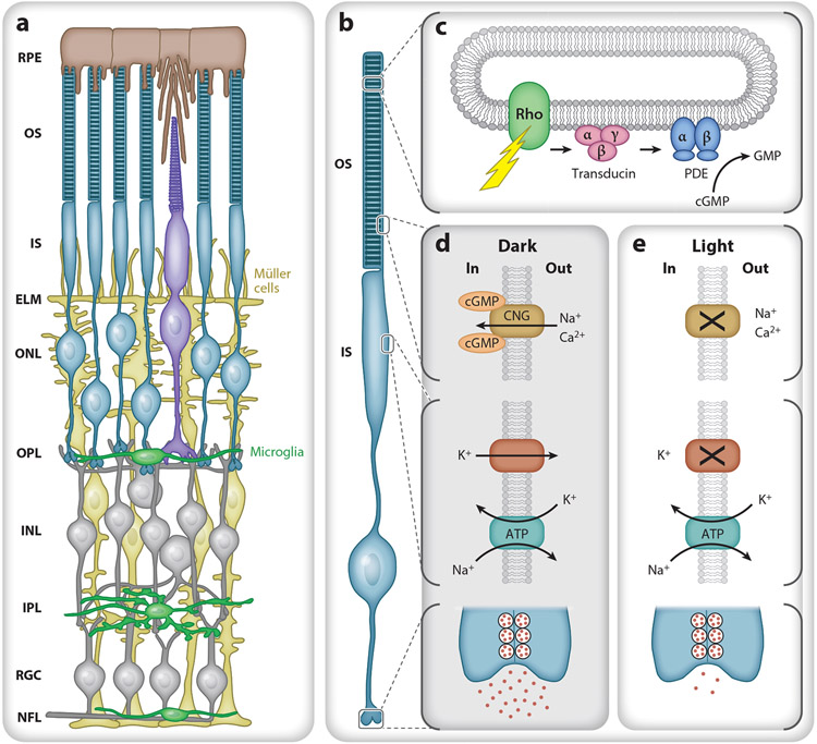
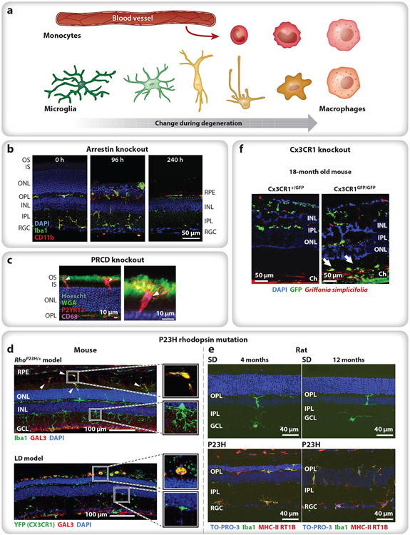
Photoreceptors are highly specialized sensory neurons with unique metabolic and physiological requirements. These requirements are partially met by Müller glia and cells of the retinal pigment epithelium (RPE), which provide essential metabolites, phagocytose waste, and control the composition of the surrounding microenvironment. A third vital supporting cell type, the retinal microglia, can provide photoreceptors with neurotrophic support or exacerbate neuroinflammation and hasten neuronal cell death. Understanding the physiological requirements for photoreceptor homeostasis and the factors that drive microglia to best promote photoreceptor survival has important implications for the treatment and prevention of blinding degenerative diseases like retinitis pigmentosa and age-related macular degeneration.

Keywords: degeneration; macrophage; monocyte; neuroinflammation; retina; rod.


Figures

Figure 1
Figure 1
Retinal morphology and basic photoreceptor physiology. (a) Schematic of the retina from the outer (top) to inner (bottom) in cross section. The retinal pigment epithelium (RPE) (brown) lies between the vascular choriocapillaris and photoreceptor outer segments (OS). Müller cells (yellow) span the retina from the nerve fiber layer (NFL) to the photoreceptor inner segments (IS) and form the external limiting membrane (ELM) that helps to establish the subretinal space as a distinct microenvironment. Normally, microglia (green) reside in the outer plexiform layer (OPL), inner plexiform layer (IPL), retinal ganglion cell layer (RGC), and NFL. (b) Rod photoreceptor compartment. From top to bottom: OS containing stacks of discs, IS with metabolic and biosynthetic machinery, cell body, and synaptic terminal. (c) A photon causes rhodopsin to change conformation, activating transducin (Gαtβ1γ1). Each GTP-bound Gαt then binds and activates cGMP phosphodiesterase 6 (PDE), allowing it to hydrolyze cGMP, decreasing intracellular cGMP levels. (d) In the dark, cGMP opens a cyclic nucleotide gated (CNG) channel in the OS plasma membrane, allowing cationic influx (Na+, Ca2+) that is balanced by an efflux of cations, mostly potassium (K+), from the IS. The IS Na+/K+ transporter uses ATP to maintain the electrochemical gradient and depolarized membrane potential, leading to the continual release of glutamate. (e) In light, cGMP levels fall, and the CNG channels close, reducing the influx of Na+ and Ca2+ and causing the cell to hyperpolarize and reduce glutamate release. Additional abbreviations: INL, inner nuclear layer; ONL, outer nuclear layer.
Figure 2
Figure 2
Variations in the response of microglia and infiltrating monocytes during photoreceptor degeneration. (a) Microglia are typically ramified in healthy tissue, with small cell bodies and numerous thin processes (green). Activated microglia lose their branched morphology and migrate (yellow) to the afflicted region of damage, often transitioning into a macrophage phenotype (tangerine). Infiltrating bone marrow–derived monocytes (red) rapidly differentiate into macrophages (pink), becoming difficult to distinguish from resident immune cells. (b) In Arrestin-1 knockout mice, light onset causes microglia and monocytes (green, green + red) to migrate to the ONL and phagocytose photoreceptor cells bodies (96 h), eliminating the ONL within a week and leaving a handful of subretinal macrophages between the RPE and the ELM (240 h). Iba1 (green) labels microglia and macrophages; CD11b (red) labels microglia and leukocytes (e.g., monocytes); and nuclei are stained with DAPI (blue). Panel adapted from Karlen et al. (2018) (CC BY-SA 4.0). (c) In PRCD knockout mice, microglia (red) primarily target the inner (unlabeled)–outer (green) segment junction, with their processes extending toward the cilium (green). P2YR12 (red) labels microglia; CD68 (purple) labels lysosomes; WGA (green) labels the OS; and nuclei are stained with Hoescht (blue). Panel adapted with permission from Spencer et al. (2019). (d) In P23H rhodopsin mice (top) and LD mice (bottom), microglia with upregulated Gal3 (green + red) accumulate in the subretinal space, whereas infiltrating monocytes (green) remain in the retina. Iba1 (top, green) labels microglia and macrophages; YFP (Cx3CR1) (bottom, green) labels Cx3CR1+ cells (e.g., microglia, macrophages); GAL3 (red) labels subretinal macrophages; and nuclei are stained with DAPI (blue). Panel adapted with permission from O’Koren et al. (2019). (e) In P23H rhodopsin rats, microglia (green, green + red) remain in the subretinal space throughout adulthood, indicating chronic neuroinflammation after photoreceptor degeneration has been completed; compare 4 months (left) to 12 months (right) in controls (SD, top) and P23H rhodopsin mutants (bottom). Iba1 (green) labels microglia and macrophages; MHC-II RT1B (red) label macrophages; and nuclei are stained with TO-PRO-3 iodide (blue). Panel adapted from Noailles et al. (2016) (CC BY-SA 4.0). (f) In aged, 18-month Cx3CR1 knockout mice exposed to normal 12 h on/off light levels, microglia (green) appear in the inner retina (both conditions), and additional microglia accumulate in the subretinal space of the knockout (Cx3CR1GFP/GFP) (arrows). GFP (green) indicates Cx3CR1-positive cells; Griffonia simplicifolia-positive (red) labels vascular endothelial cells; and nuclei are stained with DAPI (blue). Panel adapted with permission from Combadière et al. (2007). Abbreviations: ELM, external limiting membrane; GFP, green fluorescent protein; INL, inner nuclear layer; IPL, inner plexiform layer; IS, inner segment; LD, light damage; ONL, outer nuclear layer; OPL, outer plexiform layer; OS, outer segment; PRCD, progressive rod–cone degeneration; RGC, reginal ganglion cell; RPE, retinal pigment epithelium; SD, Sprague-Dawley control rats; WGA, wheat germ agglutinin.

Similar articles

Cited by

References

    1. Ajami B, Bennett JL, Krieger C, McNagny KM, Rossi FM. 2011. Infiltrating monocytes trigger EAE progression, but do not contribute to the resident microglia pool. Nat. Neurosci, 14:1142–49 - PubMed
    1. Appelbaum T, Santana E, Aguirre GD. 2017. Strong upregulation of inflammatory genes accompanies photoreceptor demise in canine models of retinal degeneration. PLOS ONE 12:e0177224. - PMC - PubMed
    1. Aredo B, Zhang K, Chen X, Wang CX-Z, Li T, Ufret-Vincenty RL. 2015. Differences in the distribution, phenotype and gene expression of subretinal microglia/macrophages in C57BL/6N (Crb1 rd8/rd8) versus C57BL6/J (Crb1 wt/wt) mice. J. Neuroinflamm 12:6 - PMC - PubMed
    1. AREDS2 Res. Group, Chew EY, Clemons TE, Sangiovanni JP, Danis RP, et al. 2014. Secondary analyses of the effects of lutein/zeaxanthin on age-related macular degeneration progression: AREDS2 report No. 3. JAMA Ophthalmol.132:142–49 - PMC - PubMed
    1. Arshavsky VY, Burns ME. 2014. Current understanding of signal amplification in phototransduction. Cell Logist. 4:e29390. - PMC - PubMed

Publication types