. 2020 Nov 10;194(5):519-531.
doi: 10.1667/RR15480.1.

Combined EGFR1 and PARP1 Inhibition Enhances the Effect of Radiation in Head and Neck Squamous Cell Carcinoma Models

Affiliations

Combined EGFR1 and PARP1 Inhibition Enhances the Effect of Radiation in Head and Neck Squamous Cell Carcinoma Models

Barbara A Frederick et al. Radiat Res. .

Abstract

Head and neck squamous cell carcinoma (HNSCC) is a challenging cancer with little change in five-year overall survival rate of 50-60% over the last two decades. Radiation with or without platinum-based drugs remains the standard of care despite limited benefit and high toxicity. HNSCCs often overexpress epidermal growth factor receptor (EGFR) and inhibition of EGFR signaling enhances radiation sensitivity by interfering with repair of radiation-induced DNA breaks. Poly (adenosine diphosphate-ribose) polymerase-1 (PARP1) also participates in DNA damage repair, but its inhibition provides benefit in cancers that lack DNA repair by homologous recombination (HR) such as BRCA-mutant breast cancer. HNSCCs in contrast are typically BRCA wild-type and proficient in HR repair, making it challenging to apply anti-PARP1 therapy in this model. A recently published study showed that a combination of EGFR and PARP1 inhibition induced more DNA damage and greater growth control than each single agent in HNSCC cells. This led us to hypothesize that a combination of EGFR and PARP1 inhibition would enhance the efficacy of radiation to a greater extent than each single agent, providing a rationale for paradigm-shifting combinatorial approaches to improve the standard of care in HNSCC. Here, we report a proof-of-concept study using Detroit562 HNSCC cells, which are proficient for DNA repair by both HR and non-homologous end joining (NHEJ) mechanisms. We tested the effect of adding cetuximab and/or olaparib (inhibitors of EGFR and PARP1, respectively) to radiation and compared it to that of cisplatin and radiation combination, which is the standard of care. Our results demonstrate that the combination of cetuximab and olaparib with radiation was superior to the combination of any single drug with radiation in terms of induction of unrepaired DNA damage, induction of senescence, apoptosis and clonogenic death, and tumor growth control in mouse xenografts. Combined with our recently published phase I safety data on cetuximab/olaparib/radiation triple combination, the data reported here demonstrate a potential for combining biologically-based therapies that might optimize radiosensitization in HNSCC.


Figures

FIG. 1.
FIG. 1.
PARP1 and EGFR expression in HNSCC cell lines and clonogenic death in response to their inhibition. Panel A: Expression of EGFR and PARP1 mRNA in a panel of HNSCC cell lines. RMA = robust multi-array average. Panel B: Immunoblot showing EGFR and PARP1 expression of Det562 and FaDu cells. Panels C and D: Clonogenic survival of Det562 and FaDu cells, respectively, after irradiation with and without cetuximab (50 nM), olaparib (1 μM) or cisplatin (1 μM) treatment. Cell nuclei were fluorescently labeled, counted and plated. After drug treatment and irradiation, plates were placed in a live cell imager and colony formation captured every 24 h for 10 days. Data are representative of three independent replicate wells, error bars are ±S.E. Numbers in parenthesis are calculated α/β ratios.
FIG. 2.
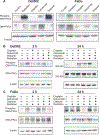
FIG. 2.
Combined PARP1 and EGFR1 inhibition corresponds to increased DNA damage. Immunofluorescent (panels A and C) and Western blots (panels B and D) for detection of γ-H2AX at 2 and 24 h postirradiation with or without cetuximab (50 nM), olaparib (1 μM), irradiation (2 Gy) and cisplatin (1 μM). Panels A and B show Det562 cells and panels C and D show FaDu cells. Panel E: The effect of single treatments at the 2 h time point. Beta-actin was used as a loading standard.
FIG. 3.
FIG. 3.
Combined PARP1 and EGFR1 inhibition downregulates DNA-PKcs autophosphorylation. Panel A: Western blots of phospho(Thr2609)- and total DNA-PKcs 2 h after single treatments: cetuximab (50 nM), olaparib (1 μM), irradiation (2 Gy) and cisplatin (1 μM) at 2 and 24 h postirradiation. Beta-actin was used as a loading standard (same images as in Fig. 2E are shown because experimental samples were the same). Panels B and C: Western blots of phospho(Thr2609)- and total DNA-PKcs in Det562 (panel B) and FaDu (panel C) cells with or without cetuximab (50 nM), olaparib (1 μM), irradiation (2 Gy) and cisplatin (1 μM) at 2 and 24 h postirradiation. Beta-actin was used as a loading standard.
FIG. 4.
FIG. 4.
Combined PARP1 and EGFR1 inhibition inhibits NHEJ. Panels A and B: Homologous recombination assay based on reconstitution of luciferase expression. Plasmid no. 1 is pGL4.14[luc2/hygro] with SV40 promoter inserted and part of the luciferase gene removed. Plasmid no. 2 is pGL4.20[luc2/puro] with part of the luciferase gene removed. The new plasmids share a region of 655 bp of the luc2 gene such that when they are co-transfected into recipient cells that are homologous recombination competent, luciferase activity is reconstituted. Panel B: Detroit562 cells were transiently transfected with pGL4.20[luc2/puro]-luc (plasmid no. 2), pGL4.14[luc2/hygro]-luc (plasmid no. 1 without the SV40 promoter), pGL4.14[luc2/hygro]-luc + pGL4.20[luc2/puro]-luc (plasmid no. 2), pGL4.14SV40[luc2/hygro]-luc (plasmid no. 1), pGL4.14SV40[luc2/hygro]-luc + pGL4.20[luc2/puro]-luc (experimental condition) or pGL4.14SV40[luc2/hygro] (positive control), then assayed for luciferase expression (Promega Luciferase assay system) 24 h after transfection. Error bars are ±S.E. of independent replicates. Panel C: NHEJ reporter based on ISceI-cleavage of an out-of-frame start codon. The sequence as cloned into pGL4.20 (luc2) between the SV40 promoter and Luc2. Panel D: Luciferase activity of cells stably transfected in Det562 cells with pGL4.20SV40SceCas alone or with pLXSN-IsceI. In the presence of both plasmids, ISceI cleaves the sites and NHEJ produces luciferase activity above the background of pGL4.20SV40SceCas alone. pGL4.20SV40 is the positive control with constitutive luciferase expression. Panel E: Luciferase activity 24 h after transfection with pLXSN-ISceI. Cells were counted by live cell imaging before luciferase assay and luciferase expression was normalized to cell counts. Baseline expression (no ISceI) was subtracted from pLXSN-ISceI transfected cells. Differences in luciferase expression from independent triplicates were determined using Tukey’s range test of all means. *P < 0.05, **P < 0.001.
FIG. 5.
FIG. 5.
Combined PARP1 and EGFR1 inhibition downregulates ERK1/2 phosphorylation and promotes PARP1 cleavage and senescence. Panel A: Det562 cells treated with or without cetuximab (50 nM), olaparib (1 μM), radiation (2 Gy) and cisplatin (1 μM) and harvested 24 or 48 h postirradiation were immunoblotted for total and phospho-ERK1/2 (Thr202/Tyr204), and PARP1. Beta-actin was used as a loading standard. Panel B: Western blot of p21 at 24 h postirradiation. The effect of single treatments at the 2 h time point are shown in panel C. Beta-actin was used as a loading standard. (Same images as in Fig. 2E are shown because experimental samples were the same). Panel D: Combined cetuximab and olaparib induces senescence after irradiation. Cells were assayed for β-galactosidase activity by cleavage of fluorescein from fluorescein di-β-D-galactopyranoside 72 h postirradiation. Fluorescein concentrations in cell supernates were determined using a fluorescein standard and fluorescent plate reader at 485 mm excitation and 525 mm emission. To calculate average fluorescein per cell, the total number of cells were counted in independent triplicate wells treated in parallel to cells used for the senescence assay. Data are means of three independent samples ±S.E.
FIG. 6.
FIG. 6.
The combination of cetuximab and olaparib with radiation significantly delays tumor growth, and induces p21 expression, accelerated senescence and increased apoptosis. Panel A: Detroit562 tumor growth after treatment. Treatments were vehicle, radiation only (2 Gy), and radiation with cisplatin (1 mg/kg), olaparib (25 mg/kg), cetuximab (1 mg/kg) or cetuximab/olaparib (1 mg/kg and 25 mg/kg, respectively). Arrows indicate treatment time points and data points are mean tumor volume (n = 10) ± S.E. Curves are shown until any animal within a group reached maximum allowable tumor volume. Panel B: Number of days that tumors in each group took to reach 1,000 mm3 in tumor volume (DTV4; mean ± S.E.). Panel C: Tumors were harvested 96 h after single-fraction irradiation (2 Gy), or cisplatin (1 mg/kg), olaparib (25 mg/kg), cetuximab (1 mg/kg) or olaparib/cetuximab with irradiation (2 Gy). Tumors were divided and fixed either in PBS-buffered formalin or frozen in OCT and stored at −80°C. p21 expression and cleaved caspase-3 were determined by IHC of sections of formalin-fixed tissue, and sections from frozen tissue were stained for β-galactosidase activity to detect senescence.

References

    1. Loo SW, Geropantas K, Roques TW. DeCIDE and PARADIGM: Nails in the coffin of induction chemotherapy in head and neck squamous cell carcinoma? Clin Transl Oncol 2013;15:248–51. - PubMed
    1. Jun HJ, Ahn MJ, Kim HS, Yi SY, Han J, Lee SK, et al. ERCC1 expression as a predictive marker of squamous cell carcinoma of the head and neck treated with cisplatin-based concurrent chemoradiation. Br J Cancer 2008; 99:167–72. - PMC - PubMed
    1. Gu F, Ma Y, Zhang Z, Zhao J, Kobayashi H, Zhang L, et al. Expression of Stat3 and Notch1 is associated with cisplatin resistance in head and neck squamous cell carcinoma. Oncol Rep 2010; 23:671–6. - PubMed
    1. Tonigold M, Rossmann A, Meinold M, Bette M, Marken M, Henkenius K, et al. A cisplatin-resistant head and neck cancer cell line with cytoplasmic p53(mut) exhibits ATP-binding cassette transporter upregulation and high glutathione levels. J Cancer Res Clin Oncol 2014; 140:1689–704. - PMC - PubMed
    1. Curran D, Giralt J, Harari PM, Ang KK, Cohen RB, Kies MS, et al. Quality of life in head and neck cancer patients after treatment with high-dose radiotherapy alone or in combination with cetuximab. J Clin Oncol 2007; 25:2191–7. - PubMed

Publication types

MeSH terms