

Review
doi: 10.1056/NEJMra1911109.
Tolerance in the Age of Immunotherapy
Affiliations
- PMID: 32937048
- PMCID: PMC7534289
- DOI: 10.1056/NEJMra1911109
Item in Clipboard
Review
Tolerance in the Age of Immunotherapy
N Engl J Med.
.
No abstract available
Conflict of interest statement
Disclosure forms provided by the authors are available with the full text of this article at
Figures
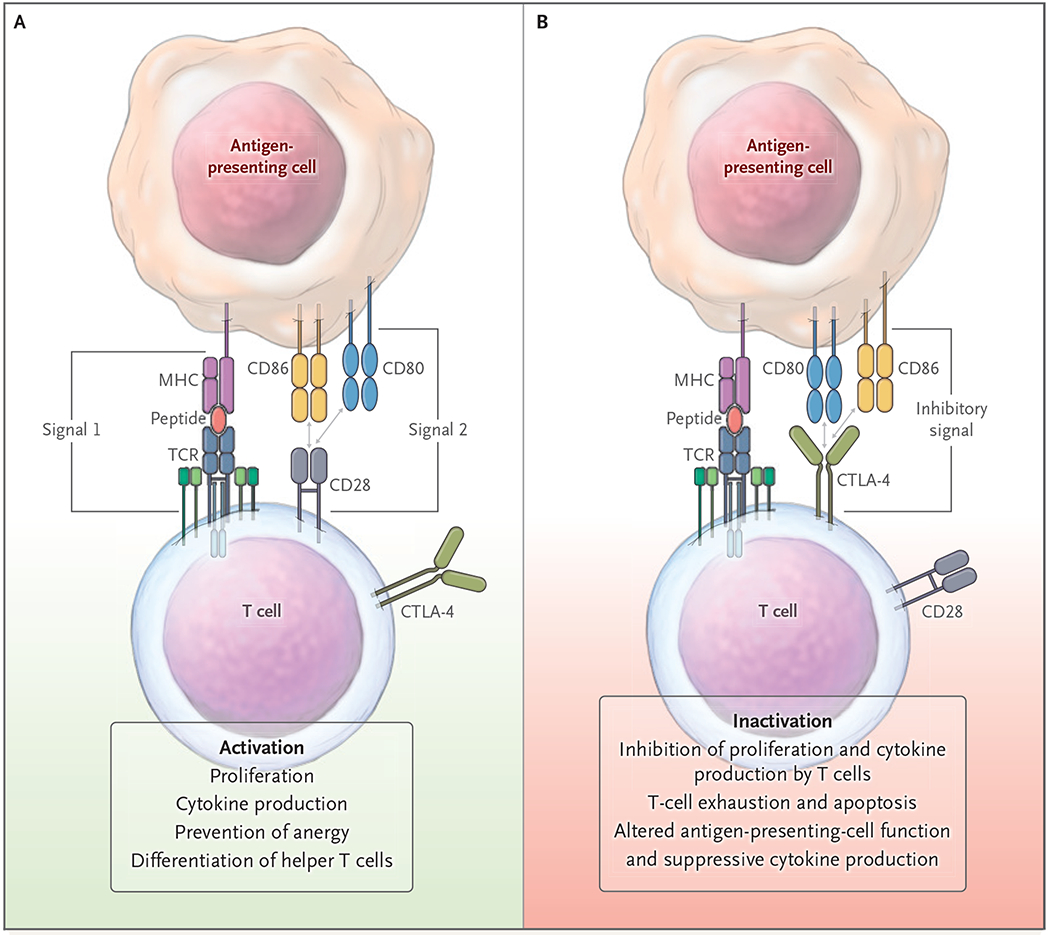
Initiation of a productive T-cell response involves integration of a primary signal delivered through the T-cell receptor (TCR) and major histocompatibility complex (MHC)–peptide, followed by a second signal delivered through the CD28–CD80 or CD28–CD86 pathway (Panel A). After initiation of T-cell activation, other inhibitory checkpoint interactions can shut down T-cell activity (Panel B). Pathways that may be affected as a consequence of both positive and negative second signals are listed at the bottom of the figure. CTLA-4 denotes cytotoxic T-lymphocyte–associated protein 4.

In addition to the two-signal models of costimulatory and checkpoint pathways, additional stimulatory and inhibitory pathways (indicated by plus and minus signs, respectively) influence the immune response, including molecules of the tumor necrosis factor (TNF)–related family, other members of the CD28 family, adhesion molecules, and T-cell immunoglobulin and mucin (TIM) molecules. The various stimulatory and inhibitory pathways can influence and be influenced by cytokines. Pep denotes peptide, and TGF-β transforming growth factor β.

The majority of cells interacting with autoimmune regulator (AIRE)–expressing medullary thymic epithelial cells (mTECs) during thymic development undergo negative selection and die (Panel A). A subset of high-affinity, self-reactive CD4+ T cells interact with the mTECs, leading to the development of regulatory T cells (Tregs). The remaining mature naive T cells migrate into the immune periphery, where they have either a pathogenic role in mediating immunity (Panel B, left) or a protective role as peripherally derived Tregs (through interaction with tolerogenic antigen-presenting cells and cytokines) that control potential autoreactive responses (Panel B, right). Additional cell types, such as extrathymic AIRE–expressing cells (eTACs), can also modify potentially autoreactive T cells.
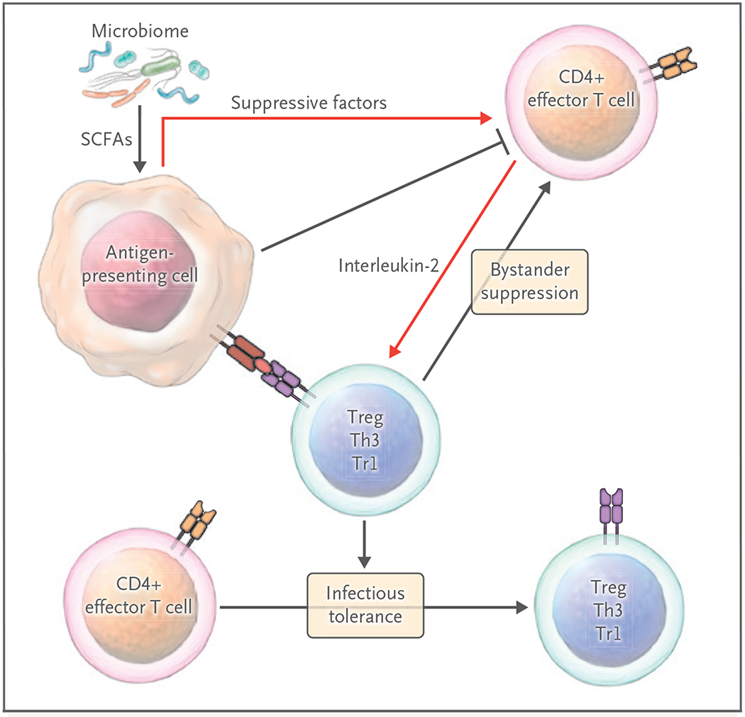
Tregs and other suppressive cells circulate and reside in lymphoid and somatic tissues to control unwanted autoimmune and inflammatory responses. Multiple cell–cell contacts, as well as soluble molecules (including the roduction of metabolites by microbiota), are generated by Tregs or antigen-presenting cells after Treg interactions to control immunity. Tregs can also act through bystander suppression, leading to dominant local immunosuppression and tolerance induction. IDO denotes indoleamine 2,3-dioxygenase, SCFA short-chain fatty acid, Th3 type 3 helper T cell, and Tr1 type 1 regulatory T cell.
References
-
- Billingham RE, Brent L, Medawar PB. Actively acquired tolerance of foreign cells. Nature 1953; 172: 603–6. - PubMed
-
- Cooper MD, Peterson RD, Good RA. Delineation of the thymic and bursal lymphoid systems in the chicken. Nature 1965; 205: 143–6. - PubMed
-
- Miller JFAP. The golden anniversary of the thymus. Nat Rev Immunol 2011; 11: 489–95. - PubMed
Publication types
MeSH terms
Grants and funding
LinkOut - more resources
Full Text Sources
Medical