. 2020 Sep 15;32(11):108153.
doi: 10.1016/j.celrep.2020.108153.

Innate Lymphoid Cell Activation and Sustained Depletion in Blood and Tissue of Children Infected with HIV from Birth Despite Antiretroviral Therapy

Affiliations

Innate Lymphoid Cell Activation and Sustained Depletion in Blood and Tissue of Children Infected with HIV from Birth Despite Antiretroviral Therapy

Alveera Singh et al. Cell Rep. .

Abstract

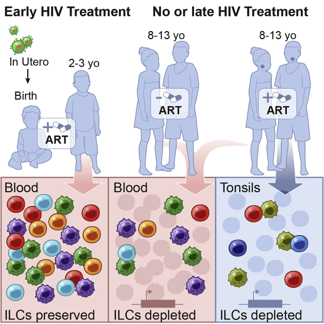
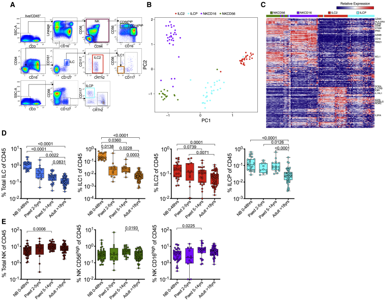
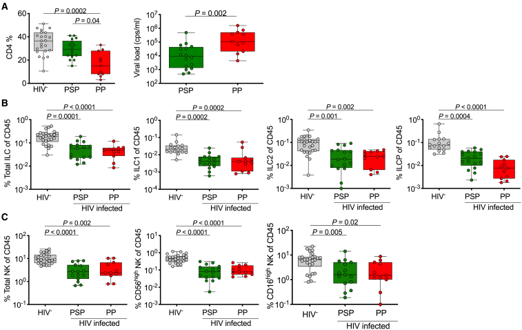
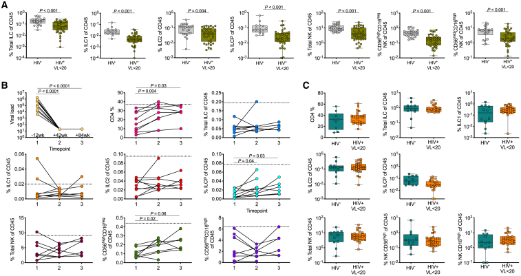
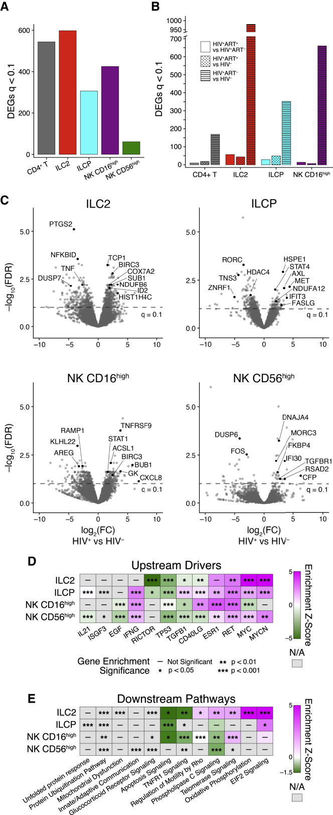
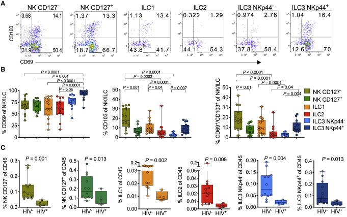
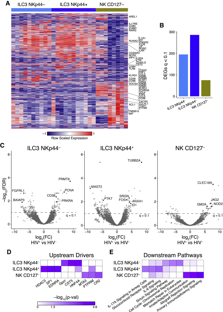
Innate lymphoid cells (ILCs) are important for response to infection and for immune development in early life. HIV infection in adults depletes circulating ILCs, but the impact on children infected from birth remains unknown. We study vertically HIV-infected children from birth to adulthood and find severe and persistent depletion of all circulating ILCs that, unlike CD4+ T cells, are not restored by long-term antiretroviral therapy unless initiated at birth. Remaining ILCs upregulate genes associated with cellular activation and metabolic perturbation. Unlike HIV-infected adults, ILCs are also profoundly depleted in tonsils of vertically infected children. Transcriptional profiling of remaining ILCs reveals ongoing cell-type-specific activity despite antiretroviral therapy. Collectively, these data suggest an important and ongoing role for ILCs in lymphoid tissue of HIV-infected children from birth, where persistent depletion and sustained transcriptional activity are likely to have long-term immune consequences that merit further investigation.

Keywords: HIV; ILCs; NK cells; pediatric infection; tonsil; vertical transmission.


Conflict of interest statement

Declaration of Interests The authors declare no competing interests.

Figures

None
Graphical abstract
Figure 1
Figure 1
Circulating ILC Populations Decrease during the Course of Immune Maturation (A) Gating strategy including lineage markers (CD3, CD4, CD11c, CD14, CD19, CD34, CD303, TCRγδ, TCRαβ) to identify two dominant NK populations defined by CD56high(green) and CD16high (purple) and three ILC subsets: ILC1 (orange), ILC2 (red), and ILCP (light blue). (B) Principal component analysis (PCA) and heatmap shown for each replicate for each participant (see Table S1). (C) DEGs among ILC2, ILCP, CD56high (NKCD56), and CD16high (NKCD16) NK cell populations from four HIV-negative and six HIV-positive pediatric subjects. (D) Frequencies of total helper ILC subsets as defined in (A), comparing HIV-negative newborn (NB) (n = 39), pediatric (2–5 years, n = 12), pediatric (>5 years, n = 25), and adult (n = 62) individuals expressed as percentage of total CD45+ lymphocytes. (E) As in (C) but showing frequencies of total NK and subset-specific differences between pediatric and adult subjects. p values by Dunn’s multiple comparisons test.
Figure 2
Figure 2
Depletion of Peripheral Helper ILC and Cytotoxic NK Cell Subsets in Treatment-Naive HIV-Infected Pediatric Subjects (A) CD4 percentage of CD45+ lymphocytes (left) and plasma viral load (right) of pediatric HIV-uninfected (HIV) subjects (n = 25), pediatric slow progressors (PSPs) (n = 15), and pediatric progressors (PPs) (n = 11) (see Table 1). (B) As in (A) but showing total ILC (left), ILC1 (center left), ILC2 (center right), and ILCP (right) subset levels as percentage of CD45+ lymphocytes. Gating as in Figure 1A. (C) As in (B) but showing total NK cells (left) and NK subsets (CD56high, middle; CD16high, right) as percentage of CD45+ lymphocytes. p values by Dunn’s multiple comparisons test.
Figure 3
Figure 3
Sustained Depletion of All Peripheral ILC and NK Subsets in Virally Suppressed Children in the Absence of Treatment Initiation at Birth (A) Cross-sectional comparisons of all blood helper ILC and NK subsets in pediatric HIV-uninfected and virally suppressed (VL <20 HIV RNA copies/ml plasma) pediatric subjects treated for a median of 88 weeks (IQR 40–218) with median CD4% of 37 (IQR 26–35) and 30 (IQR 19–36), respectively. (B) Longitudinal sampling of pediatric subjects (n = 9) before treatment initiation and at two time points after treatment intervention: time point 1 = 12 weeks before starting ART, time point 2 = 42 weeks after treatment, and time point 3 = 84 weeks after treatment when patients have fully suppressed plasma viral loads and reconstituted CD4 percentages (see also Figure S1). The dotted lines represent normal levels of HIV-negative pediatric subjects. (C) Blood CD4, helper ILC, and NK subset percentages in HIV-uninfected infants aged 2–5 years (infant HIV; n = 12; petrol) and HIV+ and viral-suppressed (VL <20 HIV RNA copies/ml plasma) infants aged 0.2–3 years (infant HIV+; n = 27; orange). p values by Dunn’s multiple comparisons test.
Figure 4
Figure 4
Blood ILC Subsets Are Transcriptionally Activated during Chronic Pediatric HIV (A and B) Number of DEGs in whole transcriptomes of CD4+ T cells, ILC2s, ILCPs, CD56high, and CD16high NK cells between HIV-infected and HIV-uninfected pediatric subjects (A) and among HIV-uninfected, HIV-infected treated and HIV-infected untreated pediatric subjects (B) (see Table S1 for subject numbers). DEGs were called using DESeq2 with a significance cut-off of FDR < 0.1. (C) Volcano plots of the DEGs between HIV-infected (positive) and HIV-uninfected (negative) pediatric subjects in (A). Genes of interest are annotated with black dots; see Table S3 for all DEGs. Dotted line annotates the significance cut-off of FDR q < 0.1. (D and E) Select upstream drivers (D) and canonical pathways (E) significant in ingenuity pathway analysis (IPA) of DEGs from each ILC subset. For directionally annotated pathways, a Z-score is calculated to represent up- or downregulation of the driver or pathway. If a driver or pathway is not directionally annotated in IPA, or there are not enough genes in the list to calculate a Z-score, N/A is reported. See Table S4 for the full IPA results. See also Figure S2.
Figure 5
Figure 5
Lymphoid Tissue-Resident ILCs Are Reduced in the Tonsils of HIV-Infected Children (A) Gating of CD69 and CD103 co-expression from each of the six innate subsets as indicated above flow plots (see Figure S3). (B) Frequencies of CD69 (left), CD103 (center), and combined CD69/CD103 (right) expression on six innate tonsil NK/ILC cell populations as defined in (A) in 15 pediatric subjects. p values by Dunn’s multiple comparisons test. (C) Frequencies of each of the six innate lymphocyte population in tonsils, comparing pediatric HIV-uninfected (HIV; n = 12) and HIV-infected (HIV+; n = 4) subjects. (See also Figure S3). P values by Mann-Whitney U test.
Figure 6
Figure 6
Coordinated Transcriptional ILC3 and NK Cell Response in the Pediatric HIV-Infected Tonsil (A) Heatmap showing DEGs among ILC3 NKp44, ILC3 NKp44+, and NK CD127 subsets from four HIV-negative pediatric tonsils performed in duplicate or triplicates with canonical genes annotated. (B) Number of DEGs in whole transcriptomes of tonsils of ILC3 NKp44, ILC3 NKp44+, and NK CD127 cells among viral-suppressed (<20 HIV RNA copies/ml plasma), HIV-infected (HIV+; n = 3), and gender- and age-matched HIV-uninfected pediatric subjects (HIV; n = 4). (C) Volcano plot showing significance plotted against log2 fold change for DEGs in the ILC3 NKp44, ILC3 NKp44+, and NK CD127 populations with genes upregulated in HIV-infected tonsils shown as positive log2 fold change (right) and genes downregulated by HIV infection shown as negative log2 fold change (left). Genes of interest are annotated with black points and with dotted line annotating the significance cut-off of FDR q < 0.1. (D and E) Upstream molecules predicted to be involved in initiating pathways shown in (E) determined by ingenuity pathway analysis of DEGs from the each of the three innate tonsil lymphocyte subsets. See also Figure S4.

References

    1. Adland E., Paioni P., Thobakgale C., Laker L., Mori L., Muenchhoff M., Csala A., Clapson M., Flynn J., Novelli V. Discordant Impact of HLA on Viral Replicative Capacity and Disease Progression in Pediatric and Adult HIV Infection. PLoS Pathog. 2015;11:e1004954. - PMC - PubMed
    1. Adland E., Millar J., Bengu N., Muenchhoff M., Fillis R., Sprenger K., Ntlantsana V., Roider J., Vieira V., Govender K. Sex-specific innate immune selection of HIV-1 in utero is associated with increased female susceptibility to infection. Nat. Commun. 2020;11:1767. - PMC - PubMed
    1. Arcone R., Arpaia G., Ruoppolo M., Malorni A., Pucci P., Marino G., Ialenti A., Di Rosa M., Ciliberto G. Structural characterization of a biologically active human lipocortin 1 expressed in Escherichia coli. Eur. J. Biochem. 1993;211:347–355. - PubMed
    1. Ardain A., Domingo-Gonzalez R., Das S., Kazer S.W., Howard N.C., Singh A., Ahmed M., Nhamoyebonde S., Rangel-Moreno J., Ogongo P. Group 3 innate lymphoid cells mediate early protective immunity against tuberculosis. Nature. 2019;570:528–532. - PMC - PubMed
    1. Azzoni L., Rutstein R.M., Chehimi J., Farabaugh M.A., Nowmos A., Montaner L.J. Dendritic and natural killer cell subsets associated with stable or declining CD4+ cell counts in treated HIV-1-infected children. J. Infect. Dis. 2005;191:1451–1459. - PubMed

Publication types

MeSH terms

Substances