

Disuse atrophy of masticatory muscles after intracranial trigeminal schwannoma resection: A case report and review of literature
- PMID: 32937220
- PMCID: PMC7498842
- DOI: 10.1016/j.ijscr.2020.08.059
Disuse atrophy of masticatory muscles after intracranial trigeminal schwannoma resection: A case report and review of literature
Abstract
Introduction: Temporomandibular disorders (TMD) are diseases of the temporomandibular joint and masticatory muscles, and are often difficult to be diagnosed because they have various symptoms, pathological conditions and causes.
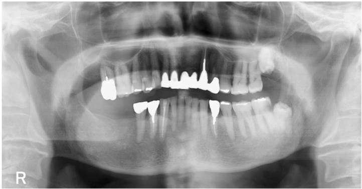
Presentation of case: Herein, we report a 78-year-old male referred to our hospital with a diagnosis of TMD and presenting with facial asymmetry, marked deviation to the right side on vertical mandibular movement and complaints of abnormal perception at the right oral and buccal region. Past medical history revealed that he had undergone a right intracranial trigeminal schwannoma resection 9 years prior. Computed tomography (CT) showed disuse atrophy of the right side of 4 masticatory muscles and 2 suprahyoid muscles controlled by the motor component of the mandibular division (V3) of the trigeminal nerve (TGN). Together with the neurosurgeon, we confirmed that there was no recurrence of the tumor and explained to the patient that the oral and maxillofacial symptoms are after-effects of the operation, and we provided oral hygiene instructions and coordinated cleaning of the inside of the oral cavity.
Discussion: Although it is difficult to compare treatment methods from case to case, we believe that in our case, the patient's understanding of the cause of his discomfort contributed significantly to the improvement of his quality of life.
Conclusion: We experienced a case of masticatory muscle disuse atrophy during long-term follow-up after resection of intracranial trigeminal schwannoma. Further studies are needed to develop the diagnostic and therapeutic protocols for disuse atrophy.
Keywords: Case report; Disuse atrophy; Masticatory muscles; Temporomandibular disorders (TMD); Trigeminal schwannoma.
Copyright © 2020 The Author(s). Published by Elsevier Ltd.. All rights reserved.
Figures


References
-
- Price S., Daly D.T. StatPearls; Treasure Island (FL): 2019. Neuroanatomy, Nucleus Trigeminal.
-
- Harriman D.G.F. The histochemistry of reactive masticatory muscle hypertrophy. Muscle Nerve. 1996;19:1447–1456. - PubMed
-
- Akita K., Sakaguchi-Kuma T., Fukino K., Ono T. Masticatory muscles and branches of mandibular nerve: positional relationships between various muscle bundles and their innervating branches. Anat. Rec. (Hoboken) 2019;302:609–619. - PubMed
Publication types
LinkOut - more resources
Full Text Sources
Research Materials
Miscellaneous
