. 2020 Aug 21;6(34):eabc0629.
doi: 10.1126/sciadv.abc0629. Print 2020 Aug.

DELTEX2 C-terminal domain recognizes and recruits ADP-ribosylated proteins for ubiquitination

Affiliations

DELTEX2 C-terminal domain recognizes and recruits ADP-ribosylated proteins for ubiquitination

Syed Feroj Ahmed et al. Sci Adv. .

Abstract

Cross-talk between ubiquitination and ADP-ribosylation regulates spatiotemporal recruitment of key players in many signaling pathways. The DELTEX family ubiquitin ligases (DTX1 to DTX4 and DTX3L) are characterized by a RING domain followed by a C-terminal domain (DTC) of hitherto unknown function. Here, we use two label-free mass spectrometry techniques to investigate the interactome and ubiquitinated substrates of human DTX2 and identify a large proportion of proteins associated with the DNA damage repair pathway. We show that DTX2-catalyzed ubiquitination of these interacting proteins requires PARP1/2-mediated ADP-ribosylation and depends on the DTC domain. Using a combination of structural, biochemical, and cell-based techniques, we show that the DTX2 DTC domain harbors an ADP-ribose-binding pocket and recruits poly-ADP-ribose (PAR)-modified proteins for ubiquitination. This PAR-binding property of DTC domain is conserved across the DELTEX family E3s. These findings uncover a new ADP-ribose-binding domain that facilitates PAR-dependent ubiquitination.


Figures

Fig. 1
Fig. 1. Structure of 2RD.
(A) Domain architecture of DTX2. (B) Cartoon representation of 2RD from crystal condition 1. The RING and DTC domains are colored cyan and green, respectively. Zn2+ ions are shown as gray spheres. (C) Cα ribbon overlay of the three molecules of forms 1 and 2 of 2RD. The DTC domains have been superposed to illustrate the flexibility between the RING and DTC domains. The molecule from condition 1 is colored green, and the two molecules from condition 2 are colored cyan and blue. (D) Model of 2RD bound to UbcH5B~Ub. The RING domain of 2RD was superposed with the RNF38 RING domain from the RNF38-UbcH5B~Ub complex (PDB 4V3L) to generate the model. Surface representation of 2RD colored green and oriented as in (A); cartoon representation of UbcH5B~Ub with UbcH5B and Ub colored gray and wheat, respectively. The red arrow indicates the position of the UbcH5B~Ub thioester. (E) Model of 2RD bound to UbcH5B~Ub. As in (D) but with surface representation of 2RD colored by charge potential. The circled area contains the charged pocket proximal to the UbcH5B~Ub thioester (indicated by the red arrow). (F) Sequence alignment of the RING-DTC fragment from the DTX family of E3s. Residues comprising the charged pocket proximal to the modeled UbcH5B~Ub thioester are highlighted in red, and conserved residues defined by an ALSCRIPT (56) level of 0.7 are highlighted in black.
Fig. 2
Fig. 2. Identification of DTX2 substrates.
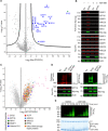
(A) Volcano plot displaying proteins that bind to Myc-DTX2 versus EV. Myc-trap pull-downs were performed in triplicate on whole-cell lysates from HEK293 cells expressing Myc-DTX2 or EV. Proteins shown as blue dots were identified as significantly enriched proteins using a t test with a 5% false discovery rate (FDR) in all three replicates and more abundant in Myc-DTX2 pull-downs compared to EV. Associated with data S1. (B) Immunoblots of Myc-trap pull-downs of whole-cell lysates from HEK293 cells expressing Myc-DTX2 or EV in the presence and absence of ABT-888 using anti-PARP1, anti-SPT16, anti-SSRP1, anti-XRCC5, anti-XRCC6, anti-RPA1, anti–Myc-tag, or anti-actin as indicated. (C) Volcano plot displaying log2 fold change in intensities in peptides modified with Ub from lysates from HEK293 cells expressing GFP-DTX2 compared to EV. Assays were performed in triplicate, and significant changes in ubiquitinated peptide intensities were determined using a Student’s t test with a 1% FDR (permutation-based). Significantly changed ubiquitinated peptides present in all three replicates, more abundant in lysates from cells expressing GFP-DTX2, and from proteins annotated in the Gene Ontology database with the term “DNA repair” are colored as indicated. Associated with data S2. (D) Immunoblots of SPT16 (left) and histone H3.1 (right) ubiquitination by DTX2 in HEK293 cells expressing His-Ub, EV or Myc-DTX2, and GFP–histone H3.1 (right only) in the presence and absence of ABT-888. The cell lysates and Ni2+–pull-down products were immunoblotted with anti-GFP, anti-SPT16, anti–Myc-tag, or anti-actin antibodies as indicated. (E) Reduced in vitro autoubiquitination assay showing the formation of GST-2RD-IR-Ubn over time in the presence and absence of ABT-888. IR-Ub, Ub labeled with near-infrared dye. Visualized with the Odyssey CLx Imaging System (LI-COR Biosciences, top) and subsequently by staining with InstantBlue (Expedeon, bottom).
Fig. 3
Fig. 3. DTC domain required for substrate ubiquitination.
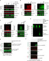
(A) Immunoblots of Myc-trap pull-downs of whole-cell lysates from HEK293 cells expressing Myc-DTX2 WWE variants or EV (−) in the presence and absence of ABT-888 using anti-SPT16, anti–histone H3.1, anti–Myc-tag, or anti-actin as indicated. (B) Immunoblots of SPT16 (left) and histone H3.1 (right) ubiquitination by DTX2 in HEK293 cells expressing His-Ub, GFP–histone H3.1 (right only), and EV (−) or full-length Myc-DTX2 variants: WT (wild type) or WDM (WWE domain mutant); cell lysates and Ni2+–pull-down products were immunoblotted with anti-GFP, anti-SPT16, anti–Myc-tag, or anti-actin antibodies as indicated. (C) Immunoblots of SPT16 (left) and histone H3.1 (right) ubiquitination by 2RD in the presence and absence of ABT-888 in HEK293 cells expressing His-Ub, GFP–histone H3.1 (right only), and Myc-2RD or EV. The cell lysates and Ni2+–pull-down products were immunoblotted with anti-GFP, anti-SPT16, anti–Myc-tag, or anti-actin antibodies as indicated. (D) Immunoblot of in vitro ADP-ribosylation of 2RD and histone H3.1 by PARP1. Biotinylated NAD+ was used as a source of ADP-ribose and immunoblotted with NeutrAvidin protein. (E) Immunoblots of in vitro ubiquitination of biotinylated ADP-ribosylated H3.1 tail-GFP (ADPrn-H3.1 tail-GFP) or H3.1 tail-GFP by 2RD. Biotinylated NAD+ was used as a source of ADP-ribose. Top: Anti-Ub (red) and NeutrAvidin protein. Bottom: Anti-Ub (red) and anti-GFP (green). Asterisk likely denotes a truncated H3.1 tail-GFP fragment (see fig. S2C). (F) Immunoblot of in vitro ubiquitination of biotinylated ADP-ribosylated His-HaPARP (ADPrn-His-HaPARP) or His-HaPARP by 2RD performed in the presence of E1, UbcH5B, and Ub. Biotinylated NAD+ was used as a source of ADP-ribose. Ni2+–pull-down products were immunoblotted with anti-His, anti-Ub, or NeutrAvidin protein as indicated to detect Ubn-ADPrn-His-HaPARP products.
Fig. 4
Fig. 4. DTC domain binds ADP-ribosylated substrate.
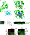
(A) Cartoon representation of complex structure of 2RD bound to ADPr, colored and oriented as in Fig. 1B. ADPr is shown in sticks, and C atoms are colored light gray, O atoms are colored red, N atoms are colored blue, and P atoms are colored pink. (B) Close-up view of 2RD-ADPr complex. Coloring as in (A) with C atoms from 2RD in the same color as the cartoon representation. Putative hydrogen bonds (2.6 to 3.2 Å) are shown as dashed black lines. (C) Close-up view colored and orientated as in (B) with a Polder map (57) in blue contoured at 3σ. (D) Single-turnover lysine discharge showing the disappearance of UbcH5B~IR-Ub over time with 2RD WT (wild type), RDM (RING double mutant), or DTM (DTC domain triple mutant), or no E3 as indicated. Associated with fig. S3 and table S1. (E) Plot showing fraction of UbcH5B~Ub remaining after 2 min of incubation with 2RD variants and lysine as indicated. Data are represented as means ± SD from five replicates. (F) Immunoblot of in vitro ubiquitination reactions of biotinylated ADP-ribosylated His-HaPARP by 2RD variants, as indicated and detected with NeutrAvidin protein. Biotinylated NAD+ was used as a source of ADP-ribose, and all reactions include E1, UbcH5B, and Ub. (G) Immunoblots of SPT16 (left) and histone H3.1 (right) ubiquitination by Myc-DTX2 variants in the presence and absence of ABT-888 in HEK293 cells expressing His-Ub, GFP–histone H3.1 (right only), and Myc-DTX2 variant or EV (−); cell lysates and Ni2+–pull-down products were immunoblotted with anti-GFP, anti-SPT16, anti–Myc-tag, or anti-actin antibodies as indicated.
Fig. 5
Fig. 5. Characterization of DTX family E3s.
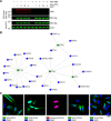
(A) Immunoblots of Myc-trap pull-downs of whole-cell lysates from HEK293 cells expressing EV (−) or Myc-tagged DTC domains from DTX1, DTX2, DTX3L, or DTX4 in the presence and absence of ABT-888 using anti-PAR, anti–Myc-tag, or anti-actin as indicated. (B) Network of DTX protein interactors generated from MS analyses of Myc-trap pull-downs of individual DTX family members. Label-free quantification (LFQ) intensities using the Hawaii plot functionality in Perseus were used to generate the networks. Associated with data S1. (C) Merged images from HeLa cells overexpressing GFP-DTX1, GFP-DTX2, mCherry-DTX2, GFP-DTX3L, or GFP-DTX4 as indicated. Cells on coverslips were fixed and mounted on glass slides using the nuclear stain DAPI (4′,6-diamidino-2-phenylindole). The scale bar (bottom right) represents 20 μm in each panel.

References

    1. Hershko A., Ciechanover A., The ubiquitin system. Annu. Rev. Biochem. 67, 425–479 (1998). - PubMed
    1. Komander D., Rape M., The ubiquitin code. Annu. Rev. Biochem. 81, 203–229 (2012). - PubMed
    1. Deshaies R. J., Joazeiro C. A., RING domain E3 ubiquitin ligases. Annu. Rev. Biochem. 78, 399–434 (2009). - PubMed
    1. Dye B. T., Schulman B. A., Structural mechanisms underlying posttranslational modification by ubiquitin-like proteins. Annu. Rev. Biophys. Biomol. Struct. 36, 131–150 (2007). - PubMed
    1. Kishi N., Tang Z., Maeda Y., Hirai A., Mo R., Ito M., Suzuki S., Nakao K., Kinoshita T., Kadesch T., Hui C.-c., Artavanis-Tsakonas S., Okano H., Matsuno K., Murine homologs of deltex define a novel gene family involved in vertebrate Notch signaling and neurogenesis. Int. J. Dev. Neurosci. 19, 21–35 (2001). - PubMed

Publication types

MeSH terms