. 2020 Jul 1;6(27):eaba7443.
doi: 10.1126/sciadv.aba7443. Print 2020 Jul.

Identification of human CD4+ T cell populations with distinct antitumor activity

Affiliations

Identification of human CD4+ T cell populations with distinct antitumor activity

Michelle H Nelson et al. Sci Adv. .

Abstract

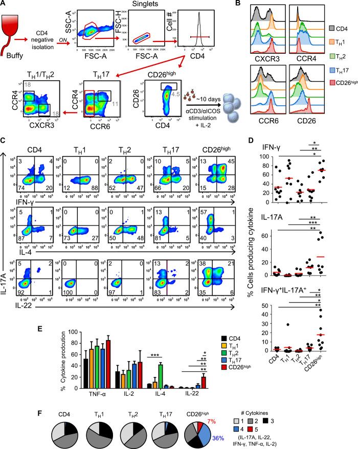
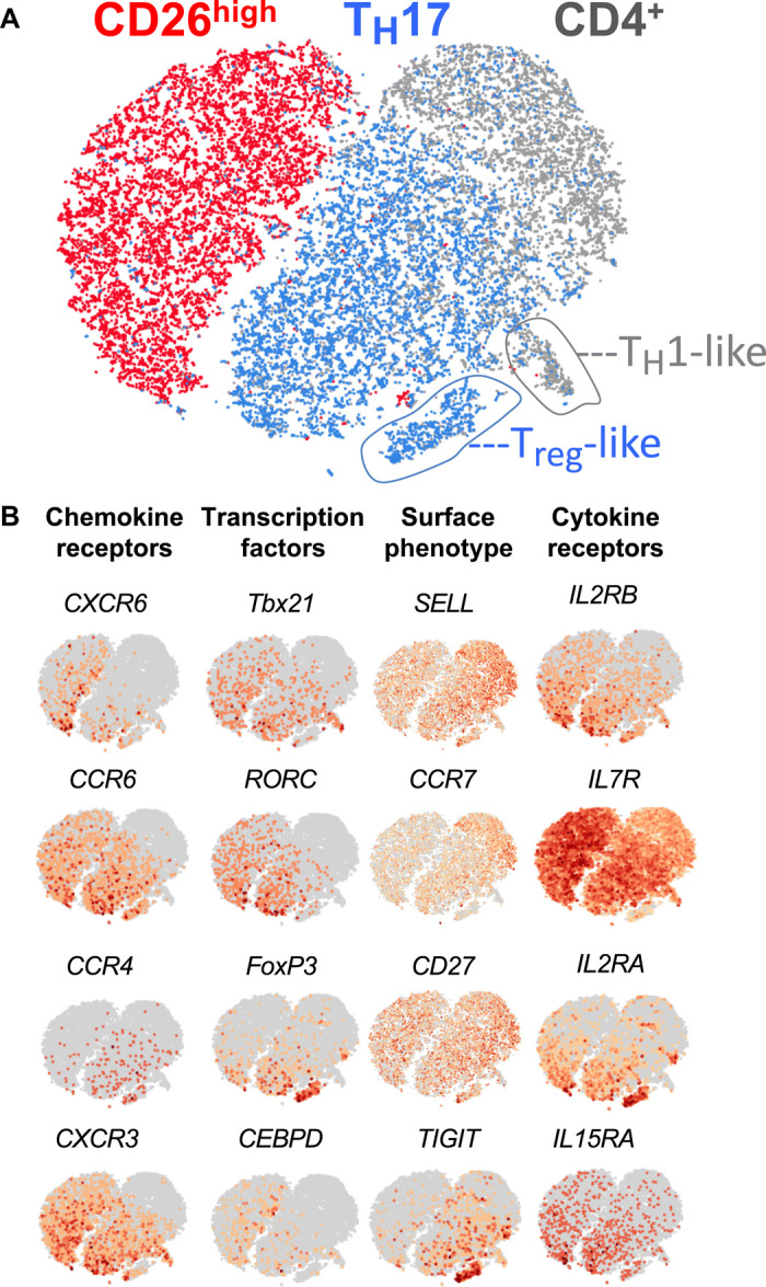
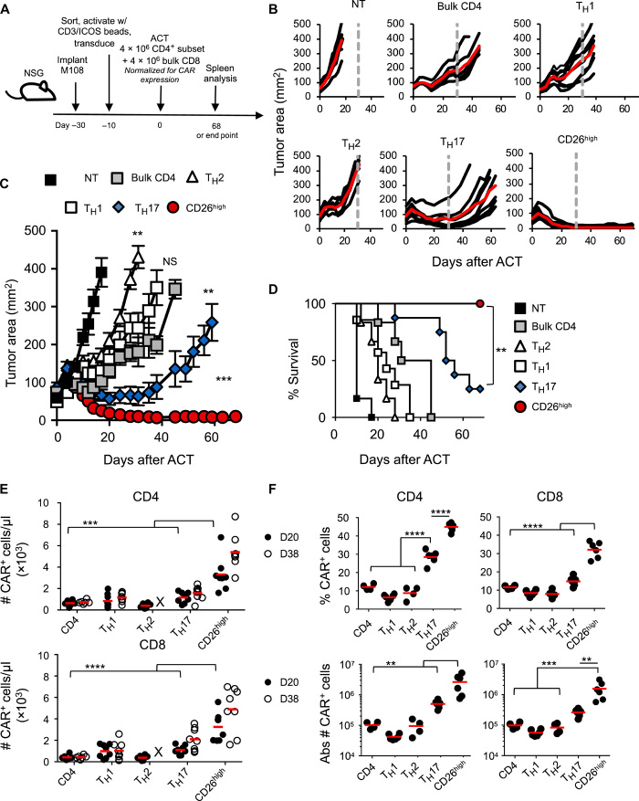
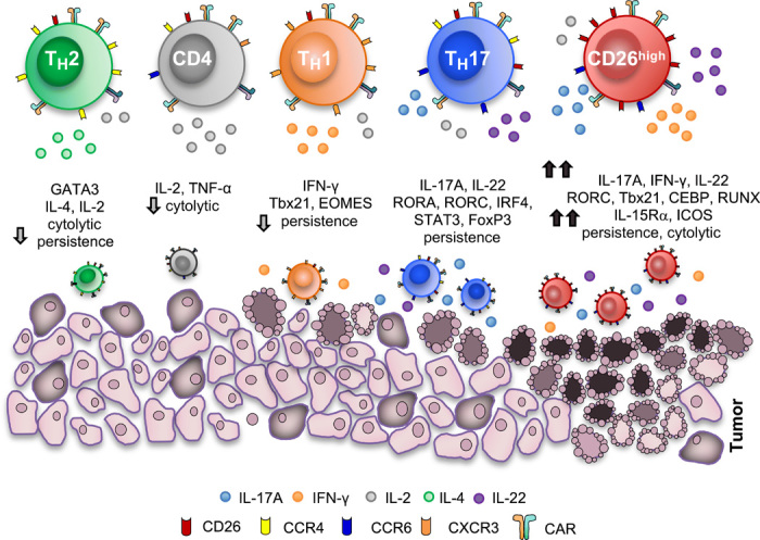
How naturally arising human CD4+ T helper subsets affect cancer immunotherapy is unclear. We reported that human CD4+CD26high T cells elicit potent immunity against solid tumors. As CD26high T cells are often categorized as TH17 cells for their IL-17 production and high CD26 expression, we posited these populations would have similar molecular properties. Here, we reveal that CD26high T cells are epigenetically and transcriptionally distinct from TH17 cells. Of clinical importance, CD26high and TH17 cells engineered with a chimeric antigen receptor (CAR) regressed large human tumors to a greater extent than enriched TH1 or TH2 cells. Only human CD26high T cells mediated curative responses, even when redirected with a suboptimal CAR and without aid by CD8+ CAR T cells. CD26high T cells cosecreted effector cytokines, produced cytotoxic molecules, and persisted long term. Collectively, our work underscores the promise of CD4+ T cell populations to improve durability of solid tumor therapies.


Figures

Fig. 1
Fig. 1. CD4+CD26high T cells have a dynamic cytokine profile.
(A) CD4+ subset sorting scheme. CD4+ lymphocytes were negatively isolated using magnetic beads from normal donor peripheral blood lymphocytes (PBLs). TH17 cells were sorted from the CCR6+CCR4+ gate. TH1 and TH2 cells are both CCR6 and subsequently sorted via CXCR3 or CCR4, respectively. CD26high cells were sorted independently on the basis of CD26 expression. (B) Chemokine receptor profile after sort. (C) CD4+ T cell subsets were stimulated with αCD3/inducible costimulator (ICOS) beads at a ratio of 1 bead:10 T cells and expanded in IL-2 (100 IU/ml). Ten days following activation, the five different cell subsets were examined for their intracellular cytokine production. Dot plot representation of IL-17, interferon-γ (IFN-γ), IL-4, and IL-22 expression by flow cytometry. (D) Graphical representation of at least eight normal donors from independent experiments demonstrating IFN-γ and IL-17 single and double producing cells by flow cytometry. (E) Graphical representation of 10 normal donors demonstrating cytokine-producing cells by flow cytometry. Two to three replicates each. Analysis of variance (ANOVA), Tukey’s post hoc comparisons; *P < 0.05, **P < 0.01, and ***P < 0.001. (F) Cells were gated on cytokine-producing cells to quantify cells that produced between one and five cytokines simultaneously. Cytokines of interest were IL-17, IFN-γ, IL-2, IL-22, and tumor necrosis factor–α (TNF-α). Representative of five experiments.
Fig. 2
Fig. 2. The epigenetic and molecular signature of CD26high T cells are unique.
(A) ATAC-seq analysis describing chromatin accessibility in FACS (fluorescence-activated cell sorting)–sorted CD4+ subsets (naïve, TH1, TH2, TH17, and CD26high) organized by TF networks known to describe TH1, TH2, TH17, and naïve subsets. Accessible transcription regions unique to CD26high T cells are also shown. Compiled from five healthy donors. (B) UCSC genome browser tracks for sorted CD4+ subsets around classical T helper TFs from ATAC-seq analysis. (C) ATAC-seq principal components analysis of sorted T cell subsets analyzed at resting state. n = 5 donors. (D) β T cell receptor (TCRβ) sequencing of CD26high, TH17, and TH1 cells sorted from peripheral blood of healthy donors demonstrates unique or shared clonotypes. Venn diagram illustrates percentage of unique or shared TCRβ sequences. The relative frequencies (standardized to sum to 1.0): CD26high only = 0.237, TH1 only = 0.487, TH17 only = 0.196, CD26high and TH1 = 0.041, CD26high and TH17 = 0.020, TH1 and TH17 = 0.015, and all three = 0.004, log-linear model.
Fig. 3
Fig. 3. CD4+CD26high T cells are distinguished from TH17 cells via single-cell sequencing.
Total CD4+, CD26high, and TH17 cell subsets were sorted from the peripheral blood of healthy donors, and ~3000 cells were assayed by single-cell RNA sequencing. (A) Data were analyzed by t-distributed stochastic neighbor embedding (t-SNE). (B) t-SNE plot overlaid with mRNA expression of chemokine receptors, TFs, memory markers, and cytokine receptors. Representative of three healthy donors.
Fig. 4
Fig. 4. Human CD26high T cells ablate large human tumors and persist relative to other CD4+ T cell subsets.
(A) ACT schematic. TH1 (CXCR3+), TH2 (CCR4+), TH17 (CCR4+/CCR6+), CD26high, or bulk CD4+ cells were sorted from normal donor PBL and expanded with αCD3/ICOS beads at a 1 bead:10 T cell ratio. Cells were transduced with a first-generation mesothelin-specific CD3ζ CAR and expanded with IL-2. NSG mice bearing mesothelioma were treated with 4 × 10⁶ transduced, sorted CD4+ cells + 4 × 10⁶ transduced CD8+ cells, and 50,000 IU of IL-2 was given to each mouse daily for 3 days. (B) Single-tumor curves overlaid with average curve (red) and (C) average tumor curves of six to nine mice per group. All groups were significantly different from non-treated mice (NT), P < 0.005. CD4 versus TH1, NS; CD4 versus TH2, **P = 0.0015; CD4 versus TH17, **P = 0.0035; CD4 versus CD26high, ***P = 0.0003; TH17 versus CD26high, **P = 0.008; polynomial regression. (D) The percentage of mice surviving with tumor size below the 200-mm2 threshold. Log-rank test. (E) Engraftment and persistence of CAR+ T cells in the peripheral blood at days 20 (D20) and 38 (D38) after ACT. n = 6 to 8 mice per group. X denotes group was at end point before bleed. (F) Spleens were analyzed by flow cytometry for the percentage and total number of CD3+CAR+CD4+ or CD8+ cells at day 68 (TH17 and CD26high) or group end point (CD4, TH1, and TH2). n = 4 to 6 mice per group. (E and F) ANOVA, Tukey’s post hoc comparisons; **P < 0.01, ***P < 0.001, and ****P < 0.0001.
Fig. 5
Fig. 5. CD4+CD26high T cells have distinct antitumor and molecular properties relative to other helper subsets.
CD26high T cells have been described herein for use in ACT therapy. These cells produce heightened levels of cytokines including IL-17, IFN-γ, IL-22, and IL-2 and can cosecrete these cytokines. CD26high T cells have a distinct chromatin landscape with accessible regions near RORC, Tbx21, CEBP, and RUNX TFs and have a unique transcriptional signature. These cells are cytotoxic, multifunctional, and inflammatory. Overall, CD26high T cells persist and regress tumors to a remarkably greater extent than other CD4+ T cells in vivo and represent a distinct CD4+ helper population with potent antitumor properties.

References

    1. Ohnuma K., Dang N. H., Morimoto C., Revisiting an old acquaintance: CD26 and its molecular mechanisms in T cell function. Trends Immunol. 29, 295–301 (2008). - PubMed
    1. Ou X., O’Leary H. A., Broxmeyer H. E., Implications of DPP4 modification of proteins that regulate stem/progenitor and more mature cell types. Blood 122, 161–169 (2013). - PMC - PubMed
    1. Fleischer B., CD26: A surface protease involved in T-cell activation. Immunol. Today 15, 180–184 (1994). - PubMed
    1. Dang N. H., Torimoto Y., Sugita K., Daley J. F., Schow P., Prado C., Schlossman S. F., Morimoto C., Cell surface modulation of CD26 by anti-1F7 monoclonal antibody. Analysis of surface expression and human T cell activation. J. Immunol. 145, 3963–3971 (1990). - PubMed
    1. Paulissen S. M. J., van Hamburg J. P., Dankers W., Lubberts E., The role and modulation of CCR6+ Th17 cell populations in rheumatoid arthritis. Cytokine 74, 43–53 (2015). - PubMed

Publication types

Substances