. 2020 Sep 14;21(18):6734.
doi: 10.3390/ijms21186734.

IKKγ/NEMO Is Required to Confer Antimicrobial Innate Immune Responses in the Yellow Mealworm, Tenebrio Molitor

Affiliations

IKKγ/NEMO Is Required to Confer Antimicrobial Innate Immune Responses in the Yellow Mealworm, Tenebrio Molitor

Hye Jin Ko et al. Int J Mol Sci. .

Abstract

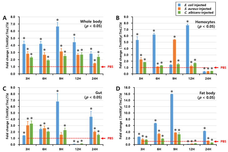
IKKγ/NEMO is the regulatory subunit of the IκB kinase (IKK) complex, which regulates the NF-κB signaling pathway. Within the IKK complex, IKKγ/NEMO is the non-catalytic subunit, whereas IKKα and IKKβ are the structurally related catalytic subunits. In this study, TmIKKγ was screened from the Tenebrio molitor RNA-Seq database and functionally characterized using RNAi screening for its role in regulating T. molitor antimicrobial peptide (AMP) genes after microbial challenges. The TmIKKγ transcript is 1521 bp that putatively encodes a polypeptide of 506 amino acid residues. TmIKKγ contains a NF-κB essential modulator (NEMO) and a leucine zipper domain of coiled coil region 2 (LZCC2). A phylogenetic analysis confirmed its homology to the red flour beetle, Tribolium castaneum IKKγ (TcIKKγ). The expression of TmIKKγ mRNA showed that it might function in diverse tissues of the insect, with a higher expression in the hemocytes and the fat body of the late-instar larvae. TmIKKγ mRNA expression was induced by Escherichia coli, Staphylococcus aureus, and Candida albicans challenges in the whole larvae and in tissues such as the hemocytes, gut and fat body. The knockdown of TmIKKγ mRNA significantly reduced the survival of the larvae after microbial challenges. Furthermore, we investigated the tissue-specific induction patterns of fourteen T. molitor AMP genes in TmIKKγ mRNA-silenced individuals after microbial challenges. In general, the mRNA expression of TmTenecin1, -2, and -4; TmDefensin1 and -2; TmColeoptericin1 and 2; and TmAttacin1a, 1b, and 2 were found to be downregulated in the hemocytes, gut, and fat body tissues in the TmIKKγ-silenced individuals after microbial challenges. Under similar conditions, TmRelish (NF-κB transcription factor) mRNA was also found to be downregulated. Thus, TmIKKγ is an important factor in the antimicrobial innate immune response of T. molitor.

Keywords: NF-κB transcription factor; RNAi; Tenebrio molitor; antimicrobial peptides; insect immunity.


Conflict of interest statement

The authors declare no conflict of interest.

Figures

Figure 1
Figure 1
The complete nucleotide and deduced amino acid sequence of Tenebrio molitor (Tm)IKKγ. The nucleotides are numbered from the first base of the initiation codon to the stop codon. This represents the open reading frame region. The asterisk indicates the stop codon. The characteristic NF-κB essential modulator (NEMO) domain is enclosed by an open box, and the leucine zipper domain of coiled coil region 2 (LZCC2) of NEMO is enclosed by a yellow box.
Figure 2
Figure 2
Developmental and tissue-specific expression of TmIKKγ mRNA using qRT-PCR. (A) Relative expression of TmIKKγ mRNA in the egg (Egg), early larva (YL), late larva (LL), prepupa (PP), pupa days 1–7 (P1–P7), and adult days 1–5 (A1–A5). (B) TmIKKγ mRNA expression in last-instar larvae. (C) Distribution of TmIKKγ mRNA in adult tissues. IT, integument; GT, gut; FB, fat body; HC, hemocytes; MT, Malpighian tubules; OV, ovary, and TE, testis. L27a from T. molitor was included as an internal control to normalize RNA levels between samples. Vertical bars represent standard errors (n = 3). Different letters above bars represent significant differences between groups.
Figure 3
Figure 3
Temporal expression patterns of TmIKKγ mRNA in the whole body (A), hemocytes (B), gut (C), and fat body (D) of T. molitor post-challenge with Escherichia coli, Staphylococcus aureus, or Candida albicans. The expression was analyzed by qRT-PCR using L27a (T. molitor) as the internal control. For each time point, the expression level in the phosphate-buffered saline (PBS)-injected control (mock control) was set to 1; this is represented by a dotted line. Values represent the mean of triplicate experiments, mean ± SE (n = 20). Asterisk denotes significant differences from the mock control at 95% confidence level (Student’s t-test).
Figure 4
Figure 4
RNA interference (RNAi)-based silencing assay for T. molitor larval survival analysis after E. coli, S. aureus, and C. albicans injections. (A) RNAi silencing efficiency of TmIKKγ was about 99%. Time-dependent survival of double-stranded (ds)TmIKKγ-injected T. molitor larvae after challenge with E. coli (B), S. aureus (C), and C. albicans (D). The survival was studied for 10 d after the microbial challenge, with dsEGFP)-treated larvae acting as negative controls. Results are shown as an average of three independent biological replicates with standard errors. Asterisks denote significant differences at 95% confidence level (Wilcoxon-Mann Whitney test).
Figure 5
Figure 5
Induction patterns of fourteen antimicrobial peptide (AMP) genes in TmIKKγ-silenced T. molitor larval hemocytes in response to E. coli, S. aureus, and C. albicans infections. This includes TmTene1 (A), TmTene2 (B), TmTene3 (C), TmTene4 (D), TmDef1 (E), TmDef2 (F), TmCole1 (G), TmCole2 (H), TmCec2 (I), TmAtta1a (J), TmAtta1b (K), TmAtta2 (L), TmTLP1 (M), and TmTLP2 (N). The experimental samples were divided into three groups (E. coli, S. aureus, or C. albicans-challenged groups) and a wounded control (PBS group). AMP expression was studied 24 h after microbial challenge. dsEGFP was used as a negative control, and TmL27a served as an internal control. The numbers above the bars represent the AMP transcription levels. All experiments were repeated three times, and similar results were obtained. Asterisks denote significance at 95% confidence levels.
Figure 6
Figure 6
Induction patterns of fourteen antimicrobial peptide (AMP) genes in the larval gut of TmIKKγ-silenced T. molitor, in response to E. coli, S. aureus, and C. albicans infections. This includes TmTene1 (A), TmTene2 (B), TmTene3 (C), TmTene4 (D), TmDef1 (E), TmDef2 (F), TmCole1 (G), TmCole2 (H), TmCec2 (I), TmAtta1a (J), TmAtta1b (K), TmAtta2 (L), TmTLP1 (M), and TmTLP2 (N). The experimental samples were divided into three groups (E. coli, S. aureus, or C. albicans-challenged groups) and a wounded control (PBS group). AMP expression was studied 24 h after microbial challenge. dsEGFP was used as a negative control, and TmL27a served as an internal control. The numbers above the bars represent the AMP transcription levels. All experiments were repeated three times, and similar results were obtained. Asterisks denote significance at 95% confidence levels.
Figure 7
Figure 7
Induction patterns of fourteen antimicrobial peptide (AMP) genes in the larval fat body of TmIKKγ-silenced T. molitor in response to E. coli, S. aureus, and C. albicans infections. This includes TmTene1 (A), TmTene2 (B), TmTene3 (C), TmTene4 (D), TmDef1 (E), TmDef2 (F), TmCole1 (G), TmCole2 (H), TmCec2 (I), TmAtta1a (J), TmAtta1b (K), TmAtta2 (L), TmTLP1 (M), and TmTLP2 (N). The experimental samples were divided into three groups (E. coli, S. aureus, or C. albicans-challenged groups) and a wounded control (PBS group). AMP expression was studied 24 h after the microbial challenge. dsEGFP was used as a negative control, and TmL27a served as an internal control. The numbers above the bars represent the AMP transcription levels. All experiments were repeated three times, and similar results were obtained. Asterisks denote significance at 95% confidence levels.
Figure 8
Figure 8
Tissue-specific induction patterns of NF-κB genes in dsTmIKKγ-treated T. molitor larvae after injections of E. coli, S. aureus, and C. albicans. mRNA expression levels of TmRelish1 (A), TmDorX1 (B), and TmDorX2 (C). T. molitor larvae injected with PBS acted as the mock control group. dsEGFP was used as a negative control, and TmL27a served as an internal control. Asterisk denotes significant differences from the mock control at 95% confidence levels (Student’s t-test).
Figure 9
Figure 9
Graphical summary of TmIKKγ on the AMP production in T. molitor. Our results suggested that TmIKKγ regulates the AMP gene expression in response to three pathogens, including E. coli, S. aureus, and C. albicans, through both NF-κB transcription factors, TmRelish and TmDorX2.

Similar articles

Cited by

References

    1. Zambon R.A., Nandakumar M., Vakharia V.N., Wu L.P. The Toll pathway is important for an antiviral response in Drosophila. Proc. Natl. Acad. Sci. USA. 2005;102:7257–7262. doi: 10.1073/pnas.0409181102. - DOI - PMC - PubMed
    1. Vercammen E., Staal J., Beyaert R. Sensing of Viral Infection and Activation of Innate Immunity by Toll-Like Receptor 3. Clin. Microbiol. Rev. 2008;21:13–25. doi: 10.1128/CMR.00022-07. - DOI - PMC - PubMed
    1. Kotwal G.J., Hatch S., Marshall W.L. Viral Infection: An Evolving Insight into the Signal Transduction Pathways Responsible for the Innate Immune Response. Adv. Virol. 2012;2012:1–11. doi: 10.1155/2012/131457. - DOI - PMC - PubMed
    1. Oshiumi H., Matsuo A., Matsumoto M., Seya T. Pan-Vertebrate Toll-Like Receptors During Evolution. Curr. Genom. 2008;9:488–493. doi: 10.2174/138920208786241234. - DOI - PMC - PubMed
    1. Chatterjee A., Roy D., Patnaik E., Nongthomba U. Muscles provide protection during microbial infection by activating innate immune response pathways in Drosophila and zebrafish. Dis. Model. Mech. 2016;9:697–705. doi: 10.1242/dmm.022665. - DOI - PMC - PubMed

MeSH terms

LinkOut - more resources