Meta-Analysis
. 2020 Sep 14;21(18):6742.
doi: 10.3390/ijms21186742.

Matrix Metalloproteinases and Tissue Inhibitors of Metalloproteinases in Extracellular Matrix Remodeling during Left Ventricular Diastolic Dysfunction and Heart Failure with Preserved Ejection Fraction: A Systematic Review and Meta-Analysis

Affiliations
Meta-Analysis

Matrix Metalloproteinases and Tissue Inhibitors of Metalloproteinases in Extracellular Matrix Remodeling during Left Ventricular Diastolic Dysfunction and Heart Failure with Preserved Ejection Fraction: A Systematic Review and Meta-Analysis

Merle M Krebber et al. Int J Mol Sci. .

Abstract

Matrix metalloproteinases (MMPs) and tissue inhibitors of metalloproteinases (TIMPs) are pivotal regulators of extracellular matrix (ECM) composition and could, due to their dynamic activity, function as prognostic tools for fibrosis and cardiac function in left ventricular diastolic dysfunction (LVDD) and heart failure with preserved ejection fraction (HFpEF). We conducted a systematic review on experimental animal models of LVDD and HFpEF published in MEDLINE or Embase. Twenty-three studies were included with a total of 36 comparisons that reported established LVDD, quantification of cardiac fibrosis and cardiac MMP or TIMP expression or activity. LVDD/HFpEF models were divided based on underlying pathology: hemodynamic overload (17 comparisons), metabolic alteration (16 comparisons) or ageing (3 comparisons). Meta-analysis showed that echocardiographic parameters were not consistently altered in LVDD/HFpEF with invasive hemodynamic measurements better representing LVDD. Increased myocardial fibrotic area indicated comparable characteristics between hemodynamic and metabolic models. Regarding MMPs and TIMPs; MMP2 and MMP9 activity and protein and TIMP1 protein levels were mainly enhanced in hemodynamic models. In most cases only mRNA was assessed and there were no correlations between cardiac tissue and plasma levels. Female gender, a known risk factor for LVDD and HFpEF, was underrepresented. Novel studies should detail relevant model characteristics and focus on MMP and TIMP protein expression and activity to identify predictive circulating markers in cardiac ECM remodeling.

Keywords: animal models; extracellular matrix; fibrosis; heart failure with preserved ejection fraction; left ventricular diastolic dysfunction; matrix metalloproteinase; systematic review; tissue inhibitor of metalloproteinase.


Conflict of interest statement

The authors declare no conflict of interest.

Figures

Figure 1
Figure 1
Flow chart of study selection. All articles are included and excluded according to the selection criteria defined in the Materials and Method section.
Figure 2
Figure 2
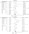
The effect of LVDD/HFpEF on cardiac parameters E/e’ (panel (A)) and E/A (panel (B)). Forrest plot; the right side shows an increased ratio in LVDD/HFpEF animals, the left side shows a decreased ratio in LVDD/HFpEF animals. Data are presented as standard mean differences (SMDs) with 95% CI. Arrows indicate increased and decreased E/e’ ratio (A), and increased and decreased E/A ratio (B) respectively. Only the first author of each study is shown; multiple comparisons within one study are shown with a, b, c or d and correspond with the study overview (Table S1). CI, confidence interval; E/A, ratio between peak early diastolic transmitral velocity (E) and late (atrial) transmitral flow velocity (A); E/e’, ratio between peak early diastolic transmitral velocity (E) and early diastolic mitral annular velocity (e’); I2, measurement of heterogeneity; N, cumulative sample size; SMD, standardized mean difference.
Figure 3
Figure 3
The effect of LVDD/HFpEF on cardiac parameters Tau (panel (A)) and dP/dtmin (panel (B)). Forrest plot; the right side shows an increased effect in LVDD/HFpEF animals, the left side shows a decreased effect in LVDD/HFpEF animals. Data are presented as SMDs with 95% CI. Arrows indicate shortened and prolonged time constant of relaxation Tau (A), and decreased and increased rate of pressure change dP/dtmin (B) respectively. Only the first author of each study is shown; multiple comparisons within one study are shown with a, b, c or d and correspond with the study overview (Table S1). CI, confidence interval; I2, measurement of heterogeneity; N, cumulative sample size; SMD, standardized mean difference; dP/dtmin, minimum rate of pressure change; Tau, time constant of ventricular relaxation.
Figure 4
Figure 4
The effect of LVDD/HFpEF on total fibrotic area. Forrest plot; the right side shows an increased effect in LVDD/HFpEF animals, the left side shows a decreased effect in LVDD/HFpEF animals. Data are presented as SMDs with 95% CI. Arrows indicate increased and decreased fibrotic percentage area respectively. Only the first author of each study is shown; multiple comparisons within one study are shown with a, b, c or d and correspond with the study overview (Table S1). CI, confidence interval; I2, measurement of heterogeneity; N, cumulative sample size; SMD, standardized mean difference.
Figure 5
Figure 5
The effect of LVDD/HFpEF on collagen type 1 (panel (A)) and collagen type 3 (panel (B)) mRNA levels. Forrest plot; the right side shows an increased effect in LVDD/HFpEF animals, the left side shows a decreased effect in LVDD/HFpEF animals. Data are presented as SMDs with 95% CI. Arrows indicate increased and decreased Collagen type 1 (A), and increased and decreased type 3 (B) mRNA expression respectively. Only the first author of each study is shown; multiple comparisons within one study are shown with a, b, c or d and correspond with the study overview (Table S1). CI, confidence interval; COL1, collagen type 1; COL3, collagen type 3; I2, measurement of heterogeneity; N, cumulative sample size; SMD, standardized mean difference.
Figure 6
Figure 6
The effect of LVDD/HFpEF on MMP2 (panel (A)) and MMP9 (panel (B)) activity. Forrest plot; the right side shows an increased effect in LVDD/HFpEF animals, the left side shows a decreased effect in LVDD/HFpEF animals. Data are presented as SMDs with 95% CI. Arrows indicate increased and decreased MMP2 (A), and increased and decreased MMP9 (B) enzyme activity respectively. Only the first author of each study is shown; multiple comparisons within one study are shown with a, b, c or d and correspond with the study overview (Table S1). CI, confidence interval; I2, measurement of heterogeneity; MMP, matrix metalloproteinase; N, cumulative sample size; SMD, standardized mean difference; TIMP, tissue inhibitor of metalloproteinase.

References

    1. Kloch-Badelek M., Kuznetsova T., Sakiewicz W., Tikhonoff V., Ryabikov A., Gonzalez A., Lopez B., Thijs L., Jin Y., Malyutina S., et al. Prevalence of left ventricular diastolic dysfunction in European populations based on cross-validated diagnostic thresholds. Cardiovasc. Ultrasound. 2012;10:10. doi: 10.1186/1476-7120-10-10. - DOI - PMC - PubMed
    1. Rasmussen-Torvik L.J., Colangelo L.A., Lima J.A.C., Jacobs D.R., Rodriguez C.J., Gidding S.S., Lloyd-Jones D.M., Shah S.J. Prevalence and predictors of diastolic dysfunction according to different classification criteria: The coronary artery risk development in young in adults study. Am. J. Epidemiol. 2017;185:1221–1227. doi: 10.1093/aje/kww214. - DOI - PMC - PubMed
    1. Palmiero P., Zito A., Maiello M., Cameli M., Modesti P.A., Muiesan M.L., Novo S., Saba P.S., Scicchitano P., Pedrinelli R., et al. Left ventricular diastolic function in hypertension: Methodological considerations and clinical implications. J. Clin. Med. Res. 2015;7:137–144. doi: 10.14740/jocmr2050w. - DOI - PMC - PubMed
    1. Nagueh S.F., Smiseth O.A., Appleton C.P., Byrd B.F., Dokainish H., Edvardsen T., Flachskampf F.A., Gillebert T.C., Klein A.L., Lancellotti P., et al. Recommendations for the evaluation of left ventricular diastolic function by echocardiography: An update from the american society of echocardiography and the european association of cardiovascular imaging. Eur. J. Echocardiogr. 2016;17:1321–1360. doi: 10.1093/ehjci/jew082. - DOI - PubMed
    1. Mitter S.S., Shah S.J., Thomas J.D. A Test in context: E/A and E/e’ to assess diastolic dysfunction and LV filling pressure. J. Am. Coll Cardiol. 2017;69:1451–1464. doi: 10.1016/j.jacc.2016.12.037. - DOI - PubMed

MeSH terms

Substances