. 2020 Nov;40(11):e296-e308.
doi: 10.1161/ATVBAHA.120.315261. Epub 2020 Sep 17.

Secreted Factors From Proinflammatory Macrophages Promote an Osteoblast-Like Phenotype in Valvular Interstitial Cells

Affiliations

Secreted Factors From Proinflammatory Macrophages Promote an Osteoblast-Like Phenotype in Valvular Interstitial Cells

Joseph C Grim et al. Arterioscler Thromb Vasc Biol. 2020 Nov.

Abstract

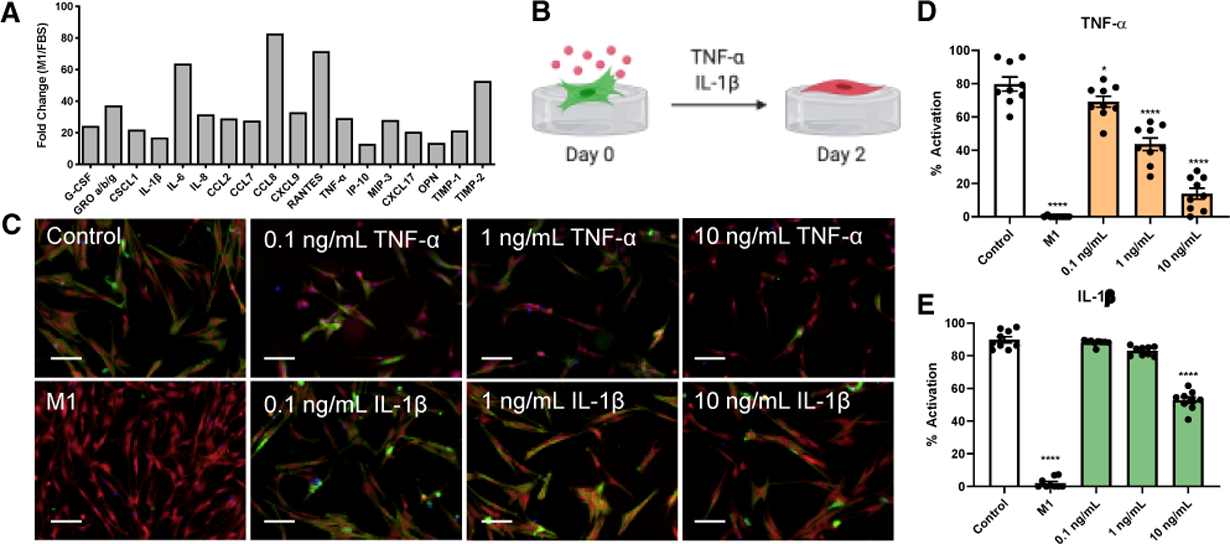
Objective: Resident valvular interstitial cells (VICs) activate to myofibroblasts during aortic valve stenosis progression, which further promotes fibrosis or even differentiate into osteoblast-like cells that can lead to calcification of valve tissue. Inflammation is a hallmark of aortic valve stenosis, so we aimed to determine proinflammatory cytokines secreted from M1 macrophages that give rise to a transient VIC phenotype that leads to calcification of valve tissue. Approach and Results: We designed hydrogel biomaterials as valve extracellular matrix mimics enabling the culture of VICs in either their quiescent fibroblast or activated myofibroblast phenotype in response to the local matrix stiffness. When VIC fibroblasts and myofibroblasts were treated with conditioned media from THP-1-derived M1 macrophages, we observed robust reduction of αSMA (alpha smooth muscle actin) expression, reduced stress fiber formation, and increased proliferation, suggesting a potent antifibrotic effect. We further identified TNF (tumor necrosis factor)-α and IL (interleukin)-1β as 2 cytokines in M1 media that cause the observed antifibrotic effect. After 7 days of culture in M1 conditioned media, VICs began differentiating into osteoblast-like cells, as measured by increased expression of RUNX2 (runt-related transcription factor 2) and osteopontin. We also identified and validated IL-6 as a critical mediator of the observed pro-osteogenic effect.

Conclusions: Proinflammatory cytokines in M1 conditioned media inhibit myofibroblast activation in VICs (eg, TNF-α and IL-1β) and promote their osteogenic differentiation (eg, IL-6). Together, our work suggests inflammatory M1 macrophages may drive a myofibroblast-to-osteogenic intermediate VIC phenotype, which may mediate the switch from fibrosis to calcification during aortic valve stenosis progression.

Keywords: aortic valve; cytokines; hydrogels; inflammation; macrophages.


Figures

Figure 1.
Figure 1.. M1 conditioned media inhibits VIC myofibroblast activation on soft and stiff hydrogels.
A, Hydrogels were fabricated via thiol-ene photopolymerization. B, Young’s modulus (E, Pa) of stiff and soft hydrogels by shear rheology. C, Schematic representation of VIC M1 conditioned media experiments. D, Representative images and E, percentage of activated VIC myofibroblast on soft and stiff hydrogels when treated with M1 conditioned media (CM). F, Representative images and G, percentage of activated VIC myofibroblasts on stiff hydrogels in response to M1 conditioned media at varying dilutions. Stained images: green, α-SMA; red, cytoplasm; blue, nuclei. Scale bars, 100 μm. For all bar graphs, n = 7 or 9 measurements per group, means ± SEM, and significance tested with one-way ANOVA for all data and indicated as ***P < 0.001 ****P < 0.0001 relative to control unless noted by brackets.
Figure 2.
Figure 2.. M1 conditioned media de-activates and protects against mechanical mediated VIC myofibroblast activation.
A, Schematic representation of experimental setup and B, representative images of VICs cultured on stiff hydrogels for 4 d with or without treatment with M1 conditioned media at 2 d. C, Schematic representation of experimental setup and D, representative images of VICs cultured on stiff hydrogels for 4 d with M1 conditioned media or removal of M1 conditioned media at 2 d. E, Quantified VIC myofibroblast activation. Stained images: green, α-SMA; red, cytoplasm; blue, nuclei. Scale bars, 100 μm. For all bar graphs, n = 9 measurements per group, means ± SEM, and significance tested with one-way ANOVA for all data and indicated as ***P < 0.001 ****P < 0.0001 to untreated unless noted by brackets.
Figure 3.
Figure 3.. TNF-α and IL-1β are responsible for de-activation of VIC myofibroblasts in M1 conditioned media.
A, Quantified cytokine array data of M1 conditioned media. B, Schematic representation of experimental setup for TNF-α and IL-1β treatment at various concentrations. C, Representative images and D, and E, quantified VIC myofibroblast activation. Stained images: green, α-SMA; red, cytoplasm; blue, nuclei. Scale bars, 100 μm. For all bar graphs, n = 9 measurements per group, means ± SEM, and significance tested with one-way ANOVA for all data and indicated as *P < 0.05, ***P < 0.001 ****P < 0.0001 to control.
Figure 4.
Figure 4.. M1 conditioned media induces VIC proliferation through TNF-α and IL-1β.
A, Schematic representation of EdU assay to assess proliferation. B, Representative images, and C, quantified proliferation in response to M1 conditioned media at varying dilutions. D, Quantified proliferation in response to TNF-α and IL-1β treatment at varying concentrations. Stained images: green, EdU; red, cytoplasm; blue, nuclei. Scale bars, 100 μm. For all bar graphs, n = 9 or 18 measurements per group, means ± SEM, and significance tested with one-way ANOVA for all data and indicated as **P < 0.01, ***P < 0.001 ****P < 0.0001 to control.
Figure 5.
Figure 5.. M1 conditioned media promotes an osteoblast-like phenotype in VICs on stiff hydrogels and is mediated by IL-6.
A, Schematic representation of experimental setup. B, Representative images, C, quantified VIC myofibroblast activation, and D, quantified RUNX2 nuclear localization when treated with OM or M1 conditioned media. RT-qPCR relative gene expression of E, RUNX2 and F, OPN. G, Quantified mean nuclear RUNX2 intensity in response to IL-6 treatment at various concentrations. Stained images: green, α-SMA; red, RUNX2; blue, nuclei. Scale bars, 100 μm. For immunostaining bar graphs, n = 9 measurements per group and n = 3 for RT-qPCR, means ± SEM, and significance tested with one-way ANOVA for all data and indicated as *P < 0.05, ***P < 0.001 ****P < 0.0001 to control.
Figure 6.
Figure 6.. Proposed mechanism of inflammatory factors driving AVS progression.
(A) Healthy valve tissue contains fibroblast-like VICs, which activate to myofibroblasts. (B) Myofibroblasts promote the initiation of fibrosis in valve tissue with increased ECM deposition. (C) During the valve thickening process, infiltrating M1 macrophages secrete TNF-α and IL-1β to de-activate myofibroblasts and cause their proliferation. (D) Inflammatory factors such as IL-6 promote differentiation toward osteoblast-like cells, ultimately causing calcification of valve tissue.

References

    1. Weiss RM, Miller JD, Heistad DD. Fibrocalcific aortic valve disease. Circ Res. 2013;113:209–222 - PMC - PubMed
    1. Osnabrugge RLJ, Mylotte D, Head SJ, Mieghem NMV, Nkomo VT, LeReun CM, et al. Aortic stenosis in the elderly disease prevalence and number of candidates for transcatheter aortic valve replacement: A meta-analysis and modeling study. J Am Coll Cardiol. 2013;62:1002–1012 - PubMed
    1. Leon MB, Smith CR, Mack M, Miller DC, Moses JW, Svensson LG, et al. Transcatheter aortic-valve implantation for aortic stenosis in patients who cannot undergo surgery. New Engl J Medicine. 2010;363:1597–1607 - PubMed
    1. Marquis-Gravel G, Redfors B, Leon MB, Généreux P. Medical treatment of aortic stenosis. Circulation. 2016;134:1766–1784 - PubMed
    1. Otto CM, Prendergast B. Aortic-valve stenosis — from patients at risk to severe valve obstruction. New Engl J Medicine. 2014;371:744–756 - PubMed

Publication types

MeSH terms

Supplementary concepts