

Cerebrospinal fluid confirmed COVID-19-associated encephalitis treated successfully
- PMID: 32938656
- PMCID: PMC7497137
- DOI: 10.1136/bcr-2020-237378
Cerebrospinal fluid confirmed COVID-19-associated encephalitis treated successfully
Abstract
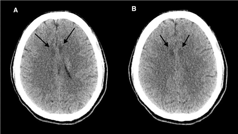
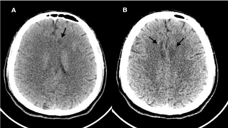
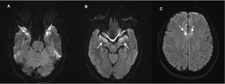
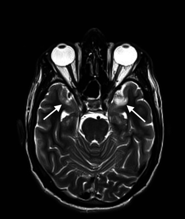
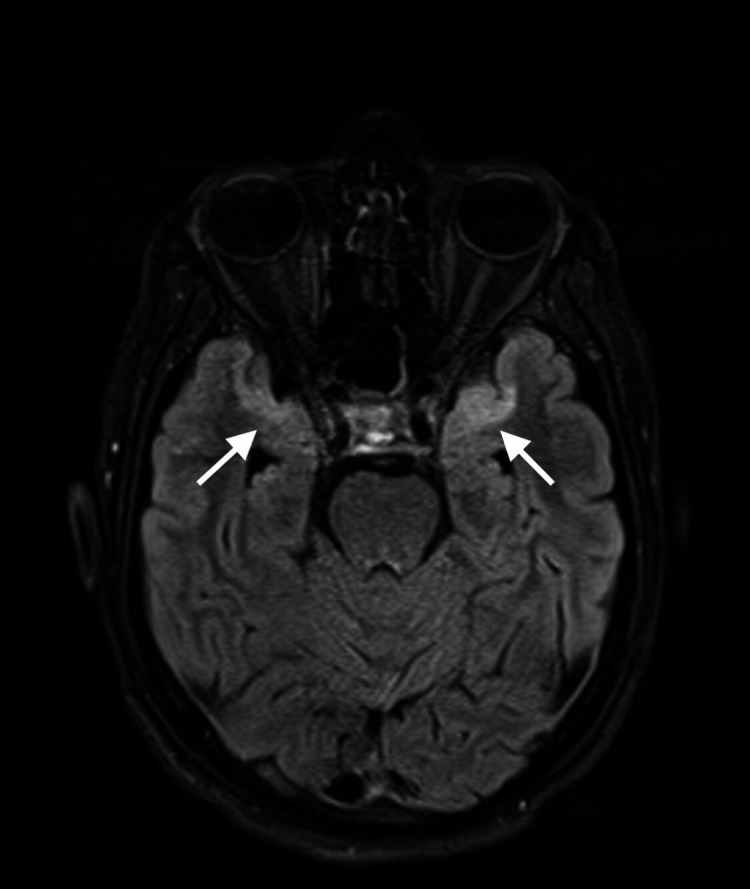
The COVID-19 pandemic that attracted global attention in December 2019 is well known for its clinical picture that is consistent with respiratory symptoms. Currently, the available medical literature describing the neurological complications of COVID-19 is gradually emerging. We hereby describe a case of a 31-year-old COVID-19-positive patient who was admitted on emergency basis. His clinical presentation was primarily neurological, rather than the COVID-19's classical respiratory manifestations. He presented with acute behavioural changes, severe confusion and drowsiness. The cerebrospinal fluid analysis was consistent with COVID-19 encephalitis, as well as the brain imaging. This experience confirms that neurological manifestations might be expected in COVID-19 infections, despite the absence of significant respiratory symptoms. Whenever certain red flags are raised, physicians who are involved in the management of COVID-19 should promptly consider the possibility of encephalitis. Early recognition of COVID-19 encephalitis and timely management may lead to a better outcome.
Keywords: global health; infection (neurology); infectious diseases; medical management; neuroimaging.
© BMJ Publishing Group Limited 2020. No commercial re-use. See rights and permissions. Published by BMJ.
Conflict of interest statement
Competing interests: None declared.
Figures
References
-
- WHO Coronavirus disease situation report. Available: https://www.who.int/docs/defaultsource/coronaviruse/situation-reports/20...
Publication types
MeSH terms
LinkOut - more resources
Full Text Sources