. 2020 Sep 17;5(18):e137646.
doi: 10.1172/jci.insight.137646.

TRPV4 antagonists ameliorate ventriculomegaly in a rat model of hydrocephalus

Affiliations

TRPV4 antagonists ameliorate ventriculomegaly in a rat model of hydrocephalus

Alexandra E Hochstetler et al. JCI Insight. .

Abstract

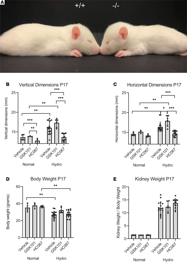
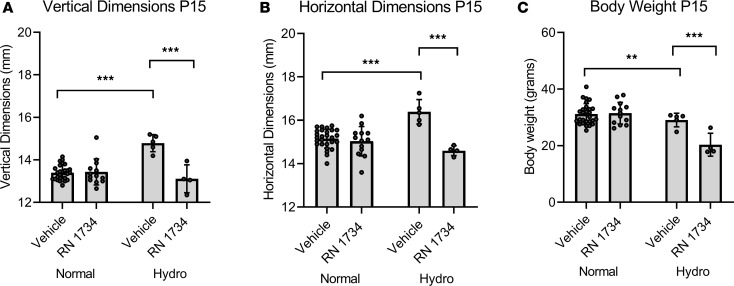
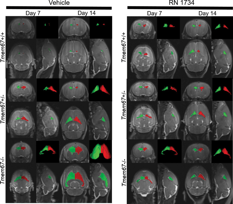
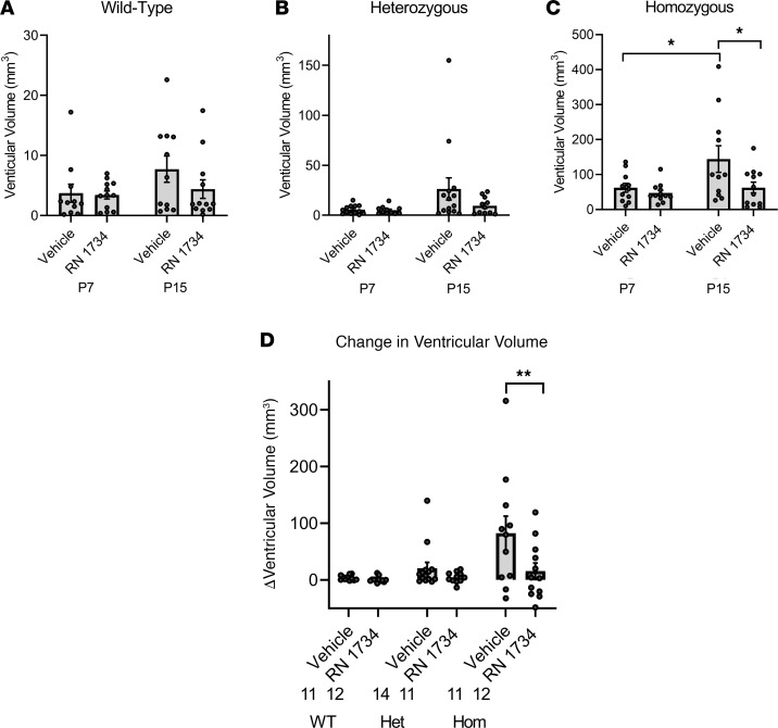
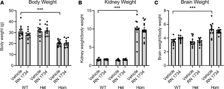
Hydrocephalus is a serious condition that impacts patients of all ages. The standards of care are surgical options to divert, or inhibit production of, cerebrospinal fluid; to date, there are no effective pharmaceutical treatments, to our knowledge. The causes vary widely, but one commonality of this condition is aberrations in salt and fluid balance. We have used a genetic model of hydrocephalus to show that ventriculomegaly can be alleviated by inhibition of the transient receptor potential vanilloid 4, a channel that is activated by changes in osmotic balance, temperature, pressure and inflammatory mediators. The TRPV4 antagonists do not appear to have adverse effects on the overall health of the WT or hydrocephalic animals.

Keywords: Epithelial transport of ions and water; Ion channels; Neurological disorders; Neuroscience; Therapeutics.


Conflict of interest statement

Conflict of interest: The authors have declared that no conflict of interest exists.

Figures

Figure 1
Figure 1. Treatment of hydrocephalic Tmem67–/– rats with TRPV4 agonist (GSK1016790A) and antagonist (HC067047).
(A) Images of P15 WT (Tmem67+/+) and hydrocephalic (Tmem67–/–) rats demonstrating enlarged horizontal and vertical cranial dimensions, and reduced BW compared with WT littermates. (B–D) Vertical and horizontal head dimensions, and BWs of normal (Tmem67+/+, Tmem67+/–) and hydrocephalic (Tmem67–/–) rats taken at P17 after 9 days of daily i.p. treatment with either vehicle, GSK101 (TRPV4 agonist), or HC067 (TRPV4 antagonist). (E) Kidney weights of animals, expressed as a function of BW, at P17 after 9 days of daily treatment with either vehicle, GSK101, or HC067, demonstrating no effect of the drugs on overt renal phenotype. Normal, vehicle (n = 4); normal, GSK101 (n = 3); normal, HC067 (n = 4). Hydro,vehicle (n = 17); hydro, GSK101 (n = 8); hydro, HC067 (n = 14). All data shown are the mean ± SEM for each group. Significance values were determined by 2-way ANOVA test in Prism using genotype and treatment as variables. Vehicle, DMSO/saline injection; GSK101, GSK1016790A, TRPV4 agonist, 0.003 mg/kg BW i.p. daily injection; HC067, HC067047, TRPV4 antagonist, 0.03 mg/kg BW i.p. daily injection.
Figure 2
Figure 2. Amelioration of cranial doming by P15 in hydrocephalic Tmem67–/– rats by treatment with TRPV4 antagonist (RN 1734).
(A and B) Vertical and horizontal dimensions of normal (Tmem67+/+, Tmem67+/–) and hydrocephalic (Tmem67–/–) rats taken at P15 after 7 days daily i.p. treatment with either vehicle or RN 1734 (TRPV4 antagonist). (C) BWs of normal and hydrocephalic rats taken at P15 after 7 days daily i.p. treatment with either vehicle or RN 1734. Normal, vehicle (n = 26); normal, RN 1734 (n = 13). Hydro, vehicle (n = 5); hydro, RN 1734 (n = 4). All data shown are the mean ± SEM for each group. Significance values were determined by 2-way ANOVA test in Prism using genotype and treatment as variables. Vehicle, DMSO/saline injection; RN 1734, RN 1734, TRPV4 antagonist, 4 mg/kg BW i.p. daily injection.
Figure 3
Figure 3. Representative MRI Scans Before and After Treatment with RN 1734.
MRI Images of P7 and P14 WT (Tmem67+/+), heterozygous (Tmem67+/–), and homozygous/hydrocephalic (Tmem67–/–) rats demonstrating the size of the lateral ventricles before and after treatment with vehicle or RN 1734. The images are shown as coronal, sagittal, and horizontal plane images, and a 3D rendering of the lateral ventricles. Red and green are pseudocolors of the right and left lateral ventricles to provide additional definition of the fluid compartments.
Figure 4
Figure 4. Quantitative measurements of amelioration of ventriculomegaly in hydrocephalic Tmem67–/– rats by treatment with a TRPV4 antagonist, RN 1734.
(A–C) WT (Tmem67+/+), heterozygous (Tmem67+/–), and homozygous/hydrocephalic (Tmem67–/–) ventricular volumes (mm3) at P7 and P15 in animals treated with either vehicle or RN 1734. (D) Change in ventricular volume (delta, Δ) from P7 to P15 for each genotype and treatment group. WT, vehicle (n = 11); WT, RN 1734 (n = 12); Het, vehicle (n = 14); Het, RN 1734 (n = 11); Hom, vehicle (n = 11); Hom, RN 1734 (n = 12). All data shown are the mean ± SEM for each group. Significance values were determined by 2-way ANOVA test in Prism using genotype and treatment as variables. Het, heterozygous; Hom, homozygous/hydrocephalic; vehicle = DMSO/saline daily i.p. injection; RN 1734, RN 1734, TRPV4 antagonist; 4 mg/kg BW i.p. daily injection.
Figure 5
Figure 5. Effect on BW and organ size by treatment with TRPV4 antagonist RN 1734.
(A–C) BWs (A), kidney weights (B) (combined left and right) expressed as a function of BW, and wet, intact brain weights (C) expressed as a function of BW collected at P15 for all groups. WT, vehicle (n = 11); WT, RN 1734 (n = 12); Het, vehicle (n = 14); Het, RN 1734 (n = 11); Hom, vehicle (n = 11); Hom, RN 1734 (n = 12). All data shown are the mean ± SEM for each group. Significance values were determined by 2-way ANOVA test in Prism using genotype and treatment as variables. Het, heterozygous; Hom, homozygous/hydrocephalic; vehicle = DMSO/saline daily i.p. injection; RN 1734, RN 1734, TRPV4 antagonist; 4 mg/kg BW i.p. daily injection
Figure 6
Figure 6. mRNA expression of water and electrolyte transporters and channels in native rat choroid plexus.
(A–C) RT-PCR gels showing the presence of selected apical transporters (A), basolateral transporters (B), and potassium channels (C) in native rat choroid plexus tissue. (D) qPCR of WT and hydrocephalic (Hom) untreated (Untr) and RN 1734–treated (RN) choroid plexus (n = 3, each in triplicate) with TRPV4, AQP1 (WT RN, n = 2), NKCC1, TMEM16A, IK, Na+/K+ ATPase α (NKa) subunit and Na+/K+ ATPase β (NKb) subunit primers. RN treated choroid plexus tissue from homozygous animals showed a significant decrease (***P < 0.0001) in TRPV4 mRNA expression relative to untreated WT tissue. RN and Untr tissue from homozygous animals demonstrated significant (*P < 0.05) increases in AQP1 mRNA expression relative to untreated WT tissue. RN treated WT and Untr homozygous tissue also exhibited significant (*P < 0.05) increases in NKCC1 mRNA expression relative to untreated WT tissue. RN treated homozygous tissue had a significant (**P < 0.01) decrease in IK mRNA relative to untreated WT tissue. NKa mRNA increased significantly (**P < 0.01) in RN treated WT animals relative to untreated WT tissue. TMEM16A and NKb did not have any significant changes in mRNA regardless of genotype or treatment. Significance values were determined by unpaired t test calculated in Prism. AQP1, aquaporin 1; ATP1A1/B2, ATPase Na+/K+ Transporting Subunits α1/β2; LRRC8A, volume regulated anion channel; NKCC1, sodium, potassium, chloride cotransporter 1; TMEM16A, anoctamin-1 chloride channel; TRPV4, transient receptor potential vanilloid 4; AE2, acid exchanger 2; NBCe2, sodium bicarbonate cotransporter; NCBE, electrogenic sodium bicarbonate exchanger 1; BK, large conductance potassium channel; IK, intermediate conductance potassium channel; SK1/2/3, small conductance potassium channels 1/2/3. Primer information for can be found in Supplemental Table 1.
Figure 7
Figure 7. Expression of TRPV4 channel in native rat choroid plexus.
(A) Immunoblotting of WT choroid plexus (WT CPe) and hydrocephalic choroid plexus (Hom CPe) for TRPV4 protein using β-actin and Ponceau S loading controls, showing no substantial change in protein expression of TRPV4 due to genotype. (B) Immunoblotting of WT kidney (WT Kid), hydrocephalic kidney (Hom Kid), WT choroid plexus (WT CPe), and hydrocephalic choroid plexus (Hom CPe) for TRPV4. (C) Deglycosylation of TRPV4 with PNGaseF enzyme with matched untreated inputs demonstrating that there are 2 isoforms of TRPV4, both of which are glycosylated in the choroid plexus. There is only 1 isoform of TRPV4 in the kidney, but it is also glycosylated. Significance values were determined by paired t test in Prism between experimental groups, and no significant differences were found.
Figure 8
Figure 8. TRPV4 activation elicits functional sodium and calcium influx into ex vivo rat choroid plexus.
(A) Ex vivo choroid plexus was incubated in Fluo-4 calcium indicator dye to study calcium influx into the epithelial cells. Ionomycin (100 μM) was used as a positive control for calcium influx, and GSK1016790A (3 nM) was used to agonize TRPV4. TRPV4 activation generated a qualitative increase in fluorescent signal, consistent with allowing calcium influx into the cells. (B) Ex vivo choroid plexus was incubated in CoroNa Green Sodium indicator dye to study sodium influx into the epithelial cells. Nystatin (100 μM) was used as a positive control for sodium influx, and GSK1016790A (3 nM) was used to agonize TRPV4. TRPV4 activation generated a qualitative increase in fluorescent signal, consistent with allowing sodium influx into the cells. TRPV4, transient receptor potential vanilloid 4; GSK, GSK1016790A. Original magnification, 40×.

References

    1. Simon TD, et al. Hospital care for children with hydrocephalus in the United States: utilization, charges, comorbidities, and deaths. J Neurosurg Pediatr. 2008;1(2):131–137. doi: 10.3171/PED/2008/1/2/131. - DOI - PubMed
    1. Dewan MC, et al. Global hydrocephalus epidemiology and incidence: systematic review and meta-analysis. J Neurosurg. 2019;130(4):1065–1079. doi: 10.3171/2017.10.JNS17439. - DOI - PubMed
    1. Williams MA, Malm J. Diagnosis and Treatment of Idiopathic Normal Pressure Hydrocephalus. Continuum (Minneap Minn) 2016;22(2 Dementia):579–599. - PMC - PubMed
    1. Guyot LL, Michael DB. Post-traumatic hydrocephalus. Neurol Res. 2000;22(1):25–28. doi: 10.1080/01616412.2000.11741034. - DOI - PubMed
    1. Kammersgaard LP, Linnemann M, Tibæk M. Hydrocephalus following severe traumatic brain injury in adults. Incidence, timing, and clinical predictors during rehabilitation. NeuroRehabilitation. 2013;33(3):473–480. doi: 10.3233/NRE-130980. - DOI - PubMed

Publication types

MeSH terms