. 2020 Sep 16;11(9):763.
doi: 10.1038/s41419-020-02945-5.

LncRNA MM2P-induced, exosome-mediated transfer of Sox9 from monocyte-derived cells modulates primary chondrocytes

Affiliations

LncRNA MM2P-induced, exosome-mediated transfer of Sox9 from monocyte-derived cells modulates primary chondrocytes

Jinyu Bai et al. Cell Death Dis. .

Abstract

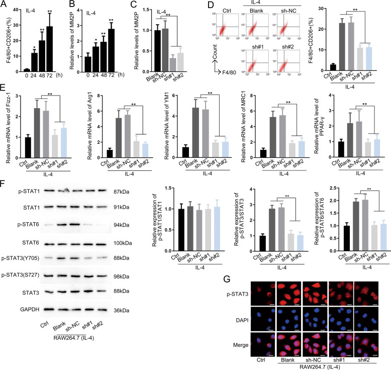
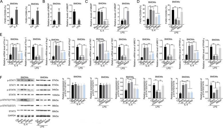
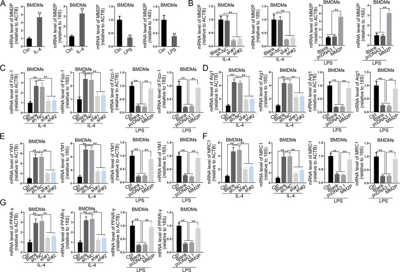
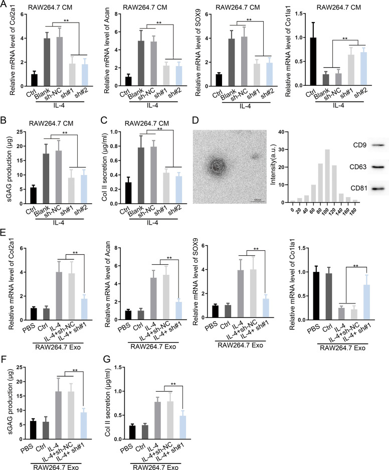
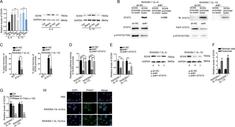
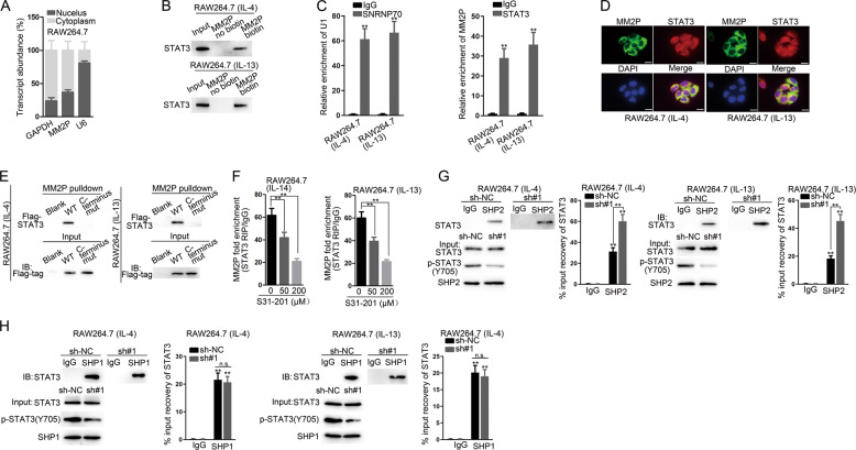
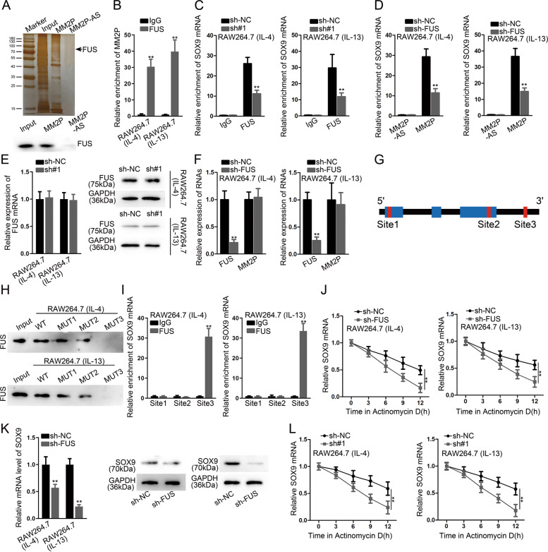
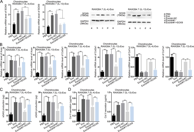
Monocyte-derived cells were shown to promote cartilage repair in osteoarthritis. The role of the long non-coding RNA (lncRNA) MM2P in this function of monocyte-derived cells remained unexplored. Treatment of RAW264.7 murine macrophages and mouse bone marrow-derived macrophages with IL-4 or IL-13 upregulated MM2P expression, upstream of STAT3 and STAT6 phosphorylation. Specifically, MM2P blocked SHP2-mediated dephosphorylation of STAT3 at Try705 and interacted with the RNA-binding protein FUS. In turn, p-STAT3 increased the Sox9 gene expression. These cells released Sox9 mRNA and protein-containing exosomes, as demonstrated by a transmission electron microscope, nanoparticle tracking analysis, and detection of typical surface markers. Their culture supernatant promoted the differentiation of mouse primary chondrocytes, i.e., upregulated the expression of Col1a2 and Acan genes and promoted the secretion of extracellular matrix components proteoglycan and type II collagen. These effects were mediated by Sox9 mRNA and protein delivered to chondrocytes by exosomes. Together, ex vivo treatment of monocyte-derived cells with IL-4 or IL-13 promoted chondrocyte differentiation and functions through exosome-mediated delivery of Sox9 mRNA and protein.


Conflict of interest statement

The authors declare that they have no conflict of interest.

Figures

Fig. 1
Fig. 1. MM2P facilitated M2 polarization.
a Flow cytometry analysis for the ratio of F4/80+CD206+ in RAW264.7 cells after treatment with IL-4. b Level of MM2P following the induction of M2 polarization at the indicated time. c The effect of sh-MM2P#1/2 transfection on the expression of MM2P in IL-4-induced M2 macrophages was measured by RT-qPCR. d Pictures of flow cytometry of F4/80+CD206+ cells and quantification of F4/80+CD206+ cell ratio under MM2P depletion in IL-4-induced cells. e RT-qPCR data for the levels of M2-related genes (Fizz-1, Arg1, YM1, MRC1, PPAR-γ) under the treatment of IL-4 with MM2P depletion in RAW264.7 cells. f Western blot for the total and phosphorylated STAT1, STAT6, and STAT3 in RAW264.7 cells treated with IL-4. g IF staining for the fluorescence intensity of p-STAT3 under IL-14 stimulation, as well as depletion of MM2P. Scale bar = 20 μm. **P < 0.01. Error bars are said to express as mean ± SD of three independent experiments in triplicates.
Fig. 2
Fig. 2. MM2P promoted switch from M1 to M2 phenotype to facilitate cartilage repair.
BMDMs were treated with IL-4 or LPS to induce M2 or M1 polarization. a Quantification of F4/80+CD206+ ratio in IL-4-treated BMDMs, and F4/80+CD86+ ratio in LPS-treated BMDMs. b RT-qPCR of MM2P levels in BMDMs of each group. c The IL-4-treated BMDMs were transfected with sh-NC or sh-MM2P#1/2, whereas the LPS-treated BMDMs were transfected with pcDNA3.1 or pcDNA3.1/MM2P. RT-qPCR of MM2P levels in BMDMs of each group. d Quantification of F4/80+CD206+ ratio in IL-4-treated BMDMs, and F4/80+CD86+ ratio in LPS-treated BMDMs. e RT-qPCR of MM2P levels in BMDMs of each group. f Western blots of p-STAT1, STAT1, p-STAT6, STAT6, p-STAT3, STAT3, and quantification of p-STAT1/STAT1, p-STAT6/STAT6, and p-STAT3/STAT3 in BMDMs treated with IL-4 and LPS. **P < 0.01. The error bar expressed as mean ± SD of three independent experiments in triplicates.
Fig. 3
Fig. 3. RT-qPCR analysis of lncRNA MM2P or the macrophage-related genes in LPS/IL-4-induced BMDMs transfected with MM2P-specific shRNA or MM2P expression vector using ACTB and 18S as housekeeping genes.
**P < 0.01. The error bar expressed as mean ± SD of three independent experiments in triplicates.
Fig. 4
Fig. 4. MM2P silence impaired the cartilage repair induced by M2 macrophage-derived exosomes.
Chondrocytes were cultured in conditioned medium (CM) of IL-4/IL-13-induced M2 macrophages with indicated transfections. a RT-qPCR data of chondrogenic-specific genes Col2a1, Acan, and SOX9, as well as dedifferentiation-related gene Col1a1 of each group. b, c The production of sGAG and Collagen II in chondrocytes. d TEM and nanoparticle tracking analysis (NTA) analysis of exosomes from IL-4/IL-13-induced M2 macrophages. The exosome markers CD9, CD63, and CD81 were detected by western blot. e Levels of Col2a1, Acan, SOX9, and Col1a1 in chondrocytes after the incubation with exosomes from IL-13/IL-4-induced M2 macrophages with MM2P silence. f, g The production of sGAG and Collagen II in each group. **P < 0.01. The error bar expressed as mean ± SD of three independent experiments in triplicates.
Fig. 5
Fig. 5. MM2P induced STAT3-regulated transactivation of SOX9 in M2 macrophages and M2 macrophages transmitted SOX9 to chondrocytes.
a The expression of SOX9 mRNA and protein in RAW264.7 cells under IL-13/IL-4 stimulation after silencing MM2P. b, c ChIP analysis of SOX9 promoter in IL-13/IL-4 induced M2 macrophages. d Luciferase activity of SOX9 promoter in IL-13/IL-4-induced M2 macrophages transfected with sh-NC, sh-MM2P, and sh-MM2P+ pcDNA3.1-STAT3. e SOX9 expression was detected by RT-qPCR and western blot in each group. f Level of SOX9 mRNA in M2 macrophage-derived exosomes and in the parental M2 macrophages. g The extracellular expression of SOX9 in the medium of IL-13/IL-4-induced M2 macrophages under the treatment of RNase A and Triton-X 100. h The exosomes isolated from IL-13/IL-4-induced M2 macrophages were marked by PKH67. The fluorescence intensity of PKH67 in chondrocytes was detected after culturing with the exosomes. Scale bar = 20 μm. **P < 0.01. The error bar expressed as mean ± SD of three independent experiments in triplicates.
Fig. 6
Fig. 6. MM2P prevented the SHP2-regulated dephosphorylation of STAT3.
a Subcellular fractionation assay for the MM2P location in RAW264.7 cells. b Pulldown assay was performed to verify the enrichment of STAT3 protein in the products pulled down by the MM2P biotin probe compared with the non-biotin probe. c RIP analysis confirmed the interaction between MM2P and STAT3. d FISH stain of MM2P and IF stain of STAT3 in M2 macrophages. Scale bar = 20 μm. e MM2P-pulldown assay for STAT3 enrichment after transfecting Flag-tagged STAT3 into IL-13/IL-4-induced M2 macrophages. f RIP analysis was used to detect the interaction between STAT3 and MM2P after adding S31–201 with indicated dose, the inhibitor of STAT3. g Co-IP assay was carried out in IL-13/IL-4-induced M2 macrophages to identify the binding of SHP2 or SHP1 with STAT3 after silencing MM2P. **P < 0.01. The error bar expressed as mean ± SD of three independent experiments in triplicates.
Fig. 7
Fig. 7. MM2P interacted with FUS to stabilize SOX9 mRNA.
a Pulldown assay followed by mass spectrometry identified the interaction between FUS and MM2P, which was confirmed by western blot. b, c RIP assay for the abundance of MM2P in FUS precipitates, and the abundance of SOX9 mRNA in the precipitates of FUS. d Pulldown assay for the abundance of SOX9 in the pulldown of the MM2P biotin group after silencing FUS. e RT-qPCR and western blot analysis of FUS mRNA and protein levels under MM2P depletion. f Levels of FUS and MM2P in IL-13/IL-4-induced M2 macrophages after silencing FUS. g Three potential FUS binding sites on SOX9 mRNA predicted by CLIP experimental data from Starbase3.0. h Pulldown-western blot assay for the enrichment of FUS in the pulldown of SOX9 mRNA. i RIP experiments confirmed the interaction between FUS and SOX9 mRNA at site 3 rather site 1 and 2. j SOX9 mRNA stability after Actinomycin D treatment, responding to FUS knockdown. k SOX9 mRNA and protein expressions under the silence of FUS in IL-13/IL-4-induced M2 macrophages. l mRNA stability of SOX9 in IL-13/IL-4-induced M2 macrophages, in response to MM2P knockdown. **P < 0.01. The error bar expressed as mean ± SD of three independent experiments in triplicates.
Fig. 8
Fig. 8. MM2P induced exosomal SOX9 from M2 macrophages to promote cartilage repair.
Chondrocytes were cultured with IL-4/IL-13-induced M2 macrophages with indicated transfections. a RT-qPCR and western blot experiments for the SOX9 mRNA and protein levels in chondrocytes of each group. b Levels of Acan, Col2a1, and Col1a1 in chondrocytes of each group. c, d The production of sGAG and Collagen II in chondrocytes of each group. **P < 0.01. The error bar expressed as mean ± SD of three independent experiments in triplicates.

References

    1. Kronenberg HM. Developmental regulation of the growth plate. Nature. 2003;423:332–336. doi: 10.1038/nature01657. - DOI - PubMed
    1. Hiroshi K. Endochondral ossification signals in cartilage degradation during osteoarthritis progression in experimental mouse models. Mol. Cells. 2008;25:1–6. - PubMed
    1. Bijlsma JWJ, Berenbaum F, Lafeber FPJG. Osteoarthritis: an update with relevance for clinical practice. Lancet. 2011;377:2115–2126. doi: 10.1016/S0140-6736(11)60243-2. - DOI - PubMed
    1. Altman, R. D., Schemitsch, E. & Bedi, A. Assessment of clinical practice guideline methodology for the treatment of knee osteoarthritis with intra-articular hyaluronic acid. Semin. Arthritis Rheum. 10.1016/j.semarthrit.2015.04.013 (2015). - PubMed
    1. Gao X-R, Chen Y-S, Deng W. The effect of vitamin D supplementation on knee osteoarthritis: a meta-analysis of randomized controlled trials. Int. J. Surg. 2017;46:14–20. doi: 10.1016/j.ijsu.2017.08.010. - DOI - PubMed

Publication types

LinkOut - more resources