. 2020 Sep 16;10(1):15171.
doi: 10.1038/s41598-020-71519-4.

An engineered antibody binds a distinct epitope and is a potent inhibitor of murine and human VISTA

Affiliations

An engineered antibody binds a distinct epitope and is a potent inhibitor of murine and human VISTA

Nishant Mehta et al. Sci Rep. .

Abstract

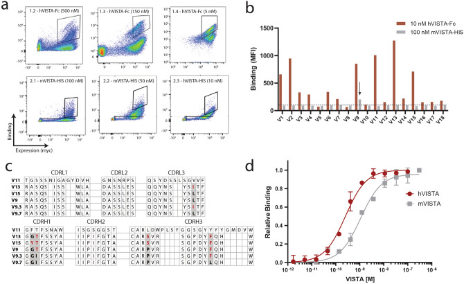
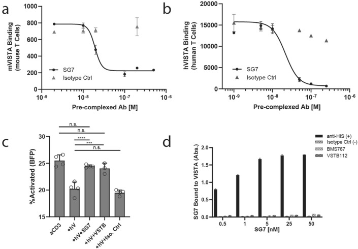
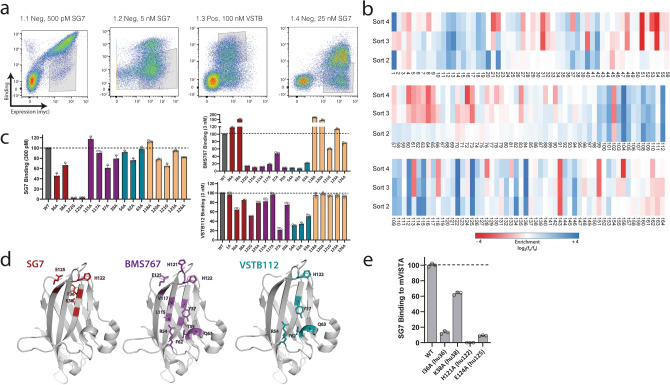
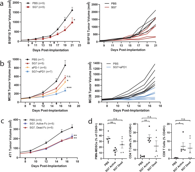
V-domain immunoglobulin (Ig) suppressor of T cell activation (VISTA) is an immune checkpoint that maintains peripheral T cell quiescence and inhibits anti-tumor immune responses. VISTA functions by dampening the interaction between myeloid cells and T cells, orthogonal to PD-1 and other checkpoints of the tumor-T cell signaling axis. Here, we report the use of yeast surface display to engineer an anti-VISTA antibody that binds with high affinity to mouse, human, and cynomolgus monkey VISTA. Our anti-VISTA antibody (SG7) inhibits VISTA function and blocks purported interactions with both PSGL-1 and VSIG3 proteins. SG7 binds a unique epitope on the surface of VISTA, which partially overlaps with other clinically relevant antibodies. As a monotherapy, and to a greater extent as a combination with anti-PD1, SG7 slows tumor growth in multiple syngeneic mouse models. SG7 is a promising clinical candidate that can be tested in fully immunocompetent mouse models and its binding epitope can be used for future campaigns to develop species cross-reactive inhibitors of VISTA.


Conflict of interest statement

N.M., S.M., R.K, and J.R.C. are included as inventors on intellectual property related to the work described in this manuscript. J.R.C. is a co-founder and J.R.C and R.K. are shareholders in xCella Biosciences, which is developing antibody therapeutics for applications in oncology. Other authors declare no competing financial interests.

Figures

Figure 1
Figure 1
Engineering a cross-reactive anti-VISTA antibody. (a) Library screening progression used to isolate scFv variants that bound human VISTA (Round 1) and mouse VISTA (Round 2). Flow cytometry gates used for screening are shown on dot plots of individual sorts. X-axis depicts expression of scFv on the yeast cell surface, y-axis depicts VISTA binding, measured by antibodies against c-myc and VISTA, respectively. (b) Binding intensity to human VISTA-Fc (red) and mouse VISTA-His (gray) of individual scFv clones isolated after Round 1 of screening. The V9 clone displayed above background binding signal to mouse VISTA-His. (c) Sequence comparison of scFv clones after the first round (V9, V11, V13, V15) and after the second round (V9.3 and V9.7) of sorting. Amino acid differences from final V9.7 clone are highlighted in red. (d) Full kinetic binding curves of SG7 (antibody form of top clone V9.7) against human VISTA-His and mouse VISTA-His monomers, as measured by KinExA. Mean ± standard deviation of duplicate measurements are shown for D.
Figure 2
Figure 2
SG7 is an inhibitor of mouse and human VISTA. (a) Mouse VISTA-Fc binding to activated mouse T cells at pH 6.0 with increasing concentrations of pre-complexed SG7 or isotype control. (b) Human VISTA-Fc binding to activated human T cells at pH 6.0 with increasing concentrations of pre-complexed SG7 or isotype control. (c) Rescuing effect of SG7 and VSTB112 on the activation of Jurkat NFAT (BFP) T cells in the presence of human VISTA-Fc. P-values obtained by one-way ANOVA (Tukey’s multiple comparison test), ***p < .005, ****p < .0005. (d) Competition ELISA to test simultaneous binding of SG7 and microtiter well-coated BMS767 or VSTB112 to soluble human VISTA. Binding signal of SG7 bound to the complex of VISTA and coated antibody is shown with increasing concentrations of SG7. In this assay, only the anti-His positive control can bind simultaneously to SG7 and VISTA. Mean ± SD for triplicate measurements are shown for all panels.
Figure 3
Figure 3
SG7 binds to a distinct epitope on VISTA. (a) Library screening progression used to isolate human VISTA mutants that lost binding to SG7 but retained binding to VSTB112. Flow cytometry gates used in screening are shown on dot plots of individual sorts. (b) Enrichment of mutations at each residue location in human VISTA obtained from NGS analysis of gated populations after each epitope mapping sort (blue—more enriched, red—less enriched). (c) Analysis of individual human VISTA mutations expressed on yeast. Binding intensity of each mutant to SG7, BMS767, or VSTB112 at the respective approximate dissociation constant (Kd) of each antibody. Bars are colored based on predicted epitope (SG7-red, BMS767-purple, VSTB112-cyan, neighboring residues-beige). Binding signal of each mutant was normalized to wild-type hVISTA. (d) Epitopes of SG7 (red), BMS767 (purple), and VSTB112 (cyan) antibodies based on single clone binding analysis. (e) Analysis of individual mouse VISTA mutations displayed on yeast. Selected mutants correspond with aligned human VISTA residues (in parentheses) that make up or are near the predicted SG7 epitope. Binding intensity of each mutant to 4 nM SG7 is shown. Binding signal of each mutant was normalized to wild-type mVISTA. Mean ± SD for triplicate measurements are shown for (c,e).
Figure 4
Figure 4
SG7 blocks VISTA binding interactions with PSGL-1 and VSIG3. (a) ELISA binding assay with microtiter well-coated hPSGL-1-Fc or hVSIG3-Fc incubated with human VISTA-Fc. (b) Competition binding ELISA with microtiter well-coated PSGL-1-Fc (left) or VSIG3-Fc (right) and increasing concentrations of SG7, BMS767, or VSTB112 in complex with 250 nM human VISTA-Fc. The binding signal of VISTA that was able to bind coated PSGL-1 or VSIG3 was detected. Competition binding ELISAs were performed at pH 6.0. (c) Single clone analysis of soluble VISTA-Fc mutants binding to well-coated PSGL-1-Fc or VSIG3-Fc in ELISA format. The binding signal of each VISTA-Fc mutant was normalized to wild-type VISTA binding to PSGL-1 or VSIG3, respectively. (d,e) Predicted epitope of VISTA binding to both cognate binding partners, PSGL-1 (red) and VSIG3 (cyan), based on single clone analysis using a panel of soluble mutants. Residue locations at which alanine mutations increased binding to PSGL-1 (d) or VSIG3 (e) are shown in orange. Mean ± SD for triplicate measurements are shown for ac.
Figure 5
Figure 5
VISTA blockade with SG7 slows tumor growth in multiple syngeneic mouse models. (a) B16F10 tumor-bearing C57BL/6 mice were treated with 10 mg/kg SG7 (dead Fc; mIgG2a-LALA/PG) bi-weekly, starting on day 10 (red arrows). n = 5 mice per group; mean tumor volume of each group (left) and individual tumor growth curves (right). All mice were euthanized on Day 21 due to volume and ulceration of untreated tumors. These data are representative of two independent experiments. Mean + SEM are shown. (b) MC38 tumor-bearing C57BL/6 mice were treated with 30 mg/kg SG7 (dead Fc) and/or 5 mg/kg anti-PD1, starting on day 9 (black arrows); n = 7 mice per group; mean tumor volume of each group (left) and individual tumor growth curves (right); all mice were euthanized on Day 17 due to volume and ulceration of untreated tumors. These data are representative of two independent experiments. Mean + SEM are shown. (c) 4T1 tumor-bearing BALB/c mice were treated with 30 mg/kg SG7 (active Fc; mIgG2a) or 30 mg/kg SG7 (dead Fc) bi-weekly, starting on day 8 (black arrows). n = 5–7 mice per group as indicated. All mice were euthanized on Day 17 for tumor extraction. Mean + SEM are shown. P-values in ac calculated by repeated measures two-way ANOVA (Tukey’s), asterisks denote significant difference from PBS (untreated), *p < 0.05, **p < 0.005, (d) Immune flow analysis of extracted 4T1 tumors on Day 17. The percentage of PMN-MDSCs, CD4 + T cells and CD8 + T cells out of total CD45 + cells in each tumor sample are shown. Mean ± SEM are shown. P-values calculated by one-way ANOVA (DMCT), *p < 0.05, **p < 0.005.

References

    1. Wang L, et al. VISTA, a novel mouse Ig superfamily ligand that negatively regulates T cell responses. J. Exp. Med. 2011;208:577–592. doi: 10.1084/jem.20100619. - DOI - PMC - PubMed
    1. ElTanbouly MA, et al. VISTA is a checkpoint regulator for naïve T cell quiescence and peripheral tolerance. Science. 2020;367:2. doi: 10.1126/science.aay0524. - DOI - PMC - PubMed
    1. Mercier IL, et al. VISTA regulates the development of protective antitumor immunity. Cancer Res. 2014;74:1933–1944. doi: 10.1158/0008-5472.CAN-13-1506. - DOI - PMC - PubMed
    1. Gao J, et al. VISTA is an inhibitory immune checkpoint that is increased after ipilimumab therapy in patients with prostate cancer. Nat. Med. 2017;23:551–555. doi: 10.1038/nm.4308. - DOI - PMC - PubMed
    1. Xie S, et al. Expression of the inhibitory B7 family molecule VISTA in human colorectal carcinoma tumors. Cancer Immunol. Immunother. 2018;67:1685–1694. doi: 10.1007/s00262-018-2227-8. - DOI - PMC - PubMed

Publication types

MeSH terms