. 2021 Jun;15(2):443-460.
doi: 10.1007/s12105-020-01216-1. Epub 2020 Sep 16.

Architectural Alterations in Oral Epithelial Dysplasia are Similar in Unifocal and Proliferative Leukoplakia

Affiliations

Architectural Alterations in Oral Epithelial Dysplasia are Similar in Unifocal and Proliferative Leukoplakia

Chia-Cheng Li et al. Head Neck Pathol. 2021 Jun.

Abstract

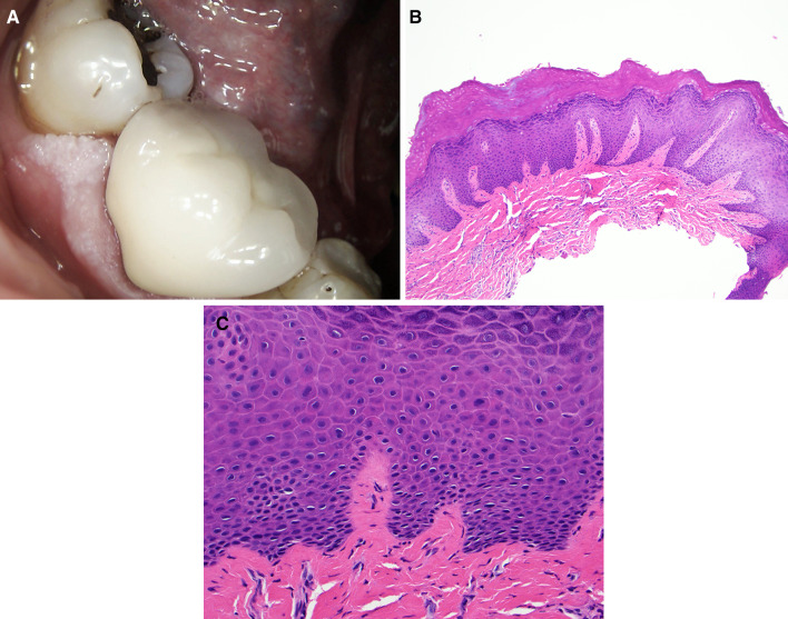
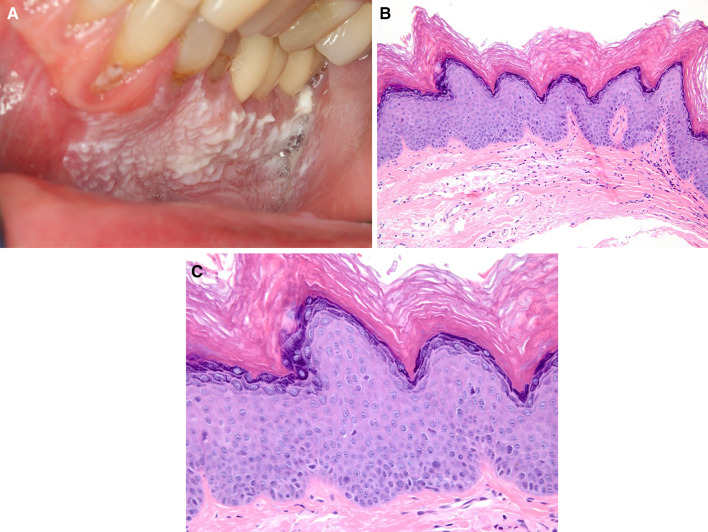
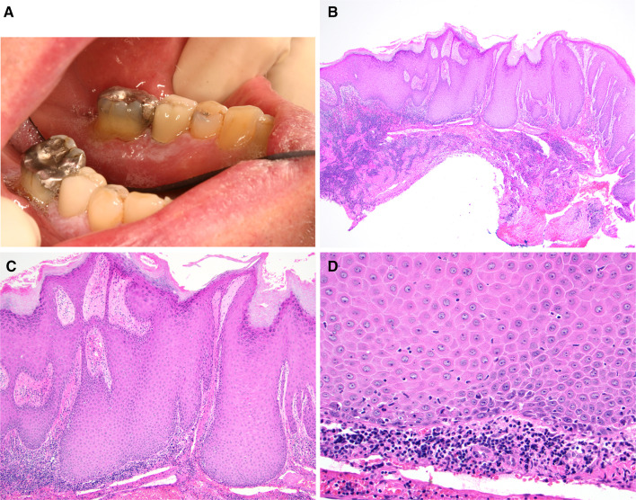
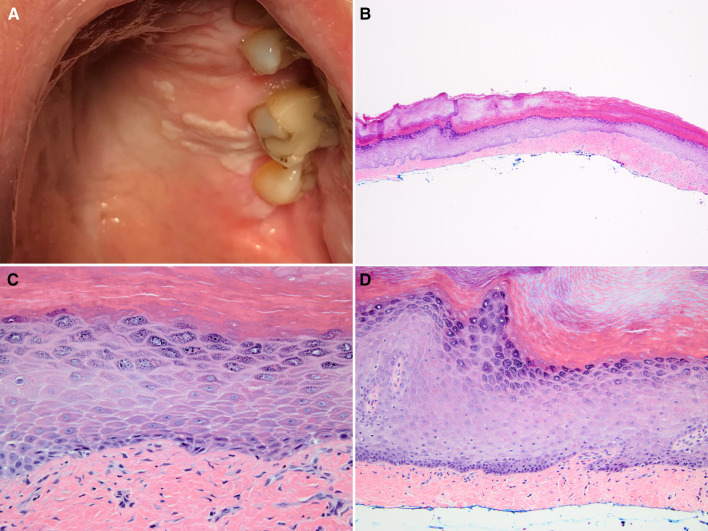
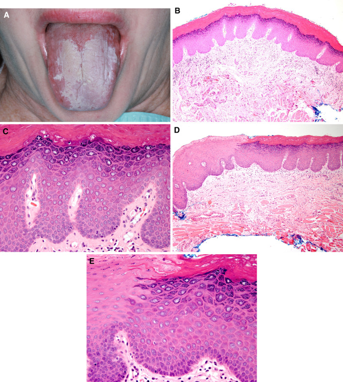
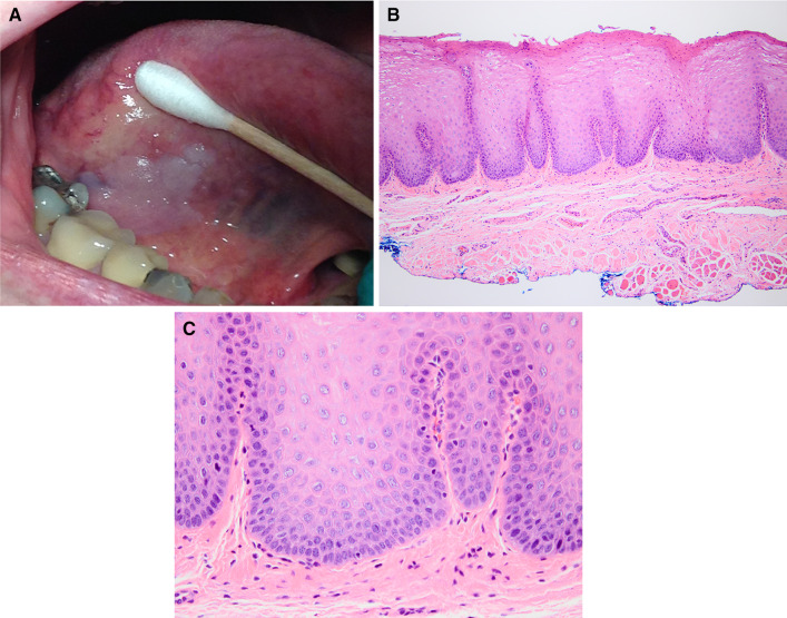
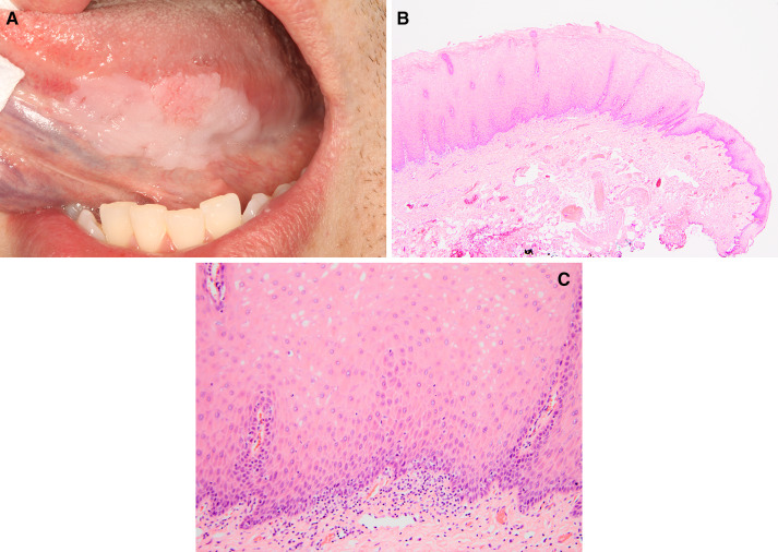
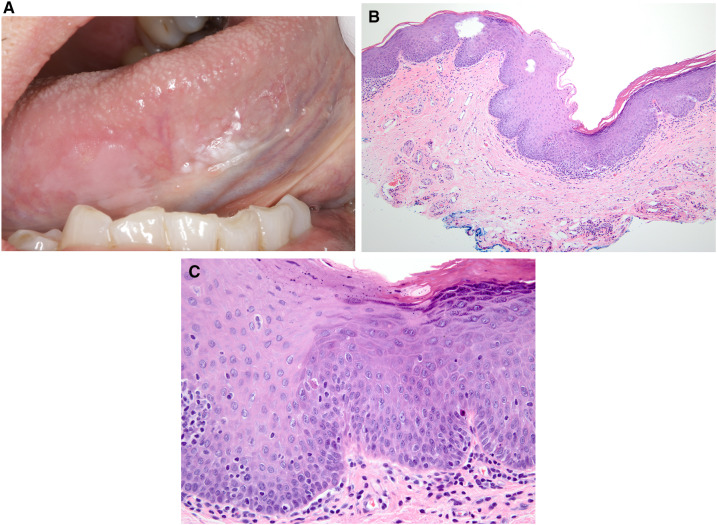
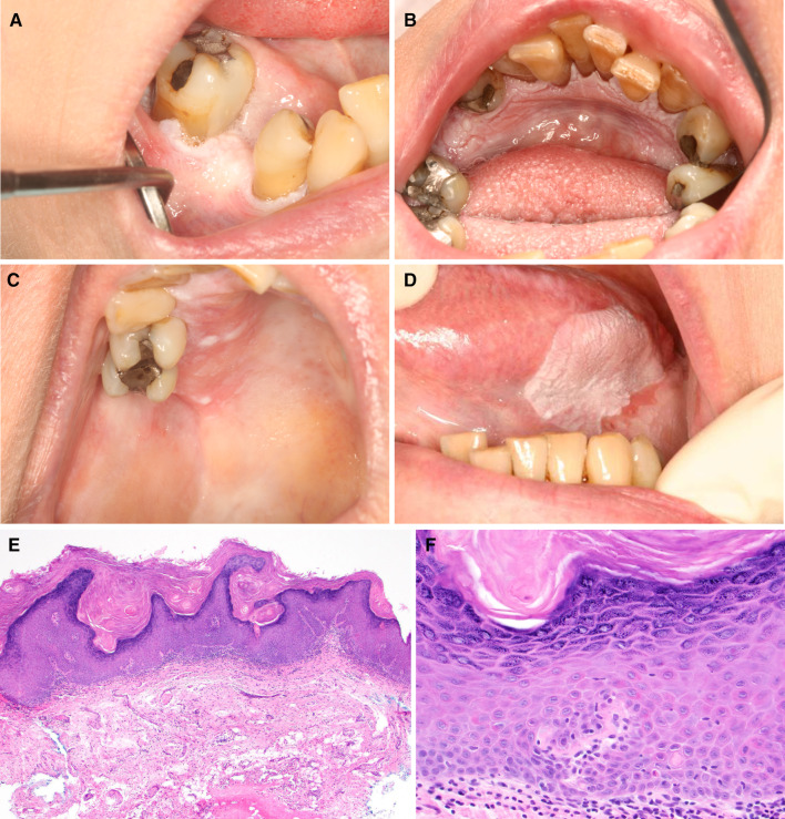
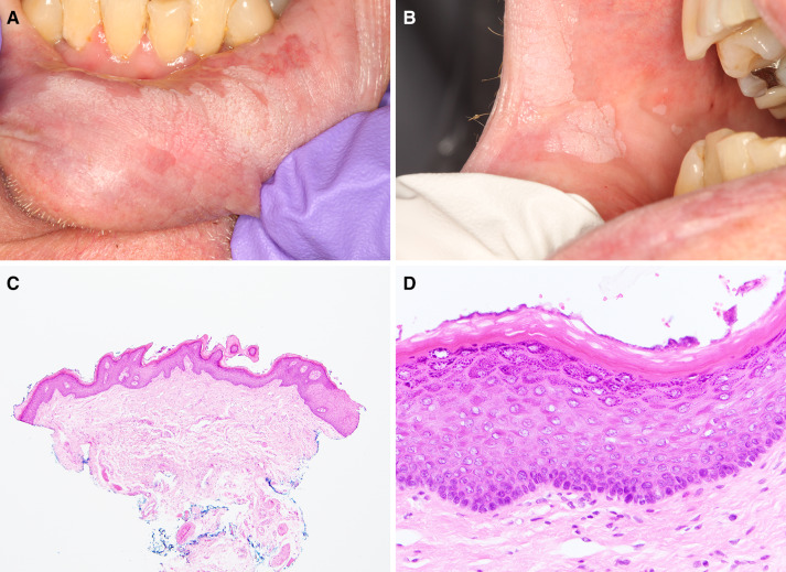
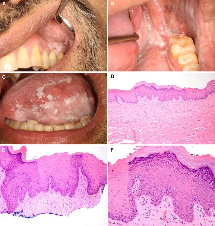
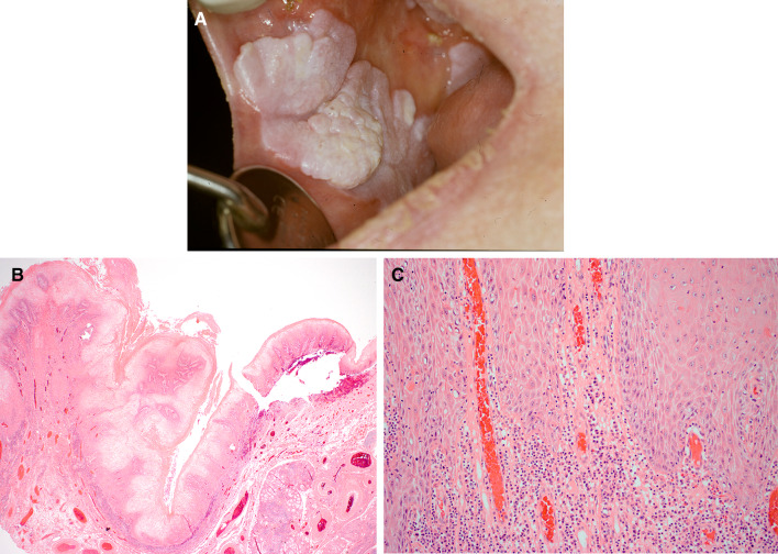
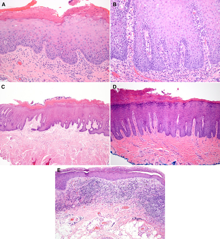
The current WHO histopathologic criteria for oral epithelial dysplasia (ED) are based on architectural and cytologic alterations, and do not address other histopathologic features of ED. Here we propose new diagnostic criteria including architectural, organizational, and cytologic features for oral ED. Cases of unifocal leukoplakia (UL) and proliferative leukoplakia (PL) with clinical photographs and follow-up information were identified. Only cases that showed minimal cytologic atypia or mild ED were used to demonstrate critical architectural changes as defined in this study. Eight biopsies from eight UL patients and 34 biopsies from four PL patients were included. The biopsies showed (a) corrugated, verrucous or papillary architecture, (b) hyperkeratosis with epithelial atrophy, (c) bulky squamous epithelial proliferation, and (d) demarcated hyperkeratosis and "skip" segments. The architectural alterations defined here are as important as the currently used criteria for the diagnosis of ED. Clinicopathologic correlation when diagnosing oral ED is also of the utmost importance in accurate diagnosis.

Keywords: Architectural alteration; Leukoplakia; Malignant transformation; Oral epithelial dysplasia; Proliferative verrucous leukoplakia.


Conflict of interest statement

No conflict of interest to disclose.

Figures

Fig. 1
Fig. 1
Case 1 a Unifocal non-homogenous leukoplakia on the mandibular gingiva exhibiting corrugated surface. b Corrugated hyperkeratosis, hypergranulosis and epithelial hyperplasia (H&E, original magnification ×40). c No ED present (H&E, original magnification ×400)
Fig. 2
Fig. 2
Case 2 a Unifocal non-homogenous leukoplakia on the mandibular gingiva exhibiting corrugated surface. b Atypical verrucous hyperkeratosis and epithelial atrophy (H&E, original magnification ×100). c Minimal cytologic atypia present (H&E, original magnification ×400)
Fig. 3
Fig. 3
Case 3 a Unifocal non-homogenous leukoplakia on the mandibular gingiva with a focally corrugated surface. b There is bulky, verrucous proliferation slightly exo- and endo-phytic forming bulbous rete ridges (H&E, original magnification ×40). c Bulbous rete ridges (H&E, original magnification ×200). d Minimal cytologic atypia and minimal chronic inflammatory infiltrate present (H&E, original magnification ×400)
Fig. 4
Fig. 4
Case 4 a Unifocal homogenous leukoplakia on the hard palatal mucosa. b Hyperkeratosis with hypergranulosis and epithelial atrophy (H&E, original magnification ×40). c No ED present (H&E, original magnification ×400). d Focal surface corrugation is identified (H&E, original magnification ×400)
Fig. 5
Fig. 5
Case 5 a Unifocal fissured homogenous leukoplakia on the dorsal tongue. b Demarcated and corrugated hyperkeratosis with hypergranulosis, and epithelial atrophy (loss of papillae) (H&E, original magnification ×100). c Minimal cytologic atypia present (H&E, original magnification ×400). d Demarcated hyperkeratosis is noted (H&E, original magnification ×100). e Minimal ED with mild chronic inflammation present (H&E, original magnification ×400)
Fig. 6
Fig. 6
Case 6 a Unifocal homogenous leukoplakia on the ventral tongue. b There is bulky squamous epithelial proliferation (H&E, original magnification ×100). c Minimal ED present (H&E, original magnification ×400)
Fig. 7
Fig. 7
Case 7 a Unifocal homogenous leukoplakia on the left ventral tongue. b There is bulky, exophytic squamous epithelial proliferation (H&E, original magnification ×100). c No ED present (H&E, original magnification ×400)
Fig. 8
Fig. 8
Case 8 a Unifocal non-homogenous leukoplakia on the right ventral tongue. b Demarcated hyperkeratosis with “skip” segments (H&E, original magnification ×100). c Mild ED and a mild chronic inflammatory infiltrate is identified (H&E, original magnification ×400)
Fig. 9
Fig. 9
Case 9 Proliferative leukoplakia on a, b the mandibular gingiva, c the hard-palatal mucosa, and d the latero-ventral tongue. e Biopsy of tongue lesion exhibits atypical verrucous hyperplasia (H&E, original magnification ×40). f Minimal cytologic atypia and minimal inflammatory infiltrate present (H&E, original magnification × 400)
Fig. 10
Fig. 10
Case 10 Proliferative leukoplakia on a the lower lip mucosa and b the buccal mucosa. c There is atypical verrucous hyperkeratosis and epithelial atrophy (H&E, original magnification ×40). d ED is not identified (H&E, original magnification ×400)
Fig. 11
Fig. 11
Case 11 Proliferative leukoplakia on a the gingiva, b retromolar trigone extending to the anterior tonsillar pillar, and c the latero-ventral and dorsal tongue. d Biopsy from the tongue exhibits demarcated hyperkeratosis (H&E, original magnification ×100). e A later biopsy from the tongue exhibits demarcated corrugated hyperkeratosis and acanthosis with skip segments (H&E, original magnification ×100). f Mild ED is focally present (H&E, original magnification ×400)
Fig. 12
Fig. 12
Case 12 a Proliferative leukoplakia on the buccal mucosa extending to the commissure area with a second lesion further posteriorly. b There is bulky squamous epithelial proliferation (H&E, original magnification ×20). c No ED present (H&E, original magnification ×400)
Fig. 13
Fig. 13
a Moderate ED characterized by drop-shaped rete ridges, cells with increased nuclear:cytoplasmic ratio and dyskeratosis involving approximately half the thickness of the epithelium (H&E, original magnification ×200). b Severe ED characterized by bulbous rete ridges, dyscohesion, cells with increased nuclear:cytoplasmic ratio, dyskeratosis and slight nuclear pleomorphism and hyperchromasia involving greater than 2/3 the thickness of the epithelium (H&E, original magnification ×200). c Chronic bite/factitial keratosis (morsicatio mucosae oris) with parakeratosis and acanthosis (H&E, original magnification ×100). d Benign alveolar ridge keratosis with hyperkeratosis, wedge-shaped hypergranulosis and acanthosis (H&E, original magnification ×100). e Lichen planus exhibiting hyperkeratosis, epithelial atrophy, and interface inflammation (H&E, original magnification ×100)

References

    1. Warnakulasuriya S, Johnson NW, van der Waal I. Nomenclature and classification of potentially malignant disorders of the oral mucosa. J Oral Pathol Med. 2007;36(10):575–580. - PubMed
    1. Reibel J, Gale N, Hille J, et al. Oral potentially malignant disorders and oral epithelial dysplasia. WHO Classif Head Neck Tumours. 2017;9:112.
    1. Woo SB, Grammer RL, Lerman MA. Keratosis of unknown significance and leukoplakia: a preliminary study. Oral Surg Oral Med Oral Pathol Oral Radiol. 2014;118(6):713–724. - PubMed
    1. Napier SS, Speight PM. Natural history of potentially malignant oral lesions and conditions: an overview of the literature. J Oral Pathol Med. 2008;37(1):1–10. - PubMed
    1. Kumar A, Cascarini L, McCaul JA, et al. How should we manage oral leukoplakia? Br J Oral Maxillofacial Surg. 2013;51(5):377–383. - PubMed

LinkOut - more resources