Review
. 2020 Jul 8;10(3):785-809.
doi: 10.1002/cphy.c190029.

Role of Skeletal Muscle in Insulin Resistance and Glucose Uptake

Affiliations
Review

Role of Skeletal Muscle in Insulin Resistance and Glucose Uptake

Karla E Merz et al. Compr Physiol. .

Abstract

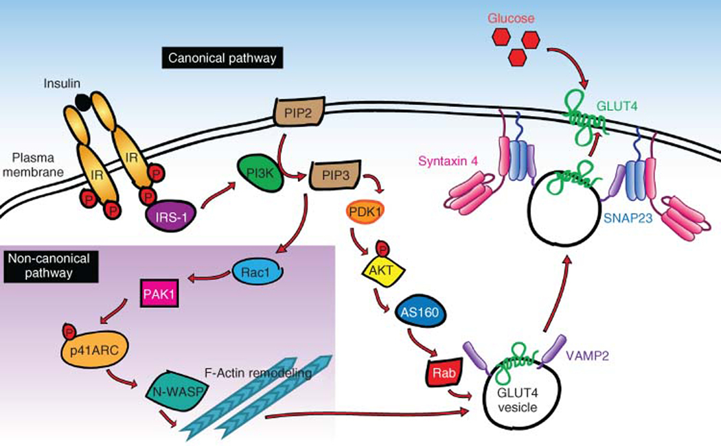
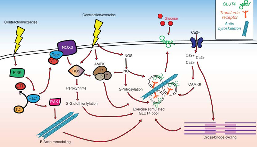
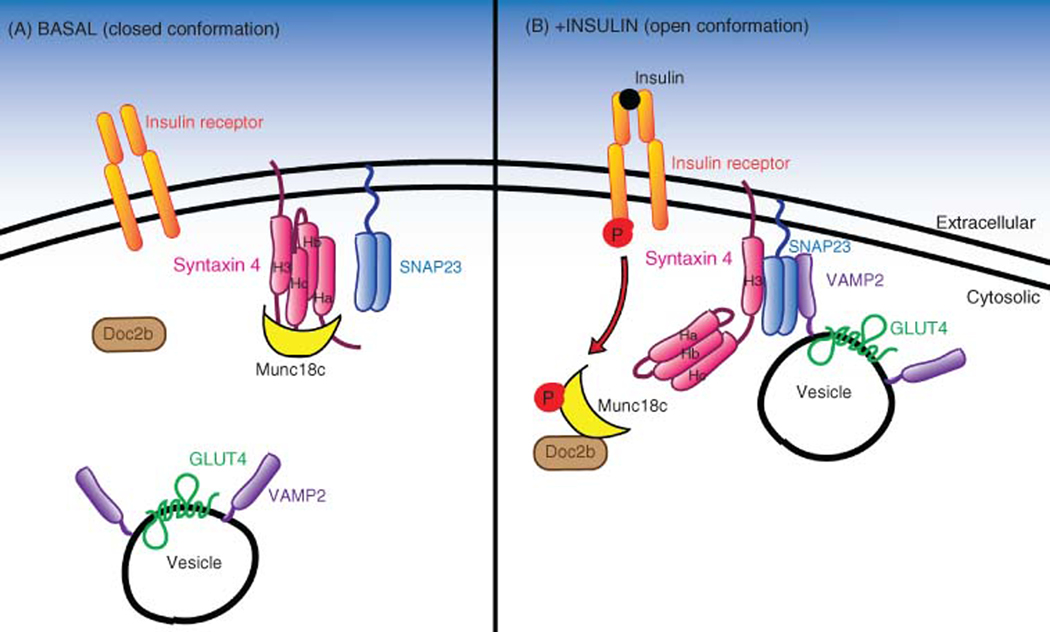
The skeletal muscle is the largest organ in the body, by mass. It is also the regulator of glucose homeostasis, responsible for 80% of postprandial glucose uptake from the circulation. Skeletal muscle is essential for metabolism, both for its role in glucose uptake and its importance in exercise and metabolic disease. In this article, we give an overview of the importance of skeletal muscle in metabolism, describing its role in glucose uptake and the diseases that are associated with skeletal muscle metabolic dysregulation. We focus on the role of skeletal muscle in peripheral insulin resistance and the potential for skeletal muscle-targeted therapeutics to combat insulin resistance and diabetes, as well as other metabolic diseases like aging and obesity. In particular, we outline the possibilities and pitfalls of the quest for exercise mimetics, which are intended to target the molecular mechanisms underlying the beneficial effects of exercise on metabolic disease. We also provide a description of the molecular mechanisms that regulate skeletal muscle glucose uptake, including a focus on the SNARE proteins, which are essential regulators of glucose transport into the skeletal muscle. © 2020 American Physiological Society. Compr Physiol 10:785-809, 2020.


Figures

Figure 1
Figure 1
A simplified model of insulin-stimulated translocation of the GLUT4 glucose transporter in skeletal muscle. Shown is the canonical insulin signaling pathway, and a noncanonical pathway (inset in purple). Both signaling pathways culminate with SNARE protein (Syntaxin 4, SNAP23, and VAMP2)-mediated vesicle trafficking and fusion with the plasma membrane. Vesicle fusion allows GLUT4 to integrate into the plasma membrane and transport glucose from the blood into the skeletal muscle.
Figure 2
Figure 2
Contraction-mediated GLUT4 translocation. When contraction occurs via exercise (depicted by yellow lightning bolts) multiple pathways are activated, which can all lead to GLUT4 translocation and glucose uptake. Left: contraction causes Rac1-GTP loading/activation, which induces PAK1 phosphorylation/activation, and F-actin remodeling to activate the exercise stimulated GLUT4 vesicle pool; it can also activate NOX2 which causes reactive oxygen species (ROS) formation. ROS can also be stimulated directly by contraction, leading to peroxynitrite and subsequent GLUT4 translocation, or can cause AMPK activation. Nitric oxide (NO) production can lead to S-nitrosylation of proteins that cause GLUT4 translocation. Muscle excitation can also cause calcium influx into the muscle cell (far right), which can activate CAMKII and lead to glucose uptake. Calcium influx can also stimulate cross-bridge cycling, the process of muscle fiber contraction, which leads to GLUT4 translocation and glucose uptake into the cell.
Figure 3
Figure 3
SNARE complex formation under basal versus insulin-stimulated conditions. (A) Under basal conditions, Syntaxin 4 (STX4, pink) is in a closed conformation, which is maintained by the accessory protein Munc18c (yellow). (B) Upon insulin stimulation, the insulin receptor (IR) is phosphorylated, after which phospho-IR binds to and phosphorylates Munc18c. Phospho-Munc18c then lets go of STX4 and binds to DOC2B (brown), allowing STX4 to adopt its open conformation. In its open conformation. In its open conformation, the STX4 H3 domain is exposed, enabling STX4 to associate with SNAP23 and VAMP2 (purple) on the GLUT4 vesicle, leading to the formation of the SNARE complex, mediating vesicle docking and fusion to the plasma membrane.
Figure 4
Figure 4
The role of mitochondrial dynamics in maintaining skeletal muscle function and preventing disease. Center: mitochondrial fission and fusion are balanced and under these conditions, there is normal mitochondrial function, ATP production, and cell death. Left: fission outweighs fusion, so mitochondria become fragmented, there is an increase in mitophagy and cell death, a reduction in mitochondrial function, and increased ROS production. This contributes to diseases including aging, obesity, and insulin resistance. Right: mitochondrial fusion outweighs fission, leading to increased mitochondrial function, reduced cell death, elongated mitochondria, and increased fatty acid oxidation. However, this can also cause cell stress and starvation due to the high demands of the large mitochondria.

Similar articles

Cited by

References

    1. Ahirwar AK, Jain A, Goswami B, Bhatnagar MK, Bhatacharjee J. Imbalance between protective (adiponectin) and prothrombotic (Plasminogen Activator Inhibitor-1) adipokines in metabolic syndrome. Diabetes Metab Syndr 8: 152–155, 2014. - PubMed
    1. Akbar H, Duan X, Piatt R, Saleem S, Davis AK, Tandon NN, Bergmeier W, Zheng Y. Small molecule targeting the Rac1-NOX2 interaction prevents collagen-related peptide and thrombin-induced reactive oxygen species generation and platelet activation. J Thromb Haemost 16: 2083–2096, 2018. - PMC - PubMed
    1. Alessi DR, Andjelkovic M, Caudwell B, Cron P, Morrice N, Cohen P, Hemmings BA. Mechanism of activation of protein kinase B by insulin and IGF-1. EMBO J 15: 6541–6551, 1996. - PMC - PubMed
    1. Alessi DR, James SR, Downes CP, Holmes AB, Gaffney PR, Reese CB, Cohen P. Characterization of a 3-phosphoinositide-dependent protein kinase which phosphorylates and activates protein kinase Balpha. Curr Biol 7: 261–269, 1997. - PubMed
    1. Almendro V, Busquets S, Ametller E, Carbo N, Figueras M, Fuster G, Argiles JM, Lopez-Soriano FJ. Effects of interleukin-15 on lipid oxidation: Disposal of an oral [(14)C]-triolein load. Biochim Biophys Acta 1761: 37–42, 2006. - PubMed

Publication types