. 2020 Oct 20;53(10):2425-2442.
doi: 10.1021/acs.accounts.0c00482. Epub 2020 Sep 17.

Sulfono-γ-AApeptides as Helical Mimetics: Crystal Structures and Applications

Affiliations

Sulfono-γ-AApeptides as Helical Mimetics: Crystal Structures and Applications

Peng Sang et al. Acc Chem Res. .

Abstract

Foldamers have defined and predictable structures, improved resistance to proteolytic degradation, enhanced chemical diversity, and are versatile in their mimicry of biological molecules, making them promising candidates in biomedical and material applications. However, as natural macromolecules exhibit endless folding structures and functions, the exploration of the applications of foldamers remains crucial. As such, it is imperative to continue to discover unnatural foldameric architectures with new frameworks and molecular scaffolds. To this end, we recently developed a new class of peptidomimetics termed ″γ-AApeptides", oligomers of γ-substituted-N-acylated-N-aminoethyl amino acids, which are inspired by the chiral peptide nucleic acid backbone. To date γ-AApeptides have been shown to be resistant to proteolytic degradation and possess limitless potential to introduce chemically diverse functional groups, demonstrating promise in biomedical and material sciences. However, the structures of γ-AApeptides were initially unknown, rendering their rational design for the mimicry of a protein helical domain impossible in the beginning, which limited their potential development. To our delight, in the past few years, we have obtained a series of crystal structures of helical sulfono-γ-AApeptides, a subclass of γ-AApeptides. The single-crystal X-ray crystallography indicates that sulfono-γ-AApeptides fold into unprecedented and well-defined helices with unique helical parameters. On the basis of the well-established size, shape, and folding conformation, the design of sulfono-γ-AApeptide-based foldamers opens a new avenue for the development of alternative unnatural peptidomimetics for their potential applications in chemistry, biology, medicine, materials science, and so on.In this Account, we will outline our journey on sulfono-γ-AApeptides and their application as helical mimetics. We will first briefly introduce the design and synthetic strategy of sulfono-γ-AApeptides and then describe the crystal structures of helical sulfono-γ-AApeptides, including left-handed homogeneous sulfono-γ-AApeptides, right-handed 1:1 α/sulfono-γ-AA peptide hybrids, and right-handed 2:1 α/sulfono-γ-AA peptide hybrids. After that, we will illustrate the potential of helical sulfono-γ-AApeptides for biological applications such as the disruption of medicinally relevant protein-protein interactions (PPIs) of BCL9-β-catenin and p53-MDM2/MDMX as well as the mimicry of glucagon-like peptide 1 (GLP-1). In addition, we also exemplify their potential application in material science. We expect that this Account will shed light on the structure-based design and function of helical sulfono-γ-AApeptides, which can provide a new and alternative way to explore and generate novel foldamers with distinctive structural and functional properties.


Conflict of interest statement

The authors declare no competing financial interest.

Figures

Figure 1.
Figure 1.
Chemical structure of α-peptide, chiral PNA, l-γ-AApeptide, l-sulfono-γ-AApeptide, d-sulfono-γ-AApeptide, 1:1 α/l-sulfono-γ-AApeptide, and 2:1 α/d-sulfono-γ-AApeptide.
Figure 2.
Figure 2.
Homogeneous sulfono-γ-AA peptidic oligomers prepared for structural and spectroscopic evaluation in the study. Reproduced with permission from ref . Copyright 2020 John Wiley& Sons, Inc.
Figure 3.
Figure 3.
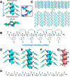
(A) Side and top views of the crystal structure of 1a. Hydrogen bonding is shown in red. (B) The intramolecular 14-hydrogen-bonding pattern of 1a detected in the crystal structure. (C) Crystal packing of 1a viewed perpendicular to and down the helix axis. (D-G) Comparison of the crystal structures of 1a, 3a, 4b, and 6a. (H) Sequence structure of oligomer 6a. Reproduced with permission from ref . Copyright 2020 John Wiley& Sons, Inc.
Figure 4.
Figure 4.
(A) Sequence structure of monomer 7 and the 13-atom-hydrogen-bonding pattern. (B) Sequence structure of dimer 8. (C) Crystal structure of monomer 7 stabilized by intramolecular hydrogen bond (magentas dashed line in inset). (D) Crystal packing model of 7. The disordered acetonitriles are excluded from the crystal lattice of 7. (E) Cartoon representation of X-ray crystal structures from mono helix 7 to the covalent-bonded zippered dimer 8. Dashed red lines highlight the intramolecular hydrogen bond in dimer 8. Reproduced with permission from ref . Copyright 2020 American Chemical Society.
Figure 5.
Figure 5.
(A) Sequence structures of foldamers 9, 10, 11, and 12. (B) Crystal structure of foldamer 9, as a helical representation. (C) Infinite 1D chain formed by the head-to-tail assembly of foldamer 9 in terms of both intramolecular and intermolecular hydrogen bonding. (D) Crystal packing of 9, including both antiparallel and perpendicular helices. (E) Crystal structure of foldamer 10. 3D supramolecular network of foldamer 10. (F) comparison of intermolecular halogen interactions between foldamer 9 (a), foldamer 10 (b), foldamer 11 (c), and foldamer 12 without halogenated side chains (d). Nonpolar hydrogens are omitted for clarity. Reproduced with permission from ref . Copyright 2020 John Wiley& Sons, Inc.
Figure 6.
Figure 6.
(A) 2:1 α/d-sulfono-γ-AA peptidic oligomers prepared for structural and spectroscopic evaluation in this study. (B) Side views of single-crystals 13, 14, 15, and 16. Hydrogen bonding is shown in cyan. (C) Top views of single-crystals 13, 14, 15, and 16 along the helix axis. (D) Structure of crystal 14 packing along the peptide axis; the intermolecular hydrogen-bonding pattern is shown in the inset for clarity. (E) Cartoon representation of 14 shown in an oval to further clarify the helix. (F) Crystal packing of oligomer 13 viewed perpendicular and then down to the helical axis. (G) 16–16-14-hydrogen-bonding pattern detected in the crystal structure of 14. Reproduced with permission from ref . Copyright 2020 American Chemical Society.
Figure 7.
Figure 7.
(A) The interaction of p53 with the crystal structure of MDM2 (PDB: 1YCR). p53 is shown as the cartoon whereas MDM2 is shown as the surface representation. (B) The chemical structure of sulfono-γ-AApeptides. a and b denote the chiral side chain and the sulfonamido side chain from the building block, respectively. (C) The crystal structure of a sulfono-γ-AApeptide. (D) Top view of panel C. (E and F) The schematic representation of the distribution of side chains from sulfono-γ-AApeptides. (E) Side view and (F) top view of the helical wheel. Reproduced with permission from ref . Copyright 2020 American Chemical Society.
Figure 8.
Figure 8.
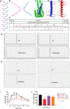
(A-D) Chemical shift mapping of 19 binding to MDM2. (A) Overlay of N heteronuclear single quantum coherence (HSQC) spectra of MDM2 before (blue resonances) and after (red resonances) the addition of 19. HSQC spectra were collected with a 2-fold stoichiometric excess of 19. (B) Average chemical shift changes, in parts per million (ppm), for the amide proton and nitrogen resonances in MDM2 p53BD residues binding to 19. (C and D) Surface image of the MDM2 p53BD structure. (E) Analytic high-performance liquid chromatography (HPLC) trace of p53 and 19 before and after incubation with Pronase (0.1 mg/mL) in 100 mM, pH 7.8 ammonium bicarbonate buffer at 37 °C. Reproduced with permission from ref . Copyright 2020 American Chemical Society.
Figure 9.
Figure 9.
(A-F) The chemical and crystal structures of the α-peptides (A and B), chemical and crystal structures of homogeneous l-sulfono-γ-AApeptides (C and D), and chemical and modeled structures of homogeneous d-sulfono-γ-AApeptides (E and F). (G and H) Schematic representation of the distribution of side chains from homogeneous d-sulfono-γ-AApeptides based on computational modeling. (G) Side view and (H) top view of the helical wheel. Reproduced with permission from ref . Copyright 2020 American Chemical Society.
Figure 10.
Figure 10.
(A) Structures of d-sulfono-γ-AApeptides investigated for the disruption the p53-MDM2 interaction. The side chains mimicking Phe19, Trp23, and Leu26 in p53 are shown in blue. (B-D) The crystal structure of the interaction of p53 with MDM2 (PDB: 1YCR) (B), modeling of the lead homogeneous l-sulfono-γ-AApeptide (C), and the designed homogeneous d-sulfono-γ-AApeptide 21 (D) interaction with MDM2. p53 and the homogeneous d-sulfono-γ-AApeptide are shown as a magenta cartoon, the homogeneous l-sulfono-γ-AApeptide is shown as a green cartoon, and MDM2 is shown as a gray cartoon. Reproduced with permission from ref . Copyright 2020 American Chemical Society.
Figure 11.
Figure 11.
(A and B) The α-helical HD2 domain of BCL9, which directly engages a surface groove of β-catenin, provided the template for structural stabilization by hydrocarbon stapling (PDB: 2GL7). (A) Cartoon representation of the residues of BCL9 (red), which are critical for binding to β-catenin, are shown as sticks. (B) BCL9 is shown in as a stick model, and β-catenin is represented with a surface model. (C-F) The schematic representation of the distribution of side chains from sulfono-γ-AApeptides. (C) Side and (D) top view of the helical wheel. (E) The position map of critical residues of the BCL9 helix. (F) The position map of side chains of sulfono-γ-AApeptides that are designed to mimic the residues in panel E. (G) The BCL9 peptide 22 and lead compounds 23–25. Reproduced with permission from ref . Copyright 2020 United States National Academy of Sciences.
Figure 12.
Figure 12.
(A) Confocal fluorescence microscopy images of SW480 cells treated with 1 μM and 10 μM of the FITC-labeled peptide 22 and sulfono-γ-AApeptides 23–25 for 2 h (magnification, 630×). (B) The SW480 cell lysate was incubated with 24-biotin or 25-biotin, followed by streptavidin pull-down experiments. (C) Co-immunoprecipitation (co-IP) experiments to evaluate the disruption of the β-catenin-BCL9 PPI by 25 in Wnt/ β-catenin hyperactive cancer cells. Reproduced with permission from ref . Copyright 2020 United States National Academy of Sciences.
Figure 13.
Figure 13.
(A and B) Schematic representation of the distribution of side chains from the sulfono-γ-AApeptide in panel F. (A) Side view and (B) top view of the helical wheel. (C) GLP-1 binds to GLP-1R (PDB: 5VAI). GLP-1 (7–36) is shown in blue, and GLP-1R is represented as a green cartoon. (D) The helical domain of GLP-1 with critical residues are presented as sticks. (E) Design of sulfono-γ-AApeptide 27, with side chains mimicking the important residues in panel B. The helix was built on the crystal structure. X, Y, and Z was designated to indicate the face of the residues on the helix. (F) The Structures and agonist activities of GLP-1(7–36) 26 and lead sulfono-γ-AApeptide 27. (G) Analytic HPLC traces of GLP-1(7–36) 26 and lead sulfono-γ-AApeptide 27 before and after incubation with pronase (0.1 mg/mL). (H) The serum stability of 26 and 27 at 37 °C for 24 h. (I and J) Pharmacodynamics of the GLP-1 mimic peptide 27 in mice. A single dose of peptides was intraperitoneally administered into mice 1 h before the oral glucose tolerance test (OGTT) (2 g/kg glucose). (I) Blood glucose concentrations were monitored for up to 120 min after oral glucose challenge. (J) The average area under the curve (AUC) was calculated from OGTT data. Results show the mean ± the standard error of the mean (SEM) of six mice per treatment group; *P < 0.05 versus vehicle; t test. Reproduced with permission from ref . Copyright 2020 the American Association for the Advancement of Science.
Figure 14.
Figure 14.
Chemical and crystal structures of the sulfono-γ-AApeptides 28–31. Reproduced with permission from ref . Copyright 2020 John Wiley & Sons, Inc.
Figure 15.
Figure 15.
Chemical and crystal structure of TPE-α/sulfono-γ-AApeptide 32. (A) Chemical structure and the 13-atom hydrogen-bonding pattern. (B) Crystal structure of the bonding pattern. (C) Helical cartoon of the crystal structure. (D) Crystal packing of 1 along the peptide axis. (E) Cartoon structure of structure in panel D. (F) Packing mode of the crystal. Reproduced with permission from ref . Copyright 2020 American Chemical Society.
Scheme 1.
Scheme 1.
General Synthetic Route for the Preparation of Sulfono-γ-AApeptide Building Blocks
Scheme 2.
Scheme 2.
General Synthetic Route for the Preparation of Sulfono-γ-AApeptides

Similar articles

Cited by

References

    1. She F; Teng P; Peguero-Tejada A; Wang M; Ma N; Odom T; Zhou M; Gjonaj E; Wojtas L; van der Vaart A; Cai J De Novo Left-Handed Synthetic Peptidomimetic Foldamers. Angew. Chem., Int. Ed 2018, 57 (31), 9916–9920. - PMC - PubMed
    1. Teng P; Niu Z; She F; Zhou M; Sang P; Gray GM; Verma G; Wojtas L; van der Vaart A; Ma S; Cai J Hydrogen-Bonding-Driven 3D Supramolecular Assembly of Peptidomimetic Zipper. J. Am. Chem. Soc. 2018, 140 (17), 5661–5665. - PMC - PubMed
    1. Sang P; Zhang M; Shi Y; Li C; Abdulkadir S; Li Q; Ji H; Cai J Inhibition of β-catenin/B cell lymphoma 9 protein-protein interaction using α-helix-mimicking sulfono-γ-AApeptide inhibitors. Proc. Natl. Acad. Sci. U. S. A. 2019, 116 (22), 10757–10762. - PMC - PubMed
    1. Sang P; Zhou Z; Shi Y; Lee C; Amso Z; Huang D; Odom T; Nguyen-Tran VTB; Shen W; Cai J The activity of sulfono-γ-AApeptide helical foldamers that mimic GLP-1. Sci. Adv 2020, 6 (20), No. eaaz4988. - PMC - PubMed
    1. Guichard G; Huc I Synthetic foldamers. Chem. Commun. 2011, 47 (21), 5933–5941. - PubMed

Publication types