. 2021 Jan 4;131(1):e137468.
doi: 10.1172/JCI137468.

Stressed erythrophagocytosis induces immunosuppression during sepsis through heme-mediated STAT1 dysregulation

Affiliations

Stressed erythrophagocytosis induces immunosuppression during sepsis through heme-mediated STAT1 dysregulation

Tolani F Olonisakin et al. J Clin Invest. .

Abstract

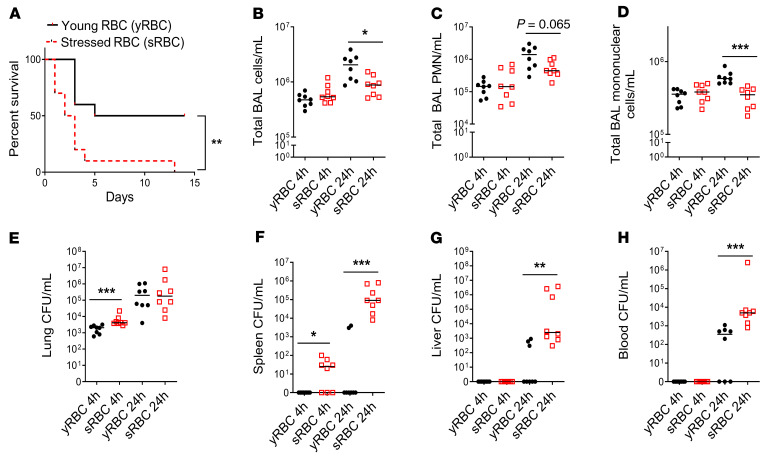
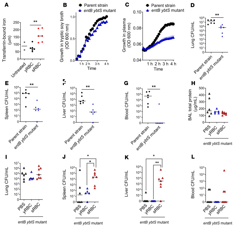
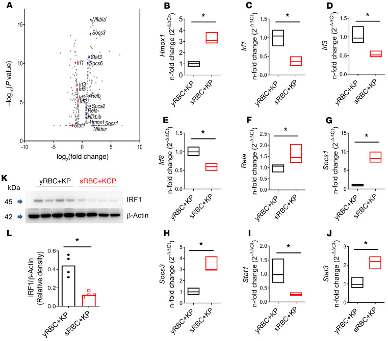
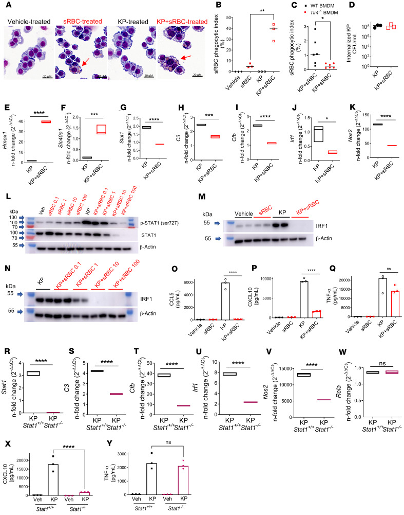
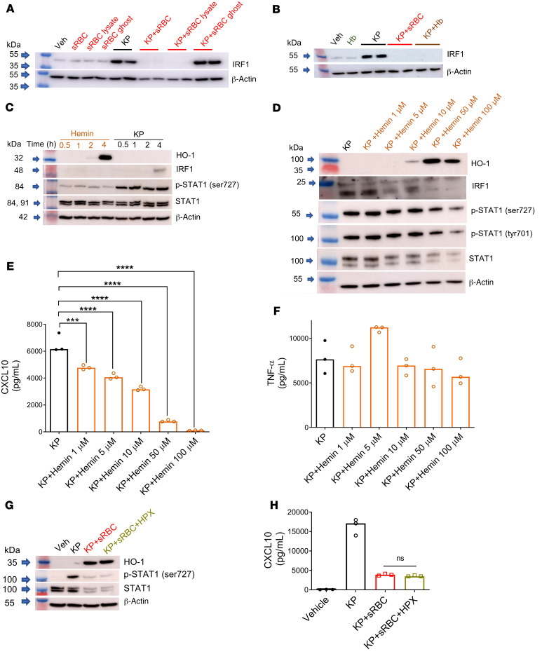
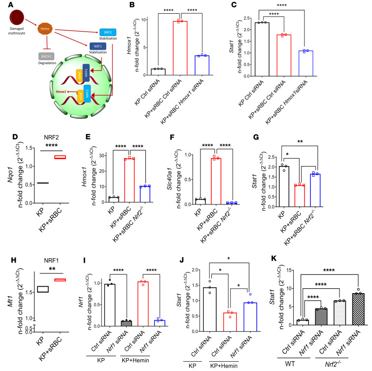
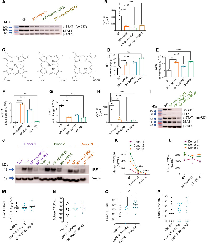
Macrophages are main effectors of heme metabolism, increasing transiently in the liver during heightened disposal of damaged or senescent RBCs (sRBCs). Macrophages are also essential in defense against microbial threats, but pathological states of heme excess may be immunosuppressive. Herein, we uncovered a mechanism whereby an acute rise in sRBC disposal by macrophages led to an immunosuppressive phenotype after intrapulmonary Klebsiella pneumoniae infection characterized by increased extrapulmonary bacterial proliferation and reduced survival from sepsis in mice. The impaired immunity to K. pneumoniae during heightened sRBC disposal was independent of iron acquisition by bacterial siderophores, in that K. pneumoniae mutants lacking siderophore function recapitulated the findings observed with the WT strain. Rather, sRBC disposal induced a liver transcriptomic profile notable for suppression of Stat1 and IFN-related responses during K. pneumoniae sepsis. Excess heme handling by macrophages recapitulated STAT1 suppression during infection that required synergistic NRF1 and NRF2 activation but was independent of heme oxygenase-1 induction. Whereas iron was dispensable, the porphyrin moiety of heme was sufficient to mediate suppression of STAT1-dependent responses in human and mouse macrophages and promoted liver dissemination of K. pneumoniae in vivo. Thus, cellular heme metabolism dysfunction negatively regulated the STAT1 pathway, with implications in severe infection.

Keywords: Bacterial infections; Infectious disease; Innate immunity; Macrophages; Pulmonology.


Conflict of interest statement

Conflict of interest: The authors have declared that no conflict of interest exists.

Figures

Figure 1
Figure 1. sRBC delivery impairs bacterial control at extrapulmonary sites and worsens survival after K. pneumoniae intrapulmonary infection.
(A) KP was instilled intratracheally into C57BL/6 mice and was followed by challenge with young RBCs (yRBCs) or senescent RBCs (sRBCs). n = 10 mice per group. **P = 0.005 by log-rank test. (B) Total BAL cell count/mL. (C) Total BAL PMN count/mL. (D) Total BAL mononuclear cell count/mL. (E) Lung bacterial burden was estimated by CFU count/mL of tissue homogenates. (F) Spleen CFU/mL. (G) Liver CFU/mL. (H) Blood CFU/mL. (BH) each point indicates individual mice, n = 7–8 mice/group, line indicates the median. *P < 0.05, **P < 0.01, ***P < 0.001 by 2-tailed Mann-Whitney U test.
Figure 2
Figure 2. Impaired K.
pneumoniaecontrol at extrapulmonary sites after sRBC delivery is independent of iron acquisition by bacterial siderophores. (A) Plasma transferrin-bound iron from mice 2 hours after challenge with either yRBCs or sRBCs, with unchallenged mice serving as baseline control. Growth curve of entB ybtS isogenic KP mutant and WT parent strain in (B) tryptic soy broth and (C) plasma obtained from mice challenged with sRBCs. (DG) EntB ybtS isogenic KP mutant and WT KP were instilled intratracheally into C57BL/6 mice (104 CFU inoculum each). Bacterial burden was obtained from homogenates of (D) lung, (E) spleen, (F) liver, and (G) blood as CFU/mL at 24 hours after infection. Each point indicates individual mice, n = 6 mice per group, line indicates median. **P < 0.01 by 2-tailed Mann-Whitney U test. (HL) EntB ybtS isogenic KP mutant bacteria were instilled into C57BL/6 mice (103 CFU inoculum), followed by challenge with either PBS, yRBCs, or sRBCs. (H) BAL total protein and bacterial burden were obtained from (I) lung, (J) spleen, (K) liver tissue homogenates, and (L) blood as CFU/mL at 24 hours. Each point indicates individual mice, n = 5–7 mice per group, line indicates median. *P < 0.05, **P < 0.01 by Kruskal-Wallis test with Dunn’s multiple comparisons test.
Figure 3
Figure 3. RNA-Seq of the liver in mice after sRBC delivery reveals a unique transcriptomic profile notable for suppression of Stat1 and IFN-related responses during K. pneumoniae infection.
KP was instilled intratracheally into C57BL/6 mice followed by challenge with either yRBCs or sRBCs. Mice were euthanized at 24 hours. (A) Volcano plot of innate immune genes. Red depicts downregulated genes. Blue depicts upregulated genes. (BJ) qPCR validation of RNA-Seq data. Floating bar plots indicate median and 25% to 75% quartiles. n = 4 mice per group. *P < 0.05 by 2-tailed Mann-Whitney U test. Fold change relative to yRBC + KP. (K) IRF1 immunoblot in livers of KP-infected mice challenged with either yRBCs or sRBCs. Each line indicates individual mice. (L) Relative density of blot depicted in K. n = 4 mice per group, line indicates the median. *P < 0.05 by 2-tailed Mann-Whitney U test.
Figure 4
Figure 4. K. pneumoniae enhances erythrophagocytosis, leading to upregulation of heme iron transcriptional responses and suppression of STAT1.
(A) sRBC internalization in RAW cells incubated with vehicle (PBS), sRBCs (50 sRBC:1 Mφ), KP (MOI 10:1), or KP + sRBC for 90 minutes. (B) Quantification of sRBC uptake shown in A. (C) Bone marrow–derived macrophages (BMDMs) obtained from WT and Tlr4–/– mice were challenged with KP + sRBC (10sRBC:1 Mφ) for 2 hours. (B and C) n = 3 technical replicates per group and data are indicative of 2 independent experiments. *P < 0.05, **P < 0.01 by 2-tailed t test. (D) Intracellular CFU/mL in RAW cells that were challenged with KP or KP + sRBC for 90 minutes. (E and F) Heme iron transcriptional genes Hmox1, Scl40a1, and (GK) Stat1 and STAT1 target genes C3, Cfb, Irf1, and Nos2 evaluated in RAW cells challenged with KP or KP + sRBC for 4 hours. (LN) STAT1 and IRF1 immunoblots in RAW cells challenged with vehicle (PBS), sRBCs, KP, or KP + sRBC for 4 hours. Blots are indicative of at least 3 independent experiments. (OQ) CCL5, CXCL10, and TNF-α were measured in cell culture supernatant by ELISA 4 hours after infection. (RW) Stat1, C3, Cfb, Irf1, Nos2, and Rela in Stat1+/+ and Stat1–/– BMDMs challenged with KP or KP + sRBC for 4 hours. (EK and RW) Gene expression was evaluated by qPCR analysis. Fold change is relative to PBS-treated macrophages. Floating bar plots indicate median and 25% to 75% quartiles, n = 3 technical replicates per group. *P < 0.05, ***P < 0.001, ****P < 0.0001 by 2-tailed t test. (X and Y) CXCL10 and TNF-α were measured in cell culture supernatant by ELISA 4 hours after infection. (OQ, X, and Y) n = 3 technical replicates per group. ****P < 0.0001 by 1-way ANOVA with Tukey’s multiple comparisons test.
Figure 5
Figure 5. Heme is the constituent of RBCs that mediates STAT1 suppression during K. pneumoniae infection.
IRF1 immunoblots in (A) RAW cells challenged with vehicle (PBS), sRBCs (30 sRBC:1 Mφ), sRBC lysates obtained from equivalent numbers of sRBCs, ghosts obtained from equivalent numbers of sRBCs, KP (MOI 10:1), KP + sRBC, KP + sRBC lysate, or KP + sRBC ghost for 4 hours, and (B) RAW cells challenged with vehicle, purified hemoglobin from an equivalent number of lysed RBCs (Hb), KP, KP + sRBC, or KP + Hb for 4 hours. Blot is indicative of 2 independent experiments. (C) HO-1, IRF1, p-STAT1 (Ser727), and STAT1 in RAW cells challenged with hemin (100 μM) or KP at 0.5, 1, 2, or 4 hours. (D) HO-1, IRF1, p-STAT1 (Ser727), p-STAT1 (Tyr701), and STAT1 in RAW cells challenged with KP and increasing concentrations of hemin for 4 hours. (E) CXCL10 and (F) TNF-α were measured in cell culture supernatant by ELISA 4 hours after infection. (G) HO-1, p-STAT1 (Ser727), and STAT1 in BMDMs challenged with vehicle (PBS), KP, KP + sRBC (50 sRBC:1 Mϕ), or KP + sRBC +hemopexin (HPX, 200 μg/mL) for 4 hours. (H) CXCL10 was measured in cell culture supernatant by ELISA 4 hours after infection. (E, F, and H) n = 3 technical replicates per group. ***P < 0.001, ****P < 0.0001 by 1-way ANOVA with Tukey’s multiple comparisons test.
Figure 6
Figure 6. STAT1 suppression requires NRF1 and NRF2 activation but is independent of HO-1 induction.
(A) Schematic depicting heme-BACH1-NRF1/2 interaction. Intracellular heme accumulation after erythrophagocytosis induces degradation of BACH1 and stabilization of NRF1 and NRF2, with nuclear translocation of NRF2 resulting in Hmox1 transcription. (B) Hmox1 and (C) Stat1 gene expression in RAW cells transfected with control or Hmox1 siRNA and subsequently challenged with KP or KP + sRBC for 4 hours. (B and C) Fold change relative to uninfected control siRNA-transfected RAW cells. Data are average of 3 independent experiments. (D) NRF2 target gene (Nqo1) expression in RAW cells challenged with KP or KP + sRBC for 4 hours. (EG) Hmox1, Slc40a1, and Stat1 expression in BMDMs obtained from WT and Nrf2–/– mice challenged with KP or KP+ RBC for 4 hours. Fold change relative to PBS-treated BMDMs. (H) NRF1 target gene (Mt1) expression in RAW cells challenged with KP or KP + sRBC for 4 hours. **P < 0.01, ****P < 0.0001 by 2-tailed t-test (D and H). (I) Nrf1 and (J) Stat1 expression in BMDMs transfected with control siRNA or Nrf1 siRNA and subsequently challenged with KP or KP + hemin (50 μM) for 4 hours. (DK) n = 3 technical replicates per group, and representative of at least 2 independent experiments. (K) Stat1 expression in BMDMs obtained from WT and Nrf2–/– mice, transfected with control siRNA or Nrf1 siRNA, and subsequently challenged with KP for 4 hours. (IK) Fold change relative to uninfected control siRNA-transfected BMDMs. *P < 0.05, **P < 0.01, ***P < 0.001, ****P < 0.0001 by 1-way ANOVA with Tukey’s multiple comparisons test (B, C, EG, and IK).
Figure 7
Figure 7. The porphyrin moiety of heme is necessary and sufficient for NRF1/NRF2 activation and STAT1 suppression.
(A) STAT1 immunoblot in BMDMs challenged with KP (MOI 10:1), KP + hemin (25 μM), KP + hemin + DFX (300 μM), or KP + hemin + DFO (300 μM) for 4 hours. All groups contained vehicle (DMSO, ~1%). Blot is representative of 2 independent experiments. (B) CXCL10 was measured in cell culture supernatant by ELISA 4 hours after infection. (C) Chemical structures of hemin (iron protoporphyrin IX, FePPIX), cobalt protoporphyrin IX (CoPPIX), and protoporphyrin IX (PPIX). (D) Mt1 and (E) Nqo1 expression, (F) Hmox1, and (G) Stat1 gene expression, (H) CXCL10 secretion in BMDMs challenged with KP, KP + FePPIX (50 μM), KP + CoPPIX (50 μM), or KP + PPIX (50 μM) for 4 hours. (DG) Fold change relative to PBS-treated BMDMs. (B and FH) n = 3 technical replicates per group. **P < 0.01, ***P < 0.001, ****P < 0.0001 by 1-way ANOVA with Tukey’s multiple comparisons test. (I) BACH1, HO-1, p-STAT1, and total STAT1 immunoblot in BMDMs challenged with KP and increasing concentrations of PPIX. (J) IRF1 immunoblot in human monocyte–derived macrophages (HMDMs) challenged with vehicle (PBS), KP, KP + FePPIX (25 μM), or KP + PPIX (25 μM) for 4 hours. (K) CXCL10 and (L) TNF-α secretion from HMDMs in J. n = 3 donors. ****P < 0.0001 by 2-way ANOVA with Tukey’s multiple comparisons test. KP was instilled intratracheally into C57BL/6 mice and followed by i.p. challenge with vehicle (2.5% DMSO) or CoPPIX (5 mg/kg, 25 mg/kg) 1 hour after KP instillation. Bacterial burden was obtained from (M) lung, (N) spleen, (O) liver tissue homogenates, and (P) blood of mice 24 hours after KP infection and reported as CFU/mL. Each point indicates individual mice, n = 10–11 mice per group combined from 2 independent studies. Line indicates median. *P < 0.05, **P < 0.01 by Kruskal-Wallis test with Dunn’s multiple comparisons test.

References

    1. Ganz T. Macrophages and systemic iron homeostasis. J Innate Immun. 2012;4(5-6):446–453. doi: 10.1159/000336423. - DOI - PMC - PubMed
    1. Higgins JM, Mahadevan L. Physiological and pathological population dynamics of circulating human red blood cells. Proc Natl Acad Sci U S A. 2010;107(47):20587–20592. doi: 10.1073/pnas.1012747107. - DOI - PMC - PubMed
    1. Muckenthaler MU, Rivella S, Hentze MW, Galy B. A red carpet for iron metabolism. Cell. 2017;168(3):344–361. doi: 10.1016/j.cell.2016.12.034. - DOI - PMC - PubMed
    1. Theurl I, et al. On-demand erythrocyte disposal and iron recycling requires transient macrophages in the liver. Nat Med. 2016;22(8):945–951. doi: 10.1038/nm.4146. - DOI - PMC - PubMed
    1. Nairz M, Theurl I, Swirski FK, Weiss G. “Pumping iron”—how macrophages handle iron at the systemic, microenvironmental, and cellular levels. Pflugers Arch. 2017;469(3–4):397–418. - PMC - PubMed

Publication types