Comparative Study
. 2021 Aug 29;117(10):2252-2262.
doi: 10.1093/cvr/cvaa265.

Cardiac cellularity is dependent upon biological sex and is regulated by gonadal hormones

Affiliations
Comparative Study

Cardiac cellularity is dependent upon biological sex and is regulated by gonadal hormones

Galen T Squiers et al. Cardiovasc Res. .

Erratum in

Abstract

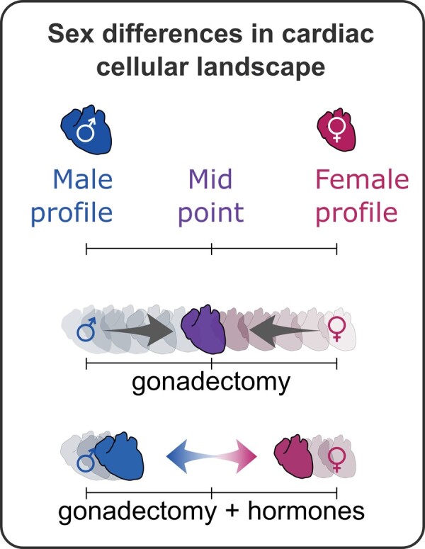
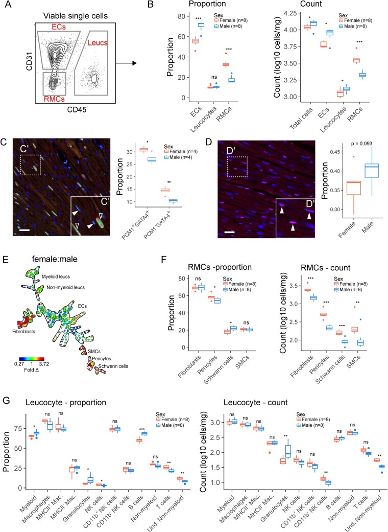
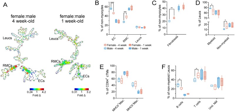
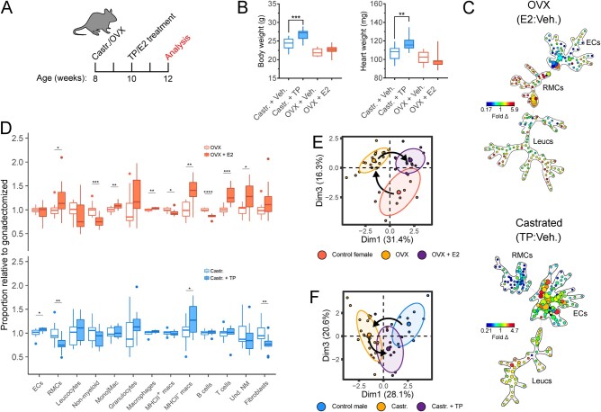
Aims: Sex differences have been consistently identified in cardiac physiology and incidence of cardiac disease. However, the underlying biological causes for the differences remain unclear. We sought to characterize the cardiac non-myocyte cellular landscape in female and male hearts to determine whether cellular proportion of the heart is sex-dependent and whether endocrine factors modulate the cardiac cell proportions.

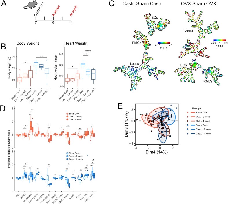
Methods and results: Utilizing high-dimensional flow cytometry and immunofluorescence imaging, we found significant sex-specific differences in cellular composition of the heart in adult and juvenile mice, that develops postnatally. Removal of systemic gonadal hormones by gonadectomy results in rapid sex-specific changes in cardiac non-myocyte cellular proportions including alteration in resident mesenchymal cell and leucocyte populations, indicating gonadal hormones and their downstream targets regulate cardiac cellular composition. The ectopic reintroduction of oestrogen and testosterone to female and male mice, respectively, reverses many of these gonadectomy-induced compositional changes.

Conclusion: This work shows that the constituent cell types of the mouse heart are hormone-dependent and that the cardiac cellular landscapes are distinct in females and males, remain plastic, and can be rapidly modulated by endocrine factors. These observations have implications for strategies aiming to therapeutically alter cardiac cellular heterogeneity and underscore the importance of considering biological sex for studies examining cardiac physiology and stress responses.

Keywords: Cardiac cell composition; Cardiac fibroblast; Cardiac macrophage; Sex differences.


Figures

None
Graphical abstract
Figure 1
Figure 1
Cellular composition of the female and male heart is distinct. (A) Flow cytometry workflow for analysis of broad non-myocyte cardiac cell populations (left) and quantification of CD31+CD45 (ECs), CD45+ (leucocytes), and CD31CD45 (RMCs) cells as a proportion of non-myocytes. (B) Proportion (left panel) and absolute numbers (right panel) of cells corresponding to ECs, leucocytes, and RMCs. (C) Immunohistochemical analysis of proportion of nuclei corresponding to cardiomyocytes (PCM+GATA4+) or RMCs (PCMGATA4+). Scale bar indicates 25 µm. (D) Immunohistochemical analysis of proportion of nuclei corresponding to endothelial cells (DACH1+ nuclei). Scale bar indicates 25 µm. (E) Ratiometric (female:male) SPADE analysis of broad cardiac cell types in 10-week-old mice. Heat map indicates fold difference (fold Δ) of cell populations in female hearts compared to male hearts. Nodes (circles) of SPADE dendrograms represent phenotypically identical/similar cell populations, size of nodes indicates relative abundances of cells represented by the node, and connectedness indicates phenotypic relation of nodes to each other. See Supplementary material online, Figure S1, for markers used for cell clustering. (F) Proportions and absolute numbers of RMCs determined by flow cytometry. (G) Proportions and absolute numbers of cardiac leucocytes. See Supplementary material online, Figure S1, for all flow cytometry gating parameters and markers used for clustering. All data analysed using Student’s t-test. Where exact values are not provided, significance indicated as *(P≤0.05), **(P≤0.01), ***(P≤0.001), and ****(P≤0.0001). Whiskers of box and whisker plots represent the highest and the lowest values, except when a value is beyond the range of 1.5 inter-quartile.
Figure 2
Figure 2
Differences based on biological sex in cardiac cellular composition principally develops after birth. (A) Ratiometric (female:male) SPADE analysis of broad cardiac cell types in 4-week-old (left) and 1-week-old (right) mice. Heat map indicates fold difference (fold Δ) of cell populations in female hearts compared to male hearts. (B) Proportions of ECs, RMCs, and Leucs (leucocytes) relative to total non-myocytes in 4- and 1-week-old males and females. (C) Proportions of fibroblasts relative to total non-myocytes in 4- and 1-week-old males and females. (D) Proportions of myeloid (CD45+CD11b+) and non-myeloid (CD45+CD11b−) Leucs relative to total Leucs in 4- and 1-week-old males and females. (E) Proportions of MHCII− and MHCII+ macrophages (Macs: CD45+CD11b+Ly6G−CD64+) relative to total macrophages in 4- and 1-week-old males and females. (F) Proportions of B cells (CD45+CD11b−MHCII+CD3ε−), T cells (CD45+CD11b−MHCII−CD3ε+), and undetermined non-myeloid cells (Und. NM; CD45+CD11b−MHCII−CD3ε−) relative to total non-myeloid Leucs in 4- and 1-week-old males and females. See Supplementary material online, Figure S3, for all flow cytometry gating parameters and markers used for SPADE clustering. n=5 per group; all statistical analysis performed using Student’s t-test with statistical significance indicated as *(P ≤ 0.05), **(P ≤ 0.01), ***(P ≤ 0.001), and ****(P ≤ 0.0001). Whiskers of box and whisker plots represent the highest and the lowest values.
Figure 3
Figure 3
Gonadal hormones maintain the homeostatic cardiac cellular signature in female and male hearts. (A) Experimental set-up for mouse ovariectomy (OVX) or castration (Castr.). (B) Body weight (left) and heart weight (right) measurements following sham surgery or gonadectomy. (C) Ratiometric (gonadectomy: sham surgery) SPADE analysis of broad cardiac cell types. Heatmap indicates fold difference (fold Δ) of cell populations in gonadectomized animals compared to sham controls. (D) Relative abundance of broad cell types (ECs, RMCs, and Leucs), broad leucocyte subsets (non-myeloid, Mono|Mac, granulocytes), macrophage subsets (MHCII+ macrophages and MHCII macrophages), and non-myeloid subsets (B cells, T cells, and Und. NM cells) between gonadectomized animals and sham surgery controls. ECs (CD31+CD45); RMCs (CD31CD45), leucocytes (Leucs; CD45+); monocytes/macrophages (Mono|Mac; CD45+CD11b+Ly6G); granulocytes (CD45+CD11b+Ly6G+); macrophages (CD45+CD11b+Ly6GCD64+); B cells (CD45+CD11bMHCII+CD3ε); T cells (CD45+CD11bMHCIICD3ε+); undetermined non-myeloid cells (Und. NM; CD45+CD11bMHCIICD3ε). (E) Principal component analysis cellular composition of sham and gonadectomized mice. Principal components that best differentiated female and male non-gonadectomized cohorts were chosen. Parenthesis on axis labels indicate variation represented by principal component. See Supplementary material online, Figure S3, for all flow cytometry gating parameters and markers used for SPADE clustering. n = 13–15; all statistical analysis performed using Student’s t-test with statistical significance indicated as *(P ≤ 0.05), **(P ≤ 0.01), ***(P ≤ 0.001), and ****(P ≤ 0.0001). NS, P-value >0.05. Whiskers of box and whisker plots represent the highest and the lowest values, except when a value is beyond the range of 1.5 inter-quartile.
Figure 4
Figure 4
Gonadal oestrogen and testosterone regulate cardiac resident mesenchymal levels (RMCs) and leucocyte subsets. (A) Experimental set-up for mouse OVX or Castr. followed by Veh. or TP/oestradiol (E2) treatment. (B) Body weight (left) and heart weight (right) measurements following gonadectomy with vehicle or TP/E2 treatment. (C) Ratiometric (TP: vehicle control and E2: vehicle control, Castr. males and OVX females, respectively) SPADE analysis of broad cardiac cell types. Heat map indicates fold difference (fold Δ) of cell populations in gonadectomized animals with TP or E2 treatment relative to gonadectomized animals with vehicle control treatment. (D) Relative abundance of broad cell types (ECs, RMCs, and Leucs), broad leucocyte subsets (non-myeloid, Mono|Mac., granulocytes), cardiac tissue macrophage subsets (MHCII+ macrophages and MHCII macrophages) and non-myeloid subsets (B cells, T cells, and Und. NM cells) between gonadectomized animals treated with TP/E2 and gonadectomized animals treated with vehicle control. (E) Principal component analysis cellular composition of non-OVX (control), OVX, and OVX+E2 treatment cohorts. Arrows indicate movement of cellular composition signatures following ovariectomy and E2 treatment following ovariectomy. (F) Principal component analysis cellular composition of non-Castr. (control), Castr., and Castr.+TP treatment cohorts. Arrows indicate movement of cellular composition signatures following castration and TP treatment following castration. See Supplementary material online, Figure S3, for all flow cytometry gating parameters and markers used for SPADE clustering. n=10–12; all statistical analysis performed using Student’s t-test with statistical significance indicated as *(P≤0.05), **(P≤0.01), ***(P≤0.001), and ****(P≤0.0001). Whiskers of box and whisker plots represent the highest and the lowest values, except when a value is beyond the range of 1.5 inter-quartile.

Comment in

References

    1. Blenck CL, Harvey PA, Reckelhoff JF, Leinwand LA.. The importance of biological sex and estrogen in rodent models of cardiovascular health and disease. Circ Res 2016;118:1294–1312. - PMC - PubMed
    1. Grandy SA, Howlett SE.. Cardiac excitation-contraction coupling is altered in myocytes from aged male mice but not in cells from aged female mice Cardiac excitation-contraction coupling is altered in myocytes from aged male mice but not in cells from aged female mice. Am J Physiol Heart Circ Physiol 2006;291:2362–2370. - PubMed
    1. Isensee J, Witt H, Pregla R, Hetzer R, Regitz-Zagrosek V, Ruiz Noppinger P.. Sexually dimorphic gene expression in the heart of mice and men. J Mol Med 2008;86:61–74. - PMC - PubMed
    1. Vijay V, Han T, Moland CL, Kwekel JC, Fuscoe JC, Desai VG.. Sexual dimorphism in the expression of mitochondria-related genes in rat heart at different ages. PLoS One 2015;10:e0117047. - PMC - PubMed
    1. Skelly DA, Squiers GT, McLellan MA, Bolisetty MTMT, Robson P, Rosenthal NA, Pinto AR.. Single-cell transcriptional profiling reveals cellular diversity and intercommunication in the mouse heart. Cell Rep Elsevier 2018;22:600–610. - PubMed

Publication types

MeSH terms