Review
. 2020 Jul 8;10(3):933-974.
doi: 10.1002/cphy.c190021.

Retinal Physiology and Circulation: Effect of Diabetes

Affiliations
Review

Retinal Physiology and Circulation: Effect of Diabetes

William S Wright et al. Compr Physiol. .

Abstract

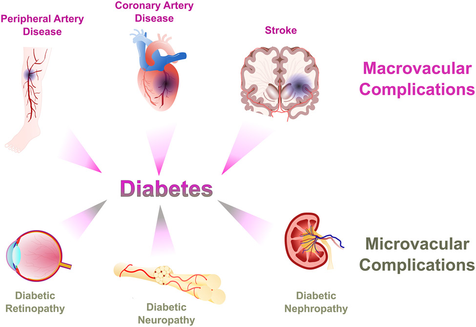
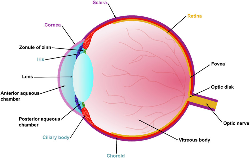
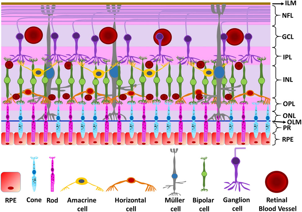
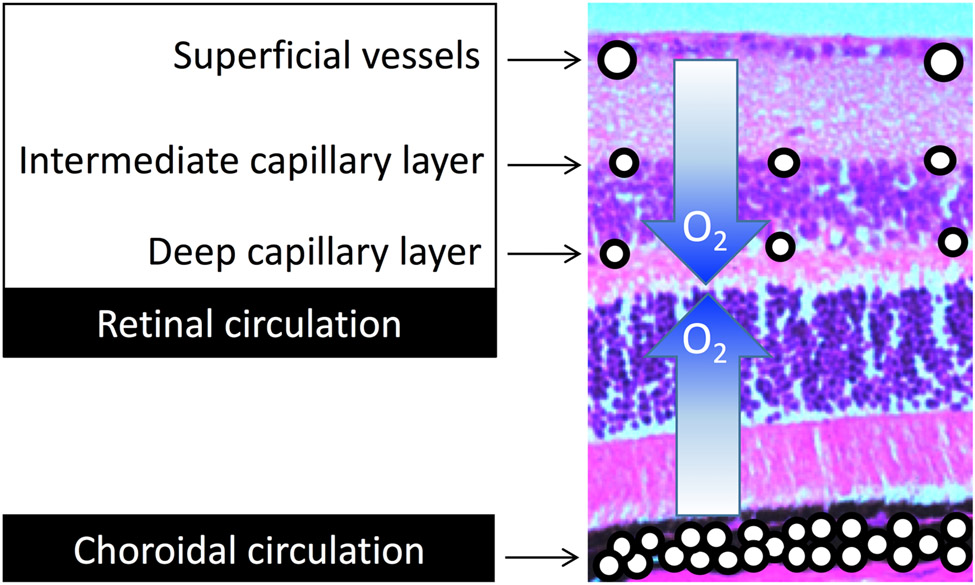
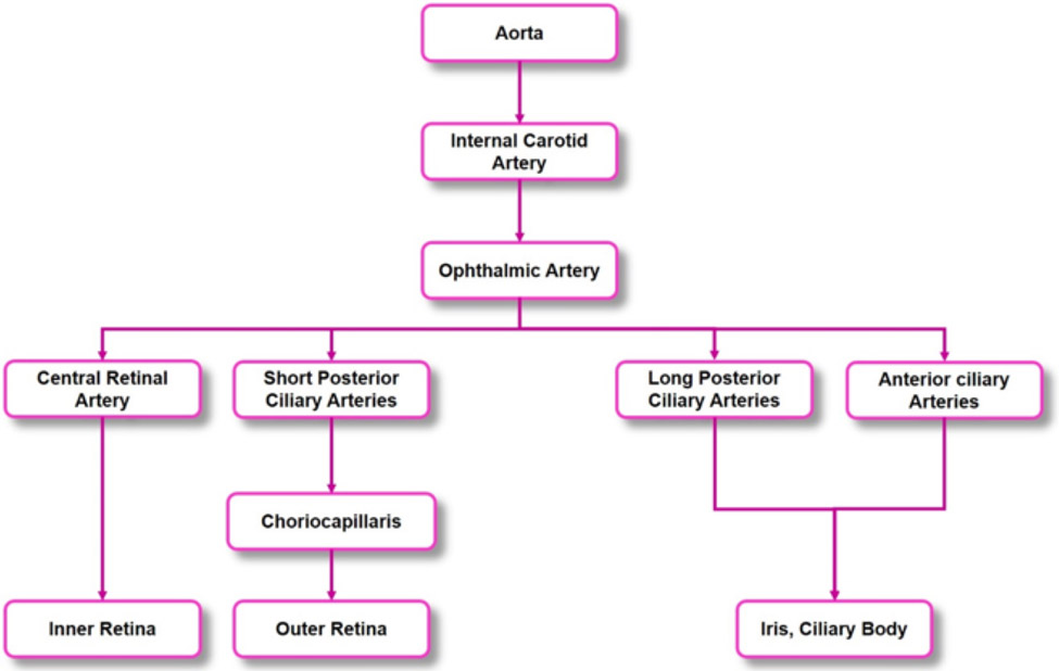
In this article, we present a discussion of diabetes and its complications, including the macrovascular and microvascular effects, with the latter of consequence to the retina. We will discuss the anatomy and physiology of the retina, including aspects of metabolism and mechanisms of oxygenation, with the latter accomplished via a combination of the retinal and choroidal blood circulations. Both of these vasculatures are altered in diabetes, with the retinal circulation intimately involved in the pathology of diabetic retinopathy. The later stages of diabetic retinopathy involve poorly controlled angiogenesis that is of great concern, but in our discussion, we will focus more on several alterations in the retinal circulation occurring earlier in the progression of disease, including reductions in blood flow and a possible redistribution of perfusion that may leave some areas of the retina ischemic and hypoxic. Finally, we include in this article a more recent area of investigation regarding the diabetic retinal vasculature, that is, the alterations to the endothelial surface layer that normally plays a vital role in maintaining physiological functions. © 2020 American Physiological Society. Compr Physiol 10:933-974, 2020.


Figures

Figure 1.
Figure 1.. Macrovascular and microvascular complications of diabetes.
Macrovascular complications include peripheral artery disease, coronary artery disease, and stroke; microvascular complications include diabetic retinopathy, neuropathy, and nephropathy.
Figure 2.
Figure 2.. Anatomy of the eye.
Ocular tissue can be divided into three layers: 1) an outer layer consisting of the sclera and the cornea, 2) an intermediate layer consisting of the iris, the ciliary body, and the choroid, and 3) an internal layer consisting of the retina.
Figure 3.
Figure 3.. Layers of the retina.
The layers of the retina starting from the outermost to the innermost layer are the 1) retinal pigmented epithelium (RPE), 2) photoreceptors (PR), 3) outer limiting membrane (OLM), 4) ) outer nuclear layer (ONL), 5) outer plexiform layer (OPL), 6) inner nuclear layer (INL), 7) inner plexiform layer (IPL), 8), ganglion cell layer (GCL), 9) nerve fiber layer (NFL), and 10) inner limiting membrane (ILM).
Figure 4.
Figure 4.. Oxygen sources to the retina.
The inner half of the retina receives oxygen from the retinal circulation, while the outer half receives oxygen from the choroidal circulation.
Figure 5.
Figure 5.. Arterial branches leading to the eye.
The blood vessels supplying blood to the inner and outer retina, the iris, and the ciliary body originate from the ophthalmic artery, which is a branch of the internal carotid artery. The ophthalmic artery further divides to give rise to the anterior ciliary arteries, the long and short posterior ciliary arteries, and the central retinal artery.
Figure 6.
Figure 6.. Retinal microvascular layers.
The retinal microcirculatory system can be divided into three distinct layers: 1) the superficial arterioles and venules, 2) the or intermediate capillary layer, and 3) the deep capillary layer.
Figure 7.
Figure 7.. Transit pathways through the retinal circulation.
Normally, most retinal blood flow travels through the deep capillary layer. In diabetic rats, it is possible that a decreased proportion of flow goes through the deep layer, with rapid transit times possibly explained by flow preferentially perfusing the superficial and/or intermediate capillary layers due to vasoconstriction of the vessels (indicated by the asterisks) leading away from the superficial layer.
Figure 8.
Figure 8.. PECAM-1 loss may contribute to the development of diabetic retinopathy.
Diabetic retinopathy is characterized by leukocyte plugging of capillaries, a decrease in endothelial cell survival, and an increase in vascular leakage. These phenomena also could occur with PECAM-1 loss, due to its important role in vascular endothelial cell functions.
Figure 9.
Figure 9.. Glycocalyx components.
The structure of the endothelial glycocalyx on the plasma side of the cell includes proteoglycans (such as syndecans and glypican-1) and glycosaminoglycans (such as heparan sulfate, chondroitin sulfate, and hyaluronic acid).

References

    1. Intensive blood-glucose control with sulphonylureas or insulin compared with conventional treatment and risk of complications in patients with type 2 diabetes (UKPDS 33). UK Prospective Diabetes Study (UKPDS) Group. Lancet 352: 837–853, 1998. - PubMed
    1. Retinal Conditions - Common Eye Disorders∣Vision Health Initiative (VHI)∣. http://www.cdc.gov/visionhealth/basic_information/eye_disorders_retinal.htm: The Vision Health Initiative (VHI), 2014.
    1. Abdouh M, Khanjari A, Abdelazziz N, Ongali B, Couture R, Hasséssian HM. Early upregulation of kinin B1 receptors in retinal microvessels of the streptozotocin-diabetic rat. British journal of pharmacology 140: 33–40, 2003. - PMC - PubMed
    1. Abdouh M, Talbot S, Couture R, Hassessian HM. Retinal plasma extravasation in streptozotocin-diabetic rats mediated by kinin B(1) and B(2) receptors. British journal of pharmacology 154: 136–143, 2008. - PMC - PubMed
    1. Abu El-Asrar AM, Desmet S, Meersschaert A, Dralands L, Missotten L, Geboes K. Expression of the inducible isoform of nitric oxide synthase in the retinas of human subjects with diabetes mellitus. American journal of ophthalmology 132: 551–556, 2001. - PubMed

Publication types

MeSH terms