. 2020 Dec:177:32-40.
doi: 10.1016/j.visres.2020.08.003. Epub 2020 Sep 15.

Eccentricity-dependent effects of simultaneous competing defocus on emmetropization in infant rhesus monkeys

Affiliations

Eccentricity-dependent effects of simultaneous competing defocus on emmetropization in infant rhesus monkeys

Earl L Smith Iii et al. Vision Res. 2020 Dec.

Abstract

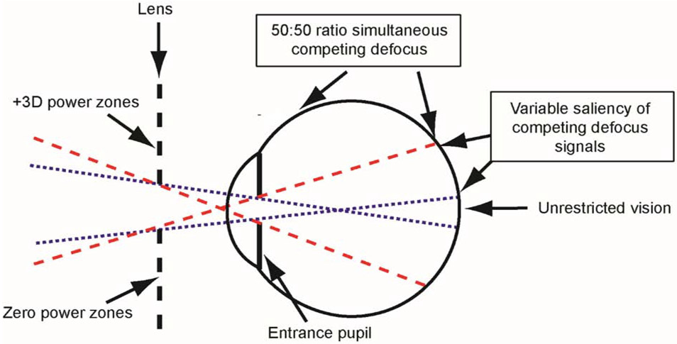
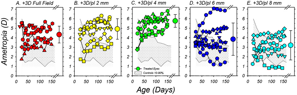
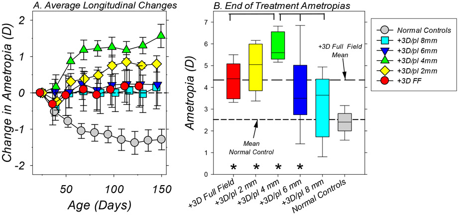
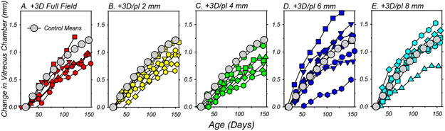
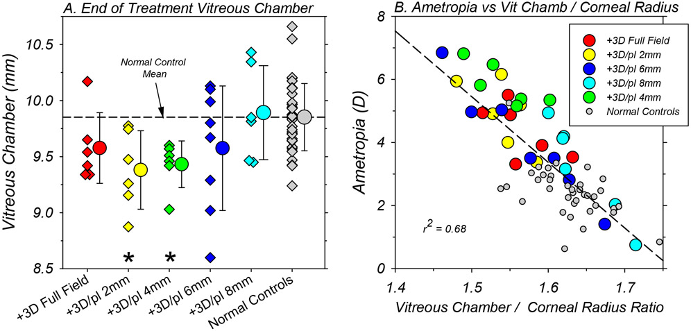
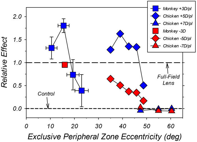
Dual-focus lenses that impose simultaneous competing myopic defocus over the entire visual field produce axial hyperopic shifts in refractive error. The purpose of this study was to characterize the effects of eccentricity on the ability of myopic defocus signals to influence central refractive development in infant monkeys. From 24 to 152 days of age, rhesus monkeys were reared with binocular, dual-focus lenses that had central, zero-powered zones surrounded by alternating concentric annular power zones of +3D and zero power. Between subject groups the diameter of the central, zero-powered zone was varied from 2 mm to 8 mm in 2 mm steps (+3D/pl 2 mm, n = 6; +3D/pl 4 mm, n = 6; +3D/pl 6 mm, n = 8, or + 3D/pl 8 mm, n = 6). For the treatment lens with 2, 4, 6 and 8 mm central zones, objects at eccentricities beyond 11°, 16°, 19° and 23°, respectively, were imaged exclusively through the dual-power peripheral zones. Refractive status (retinoscopy), corneal power (keratometry) and axial dimensions (ultrasonography) were measured at two-week intervals. Comparison data were obtained from monkeys reared with binocular, single-vision +3D full-field lenses (+3D FF, n = 6) and 41 normal control monkeys reared with unrestricted vision. At the end of the rearing period, with the exception of the +3D/pl 8 mm group (median = +3.64 D), the ametropias for the other lens-reared groups (medians: FF = +4.39 D, 2 mm = +5.19 D, 4 mm = +5.59 D, 6 mm = +3.50 D) were significantly more hyperopic than that for the normal monkeys (+2.50 D). These hyperopic errors were associated with shallower vitreous chambers. The key finding was that the extent and consistency of these hyperopic ametropias varied with the eccentricity of the dual-focus zones. The results confirm that myopic defocus in the near periphery can slow axial growth, but that imposed defocus beyond about 20° from the fovea does not consistently alter central refractive development.

Keywords: Animal model; Eccentricity; Emmetropization; Hyperopia; Multifocal lenses; Myopia; Periphery.


Conflict of interest statement

Declarations of interest

E.L. Smith III, (p) patents on optical and pharmaceutical treatment strategies for myopia, (C) consultant to Nevakar, SightGlass Vision, Treehouse Eyes, Acucela Inc. and Essilor of America; B. Arumugam, None; L.-F. Hung, None; Z. She, None; K. Beach, None; P. Sankaridudrg, (p) patents on optical and pharmaceutical treatment strategies for myopia.

Figures

Figure 1.
Figure 1.
Schematic diagram of the extent of the visual field influenced by the different power zones of the dual-focus lenses. The blue dotted lines represent the projection of the eye’s entrance pupil through the central, zero-power zone and delineate the object locations that are imaged exclusively through the central zone (i.e., unrestricted portion of the visual field). The red dashed lines delineate the object eccentricities beyond which all objects are imaged exclusively through the dual-powered portion of the lens, resulting in images that are formed simultaneously at two different focal planes. These image planes have approximately equal amounts of light refracted through the plano and +3D components of the lens. For objects between the blue and red projection lines, the resulting images are also formed at two competing image planes, with the proportion of rays entering through the central lens zone decreasing with eccentricity while those entering through the dual-focus segments increase. See the Supplementary materials.
Figure 2.
Figure 2.
Spherical-equivalent, spectacle-plane refractions plotted as a function of age for individual subjects reared with the binocular +3D full-field lenses (A) and +3D/pl dual-focus lenses with 2mm (B), 4mm (C), 6mm (D) and 8mm (E) central zone of unrestricted vision. Each of the smaller symbols represents the average ametropia for the left and right eyes of a given animal. The large symbols to the right in each plot show the group average (±SD) at the end of the treatment period. The shaded area in each plot illustrates the 10% to 90% range of ametropias for the normal control animals.
Figure 3.
Figure 3.
A. Average (±SEM) binocular change in refractive error from baseline values plotted as a function of age for each subject group. The symbol legend in the figure indicates the color/shading for the different groups. The first symbols in each function represent the start of treatment. B. Box plots of the refractive errors obtained at the end of the lens-rearing period for each treatment group. The horizontal line in each box represents the group median; the lower and upper box boundaries indicate the 25% and 75% limits, respectively; and the lower and upper error bars show the 10% and 90% limits, respectively. The long and short dashed lines represent the mean ametropias for the +3D FF controls and the normal monkeys, respectively, at ages corresponding to the end of the treatment period. The asterisks indicate treated-group refractive errors that were significantly different from that for the normal control monkeys. The horizontal brackets indicate significant differences in the average refractive errors between the 4mm treatment group and other lens-reared groups.
Figure 4.
Figure 4.
Binocular average change in vitreous chamber depth from baseline values plotted as a function of age for individual subjects reared with the +3D full-field lenses (A) and +3D/pl dual-focus lenses with 2mm (B), 4mm (C), 6mm (D) and 8mm (E) central zones of unrestricted vision. Each symbol represents the average change in vitreous chamber depth for the left and right eyes of a given animal. The grey symbols in each plot represent the mean changes for the normal control animals.
Figure 5.
Figure 5.
A. Binocular vitreous chamber depth for individual animals (diamond symbols) in each subject group obtained at ages corresponding to the end of the lens-rearing period. Each of the smaller symbols represents the average vitreous chamber depth for the left and right eyes of a given animal. The larger circular symbols next to the individual data represent the group means (±SD). The asterisks indicate mean values that were significantly different from that for the normal control monkeys. B. Refractive errors (right and left eye averages) obtained at ages corresponding to the end of the treatment period for individual animals plotted as a function of the ratio of vitreous chamber depth in mm to corneal radius in mm. The color shading for the symbols representing the different treatment groups is shown in the included symbol legend.
Figure 6.
Figure 6.
Comparisons of the average relative effects of the eccentricity of optically imposed defocus on central refractive development for monkeys (square symbols) and chickens (diamond and triangle symbols). In all cases, the bifocal/dual-focus treatment lenses had central zones that provided unrestricted vision and concentric peripheral zones that imposed either myopic (blue symbols) or hyperopic defocus (red symbols). The effect of a given treatment lens is shown relative to the normalized alterations in central refractive error produced by a single-vision treatment lens that had the same power as the peripheral zone of the bifocal/dual-focus lenses. A relative effect of 1.0 indicates that the multifocal treatment lenses produced the same alterations in refractive error as full-field single-vision lenses with powers equal to that of the peripheral zone of the bifocal/dual-focus lenses. Data points plotted above the horizontal line at 1.0 indicate that the bifocal/dual-focus lenses produced larger alterations in refractive development than full-field lenses. The horizontal dashed line at 0.0 indicates that the treatment lenses had no effect on refractive development relative to age-matched controls. The abscissa represents the object eccentricities beyond which all objects were imaged exclusively through the peripheral power zones of the treatment lenses. The vertical error bars for the dual-focus, lens-reared monkeys (blue squares) represent ±1 SEM; the horizontal error bars represent the range in eccentricity associated with a ±1 mm change in vertex distance combined with ±0.6 mm change in entrance pupil size. The data point for imposed peripheral hyperopic defocus (−3.0 D) in monkeys was derived from Smith et al. (2010). The chicken data represented by diamonds and triangles were obtained using peripheral powers of ±5.0 D (Liu and Wildsoet, 2011) and ±7.0 D (Schippert and Schaeffel, 2006), respectively.

References

    1. Arumugam B, Hung L-F, To C-H, Holden BA, & Smith EL III (2014). The effects of simultaneous dual focus lenses on refractive development in infant monkeys. Investigative Ophthalmology & Visual Science, 55, 7423–7432. - PMC - PubMed
    1. Arumugam B, Hung L-F, To C-H, Sankaridurg P, & Smith EL III (2016). The effects of the relative strength of simultaneous competing defocus signals on emmetropization in infant monkeys. Investigative Ophthalmology & Visual Science, 57, 3949–3960. - PMC - PubMed
    1. Barathi VA, Boopathi VG, Yap EPH, & Beuerman RW (2008). Two models of experimental myopia in the mouse. Vision Research, 48, 904–916. - PubMed
    1. Benavente-Perez A, Nour A, & Troilo D (2012). The effect of simultaneous negative and positive defocus on eye growth and development of refractive state in marmosets. Investigative Ophthalmology & Visual Science, 53, 6479–6487. - PMC - PubMed
    1. Benavente-Perez A, Nour A, & Troilo D (2014). Axial eye growth and refractive error development can be modified by exposing the peripheral retina to relative myopic and hyperopic defocus. Investigative Ophthalmology & Visual Science, 55, 6765–6773. - PMC - PubMed

Publication types