. 2020 Sep 16;194(3):277-287.
doi: 10.1667/RADE-20-00051.1.

Non-Human Primates Receiving High-Dose Total-Body Irradiation are at Risk of Developing Cerebrovascular Injury Years Postirradiation

Affiliations

Non-Human Primates Receiving High-Dose Total-Body Irradiation are at Risk of Developing Cerebrovascular Injury Years Postirradiation

Rachel N Andrews et al. Radiat Res. .

Abstract

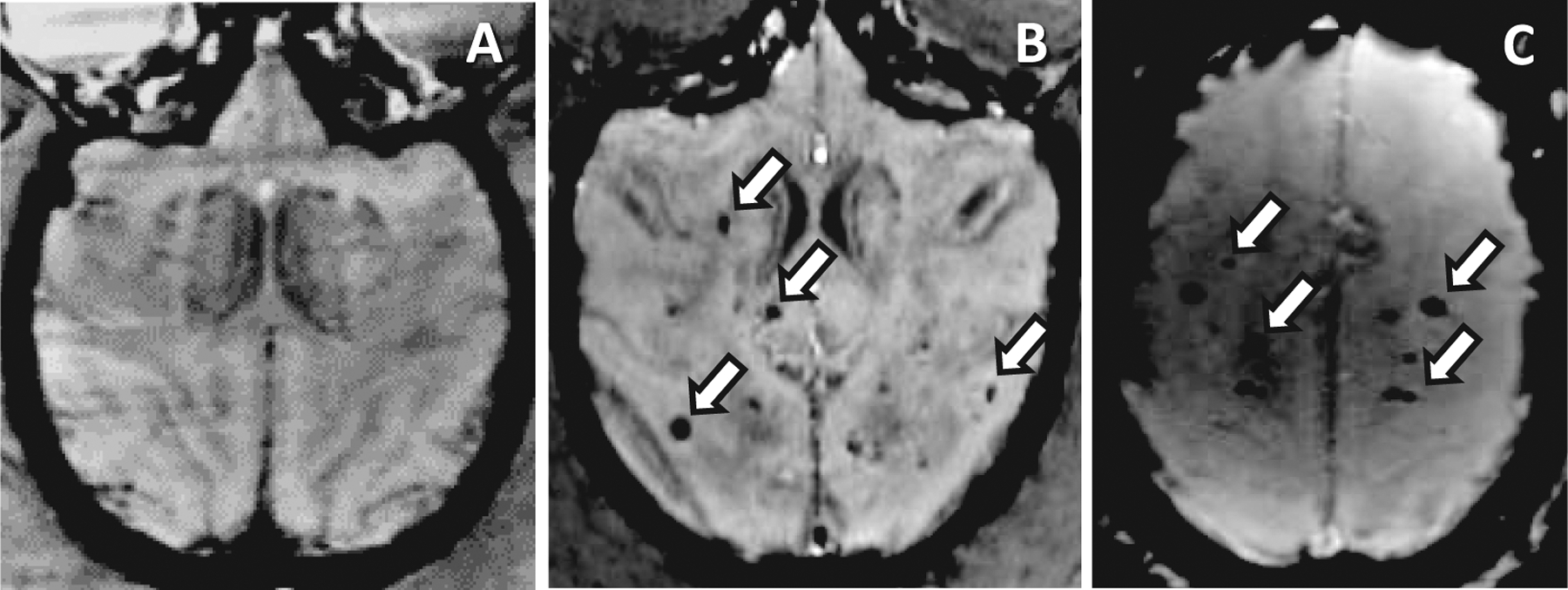
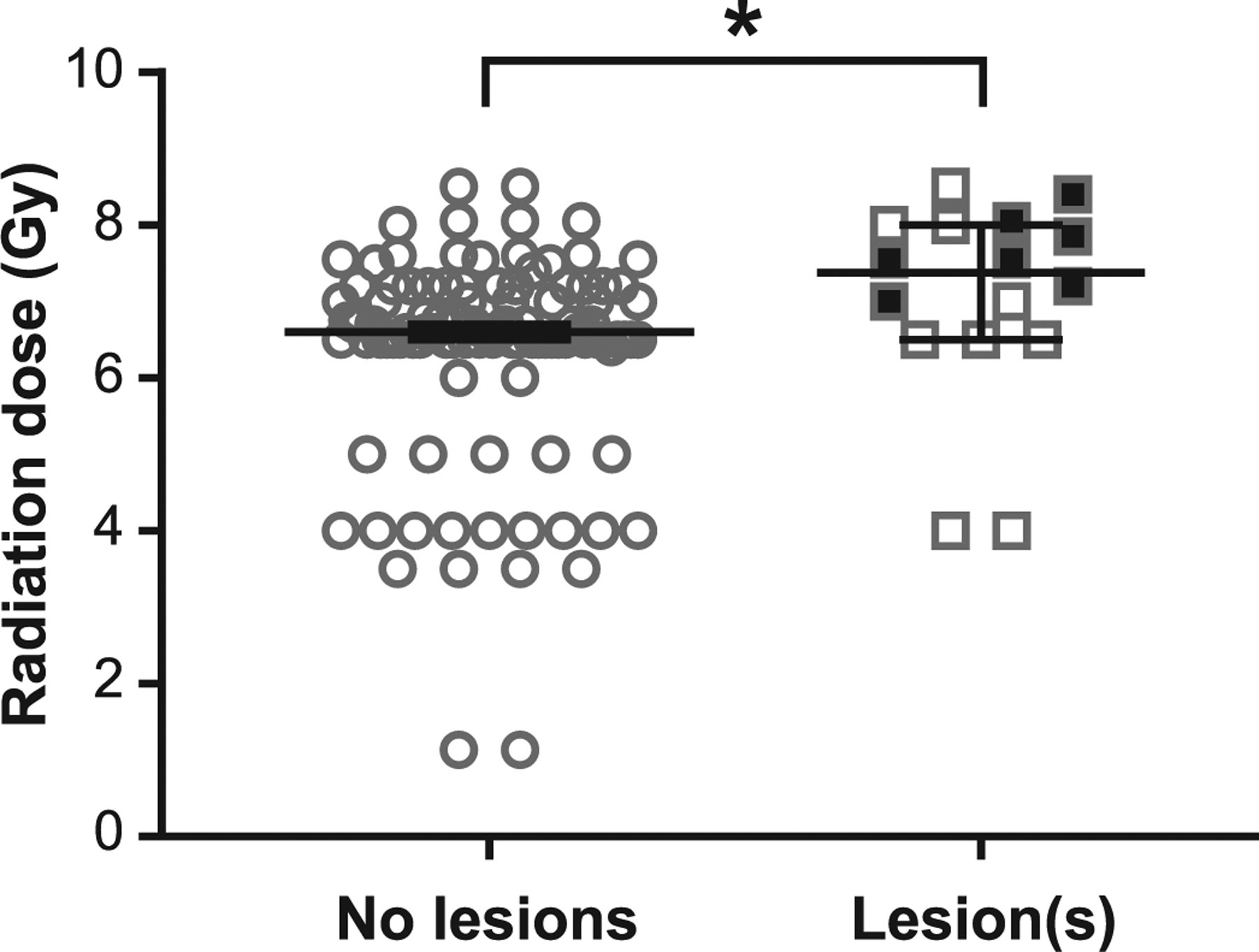
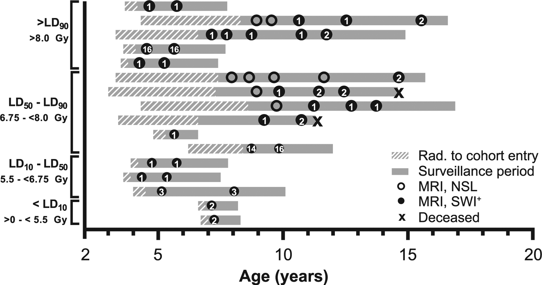
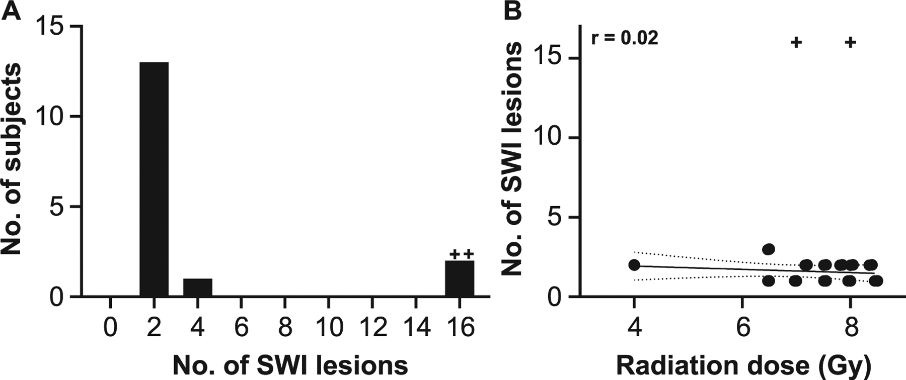
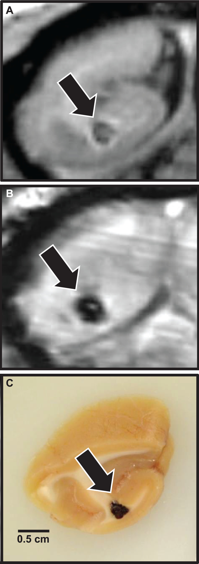
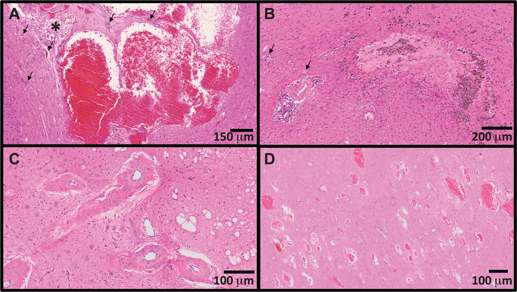
Nuclear accidents and acts of terrorism have the potential to expose thousands of people to high-dose total-body iradiation (TBI). Those who survive the acute radiation syndrome are at risk of developing chronic, degenerative radiation-induced injuries [delayed effects of acute radiation (DEARE)] that may negatively affect quality of life. A growing body of literature suggests that the brain may be vulnerable to radiation injury at survivable doses, yet the long-term consequences of high-dose TBI on the adult brain are unclear. Herein we report the occurrence of lesions consistent with cerebrovascular injury, detected by susceptibility-weighted magnetic resonance imaging (MRI), in a cohort of non-human primate [(NHP); rhesus macaque, Macaca mulatta] long-term survivors of high-dose TBI (1.1-8.5 Gy). Animals were monitored longitudinally with brain MRI (approximately once every three years). Susceptibility-weighted images (SWI) were reviewed for hypointensities (cerebral microbleeds and/or focal necrosis). SWI hypointensities were noted in 13% of irradiated NHP; lesions were not observed in control animals. A prior history of exposure was correlated with an increased risk of developing a lesion detectable by MRI (P = 0.003). Twelve of 16 animals had at least one brain lesion present at the time of the first MRI evaluation; a subset of animals (n = 7) developed new lesions during the surveillance period (3.7-11.3 years postirradiation). Lesions occurred with a predilection for white matter and the gray-white matter junction. The majority of animals with lesions had one to three SWI hypointensities, but some animals had multifocal disease (n = 2). Histopathologic evaluation of deceased animals within the cohort (n = 3) revealed malformation of the cerebral vasculature and remodeling of the blood vessel walls. There was no association between comorbid diabetes mellitus or hypertension with SWI lesion status. These data suggest that long-term TBI survivors may be at risk of developing cerebrovascular injury years after irradiation.


Figures

FIG. 1.
FIG. 1.
Animals receiving TBI develop hypointense lesions on susceptibility-weighted imaging (SWI-MRI) comparable to those which occur in RIBI after fWBI. Panel A: There are no SWI-hypointense foci in nonirradiated animals. Panel B: Multifocal SWI-hypointense foci (white arrows) are scattered throughout the cerebrum of an animal that developed RIBI after receiving 40 Gy fWBI (8 × 5 Gy fractions, 2× per week) as part of a separate study. Scan taken at 12 months postirradiation. Panel C: Comparable, multifocal SWI-hypointense foci in a long-term TBI survivor; the subject received 8.0 Gy TBI one year prior to MRI. White arrows indicate foci of hemorrhage and/or necrosis.
FIG. 2.
FIG. 2.
TBI is associated with MRI-detectable brain lesions, which occur at higher doses. Animals that developed brain lesions after TBI received higher doses than those without lesions (median: 7.4 Gy vs. 6.6 Gy, respectively; P < 0.02). Filled squares indicate incident lesions which occurred during the surveillance period; these animals received higher doses during TBI (7.8 Gy ± 0.4 SD) than those with lesions present since the time of first evaluation only (6.6 ± 1.5 SD) (P < 0.05). Bars indicate median value, error bars are 95% confidence interval. *P < 0.05.
FIG. 3.
FIG. 3.
Radiologic and life history of animals with SWI-hypointense foci. Each gray bar represents a single animal; animals are listed in order of decreasing radiation dose. Bars begin at age at irradiation; cross-hatched region indicates the period before adoption and cohort enrollment. The solid gray region indicates the period the animal was enrolled within the Radiation Survivors Cohort. Circles indicate brain MRI examinations; open circles represent MRI with no significant lesions (NSL), closed circles represent a SWI lesion that was present. Arabic numerals within closed circles indicate the number of SWI foci at the time of examination. “X” indicates the age at death of deceased animals (n = 2). Lethal dose (LD) stratifications refer to LD30 for the hematopoietic acute radiation syndrome (H-ARS) in rhesus macaques (80).
FIG. 4.
FIG. 4.
Evaluation of radiation-dose response on SWI-MRI lesion number. Panel A: Most animals had 1–2 SWI hypointense foci. Frequency histogram. Panel B: There was no statistically significant relationship between radiation dose and lesion number. Linear regression analysis, r = 0.2, P > 0.05. Solid line indicates best fit; slope = −0.1. Dotted lines indicate 95% confidence interval. +Outlier, as determined by ROUT method and excluded from regression analysis.
FIG. 5.
FIG. 5.
Localization of MRI lesions and correlative gross pathology. Coronal sections, occipital lobe, example of a focal brain lesion first noted six years postirradiation, at the time the first MRI scan was completed. Panel A: T1 MRI indicating focal parenchymal loss with central T1 isointense heterogeneity. Panel B: SWI-MRI indicating the presence of iron or calcium (blood or necrosis, respectively). Panel C: Gross tissue, demonstrating focal hemorrhage on sectioned surface.
FIG. 6.
FIG. 6.
Spectrum of cerebrovascular injury observed in long-term survivors of TBI. Panel A: Dilated and malformed vasculature forming anastomosing channels (cavernous hemangioma). There are scattered hemosiderin-laden macrophages within the vascular wall and adjacent parenchyma (evidence of chronic hemorrhage; black arrows). The adjacent parenchyma is focally edematous (asterisk). Panel B: Transmural replacement of the vascular wall by extracellular matrix with chronic hemorrhage and occlusion of the vascular lumen. Recanalization (small caliber capillaries distributed throughout the occluded lumen) and aggregates of hemosiderin-laden macrophages (brown pigment, hemorrhage) are indicative of chronic injury. The adjacent parenchyma is infiltrated by increased numbers of astrocytes and elongated, activated (reactive) microglia (rodcells). There is degeneration of the cerebral peduncle (swollen axons, dilated myelin sheathes, digestion chambers; black arrows). Panel C: Tortuous and redundant vasculature dissecting through the left optic tract with marked thickening (5x normal thickness) of the tunica media by extracellular matrix and smooth muscle hyperplasia. There is scattered vacuolation of the adjacent optic tract. Panel D: Widespread infiltration of the deep occipital white matter by abnormally dilated and haphazardly arranged capillaries (telangiectasia). The lesion was an incidental finding, and not present on susceptibility-weighted imaging six months prior to necropsy.

References

    1. Institute of Medicine (US) Committee on Medical Preparedness for a Terrorist Nuclear Event. Assessing medical preparedness to respond to a terrorist nuclear event: Workshop report. Benjamin GC, McGeary M, McCutchen SR, editors. Washington, DC: National Academies Press; 2009. - PubMed
    1. Fliedner TM, Dorr HD, Meineke V. Multi-organ involvement as a pathogenetic principle of the radiation syndromes: a study involving 110 case histories documented in SEARCH and classified as the bases of haematopoietic indicators of effect. Br J Radiol 2005; S27:1–8.
    1. Acute radiation syndrome: A brochure for physicians. Atlanta, GA: Centers for Disease Control and Prevention; 2017.
    1. Loganovsky K Do low doses of ionizing radiation affect the human brain? Data Sci J 2009; 8:PBR13–BR35.
    1. Danilov VM, Pozdeev VK. The epileptiform reactions of the human brain to prolonged exposure to low-dose ionizing radiation. (Article in Russian). Fiziol Zh Im I M Sechenova 1994; 80:88–98. - PubMed

Publication types