. 2020 Sep 15;7(9):137.
doi: 10.3390/children7090137.

Bidirectional Ductal Shunting and Preductal to Postductal Oxygenation Gradient in Persistent Pulmonary Hypertension of the Newborn

Affiliations

Bidirectional Ductal Shunting and Preductal to Postductal Oxygenation Gradient in Persistent Pulmonary Hypertension of the Newborn

Amy Lesneski et al. Children (Basel). .

Abstract

Background: The aim was to evaluate the relationship between the direction of the patent ductus arteriosus (PDA) shunt and the pre- and postductal gradient for arterial blood gas (ABG) parameters in a lamb model of meconium aspiration syndrome (MAS) with persistent pulmonary hypertension of the newborn (PPHN).

Methods: PPHN was induced by intermittent umbilical cord occlusion and the aspiration of meconium through the tracheal tube. After delivery, 13 lambs were ventilated and simultaneous 129 pairs of pre- and postductal ABG were drawn (right carotid and umbilical artery, respectively) while recording the PDA and the carotid and pulmonary blood flow.

Results: Meconium aspiration resulted in hypoxemia. The bidirectional ductal shunt had a lower postductal partial arterial oxygen tension ([PaO2] with lower PaO2/FiO2 ratio-97 ± 36 vs. 130 ± 65 mmHg) and left pulmonary flow (81 ± 52 vs. 133 ± 82 mL/kg/min). However, 56% of the samples with a bidirectional shunt had a pre- and postductal saturation gradient of < 3%.

Conclusions: The presence of a bidirectional ductal shunt is associated with hypoxemia and low pulmonary blood flow. The absence of a pre- and postductal saturation difference is frequently observed with bidirectional right-to-left shunting through the PDA, and does not exclude a diagnosis of PPHN in this model.

Keywords: oxygenation saturation; patent ductus arteriosus; pulmonary hypertension.


Conflict of interest statement

The authors declare no conflict of interest.

Figures

Figure 1
Figure 1
Left pulmonary blood flow (QP) plotted against the preductal partial arterial oxygen tension (PaO2). Left QP is significantly lower when there is bidirectional shunt through the ductus arteriosus compared to when the shunting is from left to right (aorta towards pulmonary artery).
Figure 2
Figure 2
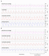
Screenshot of the left pulmonary (QP) blood flow, left carotid blood flow (QCA), and ductal arteriosus (DA) blood flow showing a transition from left-to-right shunting (top, (A)) and bidirectional shunting (bottom, (B)) through the DA. The left QP is lower when the shunt is bidirectional.
Figure 3
Figure 3
Graphical abstract illustrating hemodynamic and oxygen saturation differences comparing the bidirectional to left-to-right ductus arteriosus shunting in a lamb model of meconium aspiration syndrome and pulmonary hypertension. Lambs that experience a bidirectional ductal shunt have a significantly lower left pulmonary arterial blood flow (QP), left carotid blood flow (QCA), mean systolic blood pressure (Sys BP), cerebral oxygen delivery (C-DO2), and oxygen consumption (VO2) compared to left-to-right ductal shunting. FiO2 = fraction of inspired oxygen; PaO2/PaCO2 = partial arterial oxygen/carbon dioxide tension; SaO2 = saturation of arterial oxygen. Copyright Satyan Lakshminrusimha.

References

    1. Lakshminrusimha S., Keszler M. Persistent pulmonary hypertension of the newborn. Neoreviews. 2015;16:e680–e692. doi: 10.1542/neo.16-12-e680. - DOI - PMC - PubMed
    1. Nair J., Lakshminrusimha S. Update on PPHN: Mechanisms and treatment. Semin. Perinatol. 2014;38:78–91. doi: 10.1053/j.semperi.2013.11.004. - DOI - PMC - PubMed
    1. Kemper A.R., Mahle W.T., Martin G.R., Cooley W.C., Kumar P., Morrow W.R., Kelm K., Pearson G.D., Glidewell J., Grosse S.D., et al. Strategies for implementing screening for critical congenital heart disease. Pediatrics. 2011;128:e1259–e1267. doi: 10.1542/peds.2011-1317. - DOI - PubMed
    1. Mahle W., Koppel R. Screening with pulse oximetry for congenital heart disease. Lancet. 2011;378:749–750. doi: 10.1016/S0140-6736(11)61032-5. - DOI - PubMed
    1. Fernandes N., Lakshminrusimha S. The limitations of pulse oximetry for critical congenital heart disease screening in the neonatal intensive care units. Acta Paediatr. 2017;106:1007. doi: 10.1111/apa.13742. - DOI - PubMed