. 2020 Sep 17;21(1):38.
doi: 10.1186/s12868-020-00588-y.

NGF receptors and PI3K/AKT pathway involved in glucose fluctuation-induced damage to neurons and α-lipoic acid treatment

Affiliations

NGF receptors and PI3K/AKT pathway involved in glucose fluctuation-induced damage to neurons and α-lipoic acid treatment

Ting Yan et al. BMC Neurosci. .

Abstract

Background: Glucose fluctuation promotes neuronal apoptosis, which plays a central role in diabetic encephalopathy (DE). Nerve growth factor (NGF), and its interaction with high-affinity (TrkA) and low-affinity (p75NTR) receptors, are involved in neuronal survival. NGF/TrkA contributes to the activation of the PI3K/AKT pathway, which is beneficial for neuronal survival, and α-Lipoic acid (ALA) exerts clinically favorable neuroprotective effects in the periphery. Whether NGF receptors and the PI3K/AKT pathway are involved in glucose fluctuation-induced neuronal damage, as well as the potential molecular mechanism of ALA in protecting glucose fluctuation-induced neuronal damage, remain unclear.

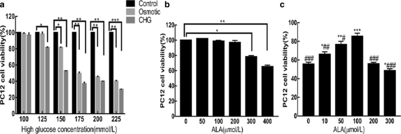
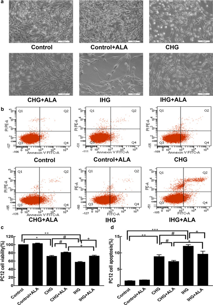
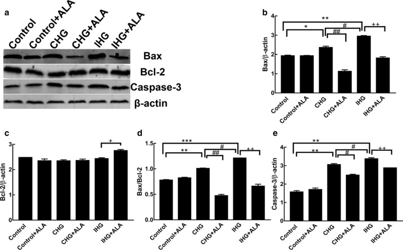
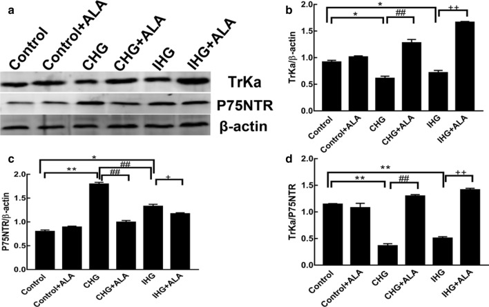
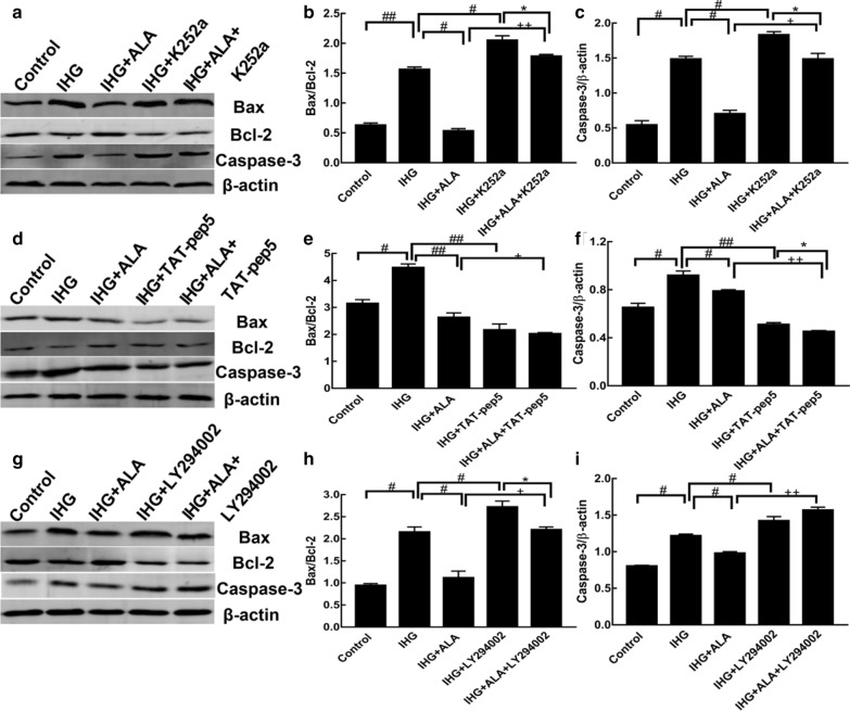
Results: The results indicated that constant high glucose (CHG) and intermittent high glucose (IHG) significantly increased the expression of Bax and caspase-3, and decreased the expression of TrkA/p75NTR and p-AKT/AKT, while ALA stimulation reversed the above proteins in PC12 cells. IHG stimulates apoptosis more effectively than CHG in PC12 cells, which is related to the PI3K/AKT pathway but not to the TrkA/p75NTR. Furthermore, neuronal apoptosis induced by IHG was aggravated by the TrkA inhibitor K252a or the PI3K/AKT inhibitor LY294002, but this effect was alleviated by the p75NTR inhibitor TAT-pep5.

Conclusion: Glucose fluctuation induced cell apoptosis by regulating the TrkA/p75NTR and PI3K/AKT pathway, meanwhile ALA exhibited neuroprotective effects in response to IHG and CHG. These observations indicated that the PI3K/AKT pathway and the balance of TrkA/p75NTR are likely to serve as potential therapeutic targets for DE. In addition, ALA could be a possible therapeutic drug for DE.

Keywords: Cell apoptosis; Diabetic encephalopathy; Intermittent high glucose; PI3K/AKT; TrkA; p75NTR.


Conflict of interest statement

The authors declare that they have no competing interests.

Figures

Fig. 1
Fig. 1
a Effect of glucose on PC12 cell viability. b Effects of ALA on PC12 cell viability under normal glucose concentration. c Effects of ALA on PC12 cell viability under glucose fluctuation. Results are presented as the mean ± SD (n = 3). ***P < 0.001, **P < 0.01,*P < 0.05, compared to the (a) control or (b, c) ALA (0 µmol/L) group. ###P < 0.001, ##P < 0.01, #P < 0.05 versus the ALA (100 µmol/L) group
Fig. 2
Fig. 2
ALA treatment attenuated IHG induced cell injuries in PC12 cells. a, c Effects of IHG on PC12 cell viability and ALA treatment. Cells were observed by microscope, and viability was determined by MTT assay. b, d Effects of IHG on PC12 cell apoptosis and ALA treatment. Apoptosis was detected by flow cytometry. Results are shown as the mean ± SD (n = 3). ***P < 0.001, **P < 0.01,*P < 0.05 versus the control; #P < 0.05 compared to the CHG group; and +P < 0.05 versus the IHG + ALA group
Fig. 3
Fig. 3
ALA treatment inhibited IHG-induced apoptosis in PC12 cells. The density of (a) Bax, Bcl-2, and Caspase-3 were measured by Western blot. Quantitative data of (b) Bax, (c) Bcl-2, (d) Bax/Bcl-2, and (e) Caspase-3 are presented. Data are shown as mean ± SD (n = 3). ***P < 0.05, **P < 0.01, *P < 0.05 versus the control; ##P < 0.01, #P < 0.05 compared to the CHG group; and ++P < 0.01, +P < 0.05 versus the IHG + ALA group
Fig. 4
Fig. 4
ALA treatment affected IHG-induced NGF receptorsin PC12 cells. a The density of TrkA and p75NTR were determined by Western blot. Quantitative data of (b) TrkA, c p75NTR, and d TrkA/p75NTR are presented. Data are presented as mean ± SD (n = 3). **P < 0.01,*P < 0.05 compared to the control; ##P < 0.01 versus the CHG group; and ++P < 0.01, +P < 0.05 compared to the IHG + ALA group
Fig. 5
Fig. 5
ALA treatment altered the IHG-inhibited PI3K/AKT pathway in PC12 cells. a The density of AKT and p-AKT were measured by Western blot. Quantitative data of b p-AKT, c AKT, and d p-AKT/AKT are presented. Data are shown as mean ± SD (n = 3). **P < 0.01 compared to the control; #P < 0.05 compared to the CHG group; and ++P < 0.01 versus the IHG + ALA group
Fig. 6
Fig. 6
ALA protected IHG-induced cell apoptosis through NGF receptors and the PI3K/AKT pathway in PC12 cells. Bax/Bcl-2 and Caspase-3 levels were evaluated in the presence or absence of K252a (a–c), TAT-pep5 (d-f), and LY294002 (g–i), respectively. Data are shown as the mean ± SD (n = 3). ##P < 0.01, #P < 0.05 versus the IHG group; ++P < 0.01, +P < 0.05 compared to the IHG + ALA group; and *P < 0.05 compared to the IHG + inhibitor (K252a, TAT-pep5, or LY294002)

References

    1. Barrett GL, Reid CA, Tsafoulis C, Zhu W, Williams DA, Paolini AG, Trieu J, Murphy M. Enhanced spatial memory and hippocampal long-term potentiation in p75 neurotrophin receptor knockout mice. Hippocampus. 2010;20(1):145–152. - PubMed
    1. Becker K, Cana A, Baumgärtner W, Spitzbarth I. p75 neurotrophin receptor: a double-edged sword in pathology and regeneration of the central nervous system. Vet Pathol. 2018;55(6):786–801. doi: 10.1177/0300985818781930. - DOI - PubMed
    1. Bibel M, Hoppe E, Barde YA. Biochemical and functional interactions between the neurotrophin receptors trk and P75NTR. EMBO J. 1999;18(3):616–622. doi: 10.1093/emboj/18.3.616. - DOI - PMC - PubMed
    1. Biessels GJ, Staekenborg S, Brunner E, Brayne C, Scheltens P. Risk of dementia in diabetes mellitus: a systematic review. Lancet Neurol. 2006;5(1):64–74. doi: 10.1016/S1474-4422(05)70284-2. - DOI - PubMed
    1. Cai F, Helke CJ. A bnormal PI3 kinase/Akt signal pathway in vagal afferent neurons and vagus nerve of streptozotocin-diabetic rats. Mol Brain Res. 2003;110:234–244. doi: 10.1016/S0169-328X(02)00652-6. - DOI - PubMed

Publication types

MeSH terms