. 2020 Sep 17;39(1):191.
doi: 10.1186/s13046-020-01676-x.

Linc00514 promotes breast cancer metastasis and M2 polarization of tumor-associated macrophages via Jagged1-mediated notch signaling pathway

Affiliations

Linc00514 promotes breast cancer metastasis and M2 polarization of tumor-associated macrophages via Jagged1-mediated notch signaling pathway

Sifeng Tao et al. J Exp Clin Cancer Res. .

Abstract

Background: Tumor-associated macrophages (TAMs) and tumor cells are important components of the tumor microenvironment. M2 polarization of TAMs, which is a major actor in breast cancer malignancy and metastasis, can be induced by breast cancer cells. However, the potential mechanisms of the interaction between breast cancer cells and TAMs remain unclear.

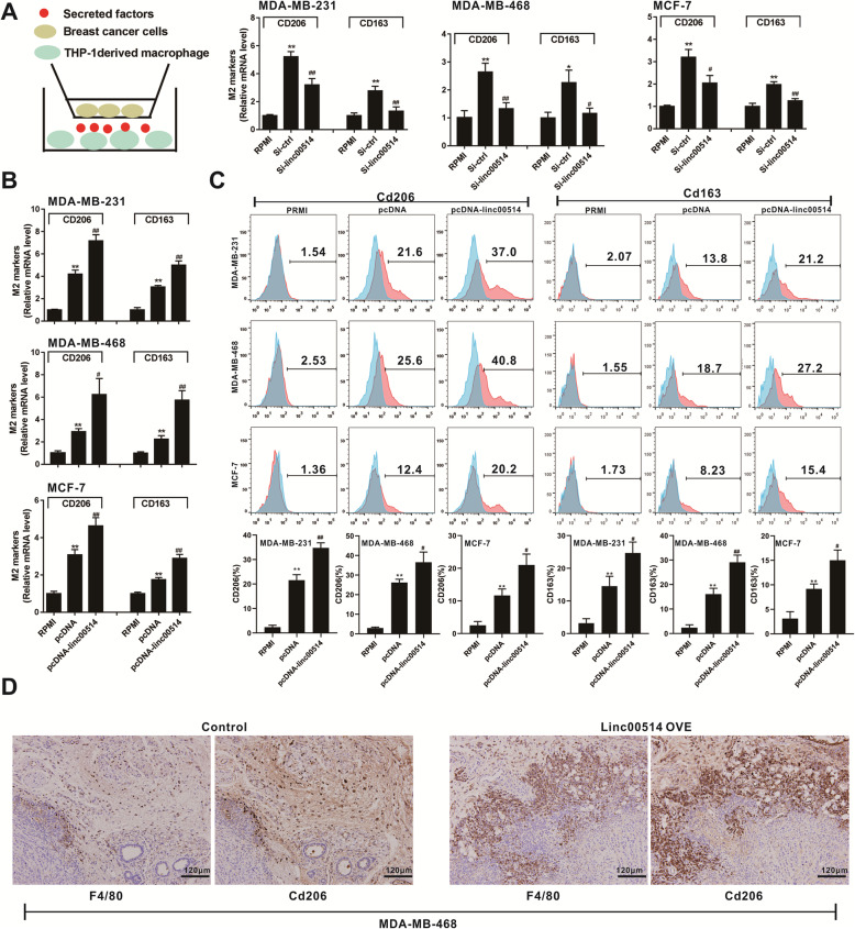
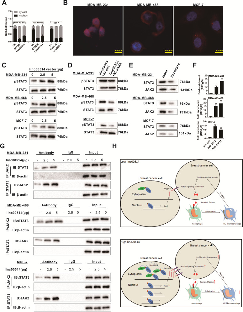
Methods: The candidate breast cancer-associated long non-coding RNAs (lncRNAs) were analyzed using the GEO database. Functional assays, including MTT assay, Transwell assay, and EdU labeling detection, were performed to investigate the oncogenic role of linc00514 in breast cancer progression. The co-culture and ELISA assays were used to assess the role of linc00514 in macrophage recruitment and M2 polarization. RNA immunoprecipitation, RNA pull-down, and luciferase reporter assays were applied to determine the mechanism of linc00514 in breast cancer metastasis. Mouse xenograft models, mouse pulmonary metastatic models, and mouse primary tumor models were used to assess the role of linc00514 in M2 macrophage polarization and breast cancer tumorigenicity.

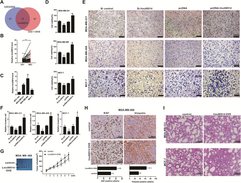
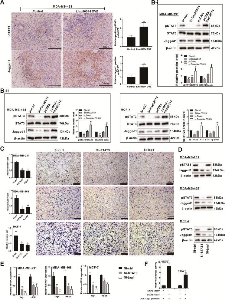
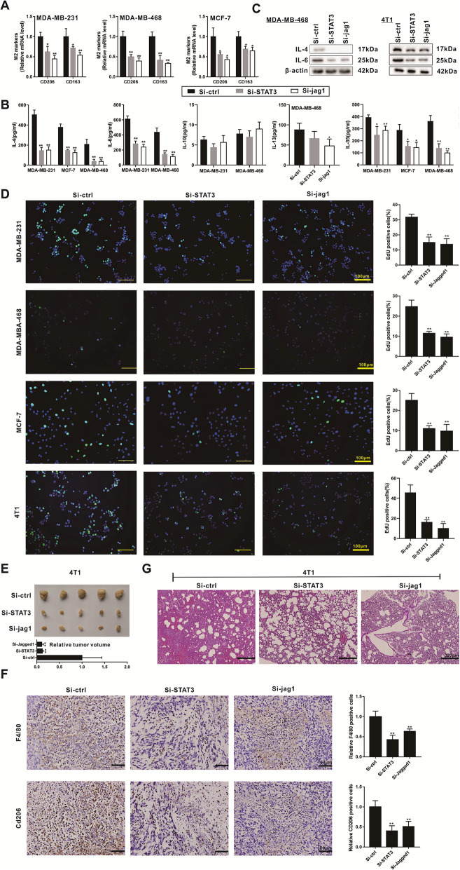
Results: Linc00514 was highly expressed in clinical breast cancer tissues and breast cancer cell lines. Overexpression of linc00514 promoted the proliferation and invasion of breast cancer cells and increased xenograft tumor volumes and pulmonary metastatic nodules. Overexpression of linc00514 also increased the percentage of macrophages expressing M2 markers CD206 and CD163. Mechanistically, linc00514 promoted Jagged1 expression in a transcriptional manner by increasing the phosphorylation of a transcription factor STAT3. Subsequently, Jagged1-mediated Notch signaling pathway promoted IL-4 and IL-6 secretions in breast cancer cells and ultimately inducing M2 polarization of macrophages.

Conclusion: Linc00514 plays an important role in regulating breast cancer tumorigenicity and M2 macrophage polarization via Jagged1-mediated Notch signaling pathway.

Keywords: Breast cancer; Jagged1; Tumor-associated macrophage; linc00514.


Conflict of interest statement

All authors declare that they have no competing interests.

Figures

Fig. 1
Fig. 1
Highly expressed linc00514 promotes breast cancer malignancy. a. Top 100 dysregulated lncRNAs whose P < 0.05, log2FC > 2, or logFC < 0.5 were compared in the GSE60689 dataset and the GSE112848 dataset from the GEO database. b. The relative expression of linc00514 in tumor tissues (BRC) and in paracancer tissues (ANT) from 46 breast cancer patients was detected using qRT-PCR. c. The relative expression of linc00514 in human breast cancer cell lines (MDA-MB-231, MDA-MB-468, and MCF-7) and in the normal breast epithelial cell line (MCF-10A) was detected using qRT-PCR. **P < 0.01 vs MCF-10A. d-f. Human breast cancer cell lines (MDA-MB-231, MDA-MB-468, and MCF-7) were transiently transfected with linc00514 siRNAs (Si-linc00514) or linc00514 plasmids (pcDNA-linc00514) for 48 h. Cell viability was detected using MTT assay (d). Cell invasion was detected using Transwell assay (e, f). Scale bar = 200 μm. g-h. Female Balb/c nude mice were subcutaneously injected with the MDA-MB-468 cells (5 × 106 cells) which were stably transfected with linc00514-overexpressing plasmids (linc00514-OVE) or the control plasmids (n = 5 in each group). The tumor volumes were detected every week (g). After 7 weeks, the Ki67 expression and the Vimentin expression in tumor tissues were detected using immunohistochemistry (h). Scale bar = 80 μm. i. Female Balb/c nude mice were intravenously injected with the MDA-MB-231/MCF-7 cells (1.5 × 106 cells) which were stably transfected with linc00514-OVE (n = 5 in each group). The pulmonary metastatic tumor tissues were observed using HE staining. Scale Bar = 160 μm. Three independent experiments. *P < 0.05, **P < 0.01 vs control (Si-ctrl). ##P < 0.01 vs control (pcDNA)
Fig. 2
Fig. 2
Overexpression of linc00514 in breast cancer cells promotes M2 polarization of macrophages. a-b. Human breast cancer cell lines (MDA-MB-231, MDA-MB-468, and MCF-7) were transiently transfected with linc00514 siRNAs (Si-linc00514) or linc00514 plasmids (pcDNA-linc00514) for 48 h before the co-culture. After co-culturing breast cancer cells with PMA-induced human monocyte THP-1 cells for 48 h using Transwell assay, we detected the relative mRNA levels of CD206 and CD163 in THP-1 derived macrophages, both of which were M2 polarization markers of macrophages. c. The percentage of CD206+ or CD163+ cells after overexpressing linc00514 was detected using flow cytometry. d. Female Balb/c nude mice were subcutaneously injected with the MDA-MB-468 cells (5 × 106 cells) which were stably transfected with linc00514-overexpressing plasmids (linc00514-OVE) or the control plasmids (n = 5 in each group). After 7 weeks, the expression of F4/80 and CD206 in tumor tissues was detected using immunohistochemistry. Scale bar = 120 μm. Three independent experiments. ** P < 0.01 vs control (RPMI). ## P < 0.01 vs control (Si-ctrl or pcDNA)
Fig. 3
Fig. 3
Linc00514 positively regulates the protein expression of pSTAT3 and Jagged1. a. Female Balb/c nude mice were subcutaneously injected with the MDA-MB-468 cells (5 × 106 cells) which were stably transfected with linc00514-overexpressing plasmids (linc00514-OVE) or the control plasmids (n = 5 in each group). After 7 weeks, the expression of pSTAT3 and Jagged1 in tumor tissues was detected using immunohistochemistry. Scale bar = 200 μm. b. Human breast cancer cell lines (MDA-MB-231, MDA-MB-468, and MCF-7) were transiently transfected with linc00514 siRNAs (Si-linc00514) or linc00514 plasmids (pcDNA-linc00514) for 48 h. The expression of pSTAT3, STAT3, and Jagged1 was detected using western blot analysis. c-e. Human breast cancer cell lines (MDA-MB-231, MDA-MB-468, and MCF-7) were transiently transfected with STAT3 siRNAs (Si-STAT3) or JAG1 siRNAs (Si-JAG1) for 48 h. Cell invasion was detected using MTT assay (c). Scale bar = 200 μm. The protein level of pSTAT3 and Jagged1 was detected using western blot analysis (d). The relative mRNA expression of JAG1 and HES1 was detected using qRT-PCR (e). f. The 293 T cells were transfected with human or mouse STAT3 vectors or empty vectors as well as JAG1 promoter plasmids. The relative luciferase activity was detected using luciferase reporter assay. Three independent experiments. *P < 0.05, ** P < 0.01 vs control (Si-ctrl). #P < 0.05, ##P < 0.01 vs control (Si-ctrl or pcDNA)
Fig. 4
Fig. 4
STAT3 and Jagged1 participates in the breast cancer metastasis and M2 polarization of macrophages. a. Human breast cancer cell lines (MDA-MB-231, MDA-MB-468, and MCF-7) were transiently transfected with STAT3 siRNAs (Si-STAT3) or JAG1 siRNAs (Si-JAG1) for 48 h before the co-culture. After co-culturing breast cancer cells with PMA-induced human monocyte THP-1 cells for 48 h using Transwell assay, we detected the relative mRNA levels of CD206 and CD163 in THP-1 derived macrophages, both of which were M2 polarization markers of macrophages. b-d. Human breast cancer cell lines (MDA-MB-231, MDA-MB-468, and MCF-7) and mouse breast cancer cell line (4 T1) were transiently transfected with STAT3 siRNAs (Si-STAT3) or JAG1 siRNAs (Si-JAG1) for 48 h. The level of IL-4, IL-6, IL-10, IL-13, and IL-35 in supernatants was detected using ELISA (b). The protein level of IL-4 and IL-6 in breast cells was detected using western blot analysis (c). The cell proliferation was detected using EdU labeling detection (d). Scale Bar =100 μm. e-f. Female Balb/c mice were subcutaneously injected with the mouse breast cancer cell line (4 T1, 5 × 105 cells) which were transfected with STAT3 siRNAs (Si-STAT3), JAG1 siRNAs (Si-JAG1), or the control siRNAs (Si-ctrl) (n = 10 in each group). The tumor tissues were collected and measured on the 20th day (e). The F4/80 and CD206 expressions in tumor tissues were detected using immunohistochemistry (f). Scale bar = 60 μm. g. Female Balb/c mice were intravenously injected with the 4 T1 cells (1.5 × 105 cells) which were transfected with STAT3 siRNAs (Si-STAT3), JAG1 siRNAs (Si-JAG1), or the control siRNAs (Si-ctrl) (n = 5 in each group). The pulmonary metastatic tumor tissues were observed using HE staining. Scale Bar = 320 μm. Three independent experiments. *P < 0.05, **P < 0.01 vs control (Si-ctrl)
Fig. 5
Fig. 5
Overexpression of linc00514 promotes proliferation and invasion of breast cancer cells and M2 polarization of macrophages via regulating STAT3. Human breast cancer cell lines (MDA-MB-231, MDA-MB-468, and MCF-7) which were stably transfected with linc00514 plasmids (linc00514-OVE) were then transfected with STAT3 siRNAs (Si-STAT3) for 48 h. a. The cell proliferation was detected using EdU labeling detection. Scale Bar =100 μm. b. The cell invasion was detected using Transwell assay. Scale bar = 200 μm. c-d. After co-culturing breast cancer cells with PMA-induced human monocyte THP-1 cells for 48 h using Transwell assay, we detected the relative percentage of CD206+ and CD163+ in THP-1 derived macrophages using flow cytometry (c). The relative mRNA expression of JAG1 and HES1 was detected using qRT-PCR (d). Three independent experiments. *P < 0.05, **P < 0.01 vs control (linc00514-OVE)
Fig. 6
Fig. 6
Linc00514 increases phosphorylation of STAT3. a-b. The distribution and localization of linc00514 in human breast cancer cell lines (MDA-MB-231, MDA-MB-468, and MCF-7) were detected using cytosol/nucleus qRT-PCR (a) and FISH (b). c. Human breast cancer cell lines (MDA-MB-231 and MCF-7) were transfected with the increasing concentration of linc00514 vectors (from 0 to 5 μg). The protein level of pSTAT3 and STAT3 was detected using western blot analysis. d. The siRNA-mediated JAK2 knockdown reduced the pSTAT3 protein level which was increased by linc00514 overexpression. e-f. The RNA pull-down experiment (e) and the RIP experiment (f) confirmed that linc00514 could bind with STAT3 and JAK2. g. Human breast cancer cell lines (MDA-MB-231 and MCF-7) were transfected with the increasing concentration of linc00514 vectors (from 0 to 5 μg). The levels of STAT3 and JAK2 pulled down by linc00514 probes were detected using the RNA pull-down experiment. h. Schema depicting the mechanisms of linc00514 in breast cancer growth and metastasis. Three independent experiments. **P < 0.01 vs control (Anti-IgG)

Similar articles

Cited by

References

    1. Geeraerts X, Bolli E, Fendt S-M, Van Ginderachter JA. Macrophage metabolism as therapeutic target for Cancer, atherosclerosis, and obesity. Front Immunol. 2017;8:289. doi: 10.3389/fimmu.2017.00289. - DOI - PMC - PubMed
    1. Biswas SK, Allavena P, Mantovani A. Tumor-associated macrophages: functional diversity, clinical significance, and open questions. Semin Immunopathol. 2013;35(5):585–600. doi: 10.1007/s00281-013-0367-7. - DOI - PubMed
    1. Komohara Y, Takeya M. CAFs and TAMs: maestros of the tumour microenvironment. J Pathol. 2017;241(3):313–315. doi: 10.1002/path.4824. - DOI - PubMed
    1. Guo Q, Jin Z, Yuan Y, Liu R, Xu T, Wei H, Xu X, He S, Chen S, Shi Z, et al. New mechanisms of tumor-associated macrophages on promoting tumor progression: recent research advances and potential targets for tumor immunotherapy. J Immunol Res. 2016;2016:9720912. - PMC - PubMed
    1. Rhee I. Diverse macrophages polarization in tumor microenvironment. Arch Pharm Res. 2016;39(11):1588–1596. doi: 10.1007/s12272-016-0820-y. - DOI - PubMed

MeSH terms