

CRISPR/Cas9-Mediated Point Mutation in Nkx3.1 Prolongs Protein Half-Life and Reverses Effects Nkx3.1 Allelic Loss
- PMID: 32943441
- PMCID: PMC7642110
- DOI: 10.1158/0008-5472.CAN-20-1742
CRISPR/Cas9-Mediated Point Mutation in Nkx3.1 Prolongs Protein Half-Life and Reverses Effects Nkx3.1 Allelic Loss
Abstract
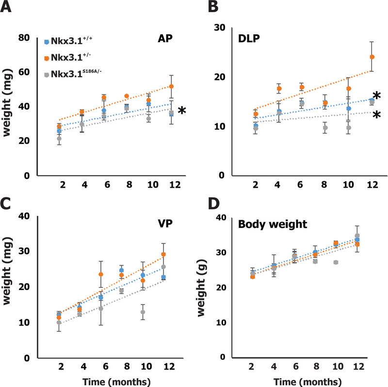
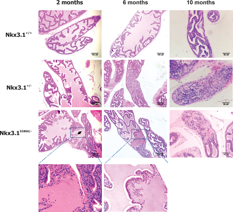
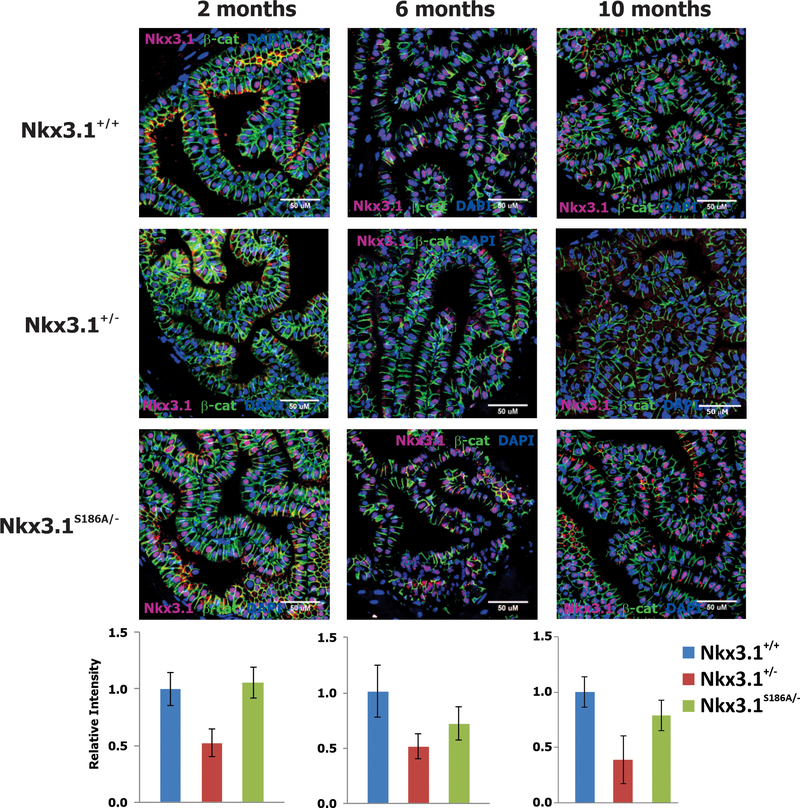
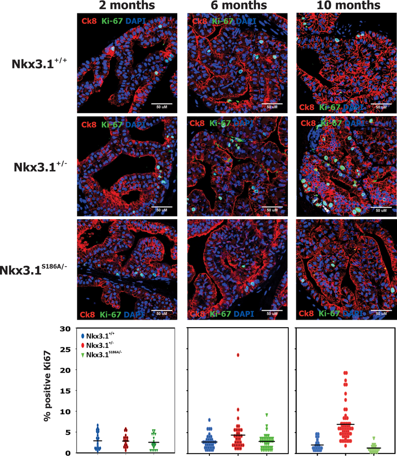
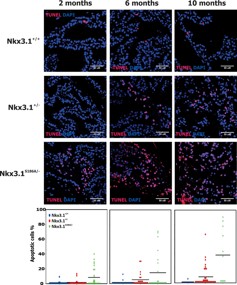
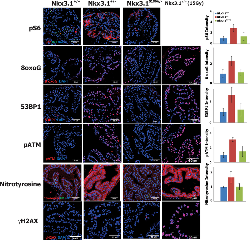
NKX3.1 is the most commonly deleted gene in prostate cancer and is a gatekeeper suppressor. NKX3.1 is haploinsufficient, and pathogenic reduction in protein levels may result from genetic loss, decreased transcription, and increased protein degradation caused by inflammation or PTEN loss. NKX3.1 acts by retarding proliferation, activating antioxidants, and enhancing DNA repair. DYRK1B-mediated phosphorylation at serine 185 of NKX3.1 leads to its polyubiquitination and proteasomal degradation. Because NKX3.1 protein levels are reduced, but never entirely lost, in prostate adenocarcinoma, enhancement of NKX3.1 protein levels represents a potential therapeutic strategy. As a proof of principle, we used CRISPR/Cas9-mediated editing to engineer in vivo a point mutation in murine Nkx3.1 to code for a serine to alanine missense at amino acid 186, the target for Dyrk1b phosphorylation. Nkx3.1S186A/-, Nkx3.1+/- , and Nkx3.1+/+ mice were analyzed over one year to determine the levels of Nkx3.1 expression and effects of the mutant protein on the prostate. Allelic loss of Nkx3.1 caused reduced levels of Nkx3.1 protein, increased proliferation, and prostate hyperplasia and dysplasia, whereas Nkx3.1S186A/- mouse prostates had increased levels of Nkx3.1 protein, reduced prostate size, normal histology, reduced proliferation, and increased DNA end labeling. At 2 months of age, when all mice had normal prostate histology, Nkx3.1+/- mice demonstrated indices of metabolic activation, DNA damage response, and stress response. These data suggest that modulation of Nkx3.1 levels alone can exert long-term control over premalignant changes and susceptibility to DNA damage in the prostate. SIGNIFICANCE: These findings show that prolonging the half-life of Nkx3.1 reduces proliferation, enhances DNA end-labeling, and protects from DNA damage, ultimately blocking the proneoplastic effects of Nkx3.1 allelic loss.
©2020 American Association for Cancer Research.
Conflict of interest statement
The authors have no competing interests and no conflicts to disclose.
Figures
Similar articles
-
Loss of PTEN Accelerates NKX3.1 Degradation to Promote Prostate Cancer Progression.Cancer Res. 2019 Aug 15;79(16):4124-4134. doi: 10.1158/0008-5472.CAN-18-4110. Epub 2019 Jun 18. Cancer Res. 2019. PMID: 31213464 Free PMC article.
-
The Tumor Suppressor NKX3.1 Is Targeted for Degradation by DYRK1B Kinase.Mol Cancer Res. 2015 May;13(5):913-22. doi: 10.1158/1541-7786.MCR-14-0680. Epub 2015 Mar 16. Mol Cancer Res. 2015. PMID: 25777618 Free PMC article.
-
Loss of Nkx3.1 leads to the activation of discrete downstream target genes during prostate tumorigenesis.Oncogene. 2009 Sep 17;28(37):3307-19. doi: 10.1038/onc.2009.181. Epub 2009 Jul 13. Oncogene. 2009. PMID: 19597465 Free PMC article.
-
Mechanisms of prostate tumorigenesis: roles for transcription factors Nkx3.1 and Egr1.Ann N Y Acad Sci. 2005 Nov;1059:33-40. doi: 10.1196/annals.1339.018. Ann N Y Acad Sci. 2005. PMID: 16382041 Review.
-
Regulating NKX3.1 stability and function: Post-translational modifications and structural determinants.Prostate. 2016 May;76(6):523-33. doi: 10.1002/pros.23144. Epub 2016 Feb 4. Prostate. 2016. PMID: 26841725 Review.
Cited by
-
Targeting mRNA-coding genes in prostate cancer using CRISPR/Cas9 technology with a special focus on androgen receptor signaling.Cell Commun Signal. 2024 Oct 17;22(1):504. doi: 10.1186/s12964-024-01833-1. Cell Commun Signal. 2024. PMID: 39420406 Free PMC article. Review.
-
Mirk/Dyrk1B Kinase Inhibitors in Targeted Cancer Therapy.Pharmaceutics. 2024 Apr 11;16(4):528. doi: 10.3390/pharmaceutics16040528. Pharmaceutics. 2024. PMID: 38675189 Free PMC article. Review.
References
-
- Vocke CD, Pozzatti RO, Bostwick DG, Florence CD, Jennings SB, Strup SE, et al. Analysis of 99 microdissected prostate carcinomas reveals a high frequency of allelic loss on chromosome 8p21–22. Cancer Res 1996;56:2411–6 - PubMed
-
- Swalwell JI, Vocke CD, Yang Y, Walker JR, Grouse L, Myers SH, et al. Determination of a minimal deletion interval on chromosome band 8p21 in sporadic prostate cancer. Genes ChromosomesCancer 2002;33:201–5 - PubMed
-
- Kim MJ, Bhatia-Gaur R, Banach-Petrosky WA, Desai N, Wang Y, Hayward SW, et al. Nkx3.1 mutant mice recapitulate early stages of prostate carcinogenesis. Cancer Res 2002;62:2999–3004 - PubMed
Publication types
MeSH terms
Substances
Grants and funding
LinkOut - more resources
Full Text Sources
Medical
Molecular Biology Databases
Research Materials
