. 2020 Sep 17;11(1):4692.
doi: 10.1038/s41467-020-18390-z.

Wood hemicelluloses exert distinct biomechanical contributions to cellulose fibrillar networks

Affiliations

Wood hemicelluloses exert distinct biomechanical contributions to cellulose fibrillar networks

Jennie Berglund et al. Nat Commun. .

Abstract

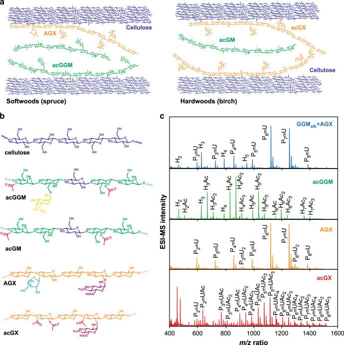
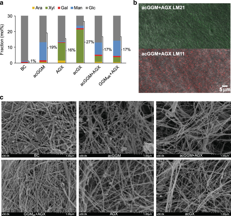
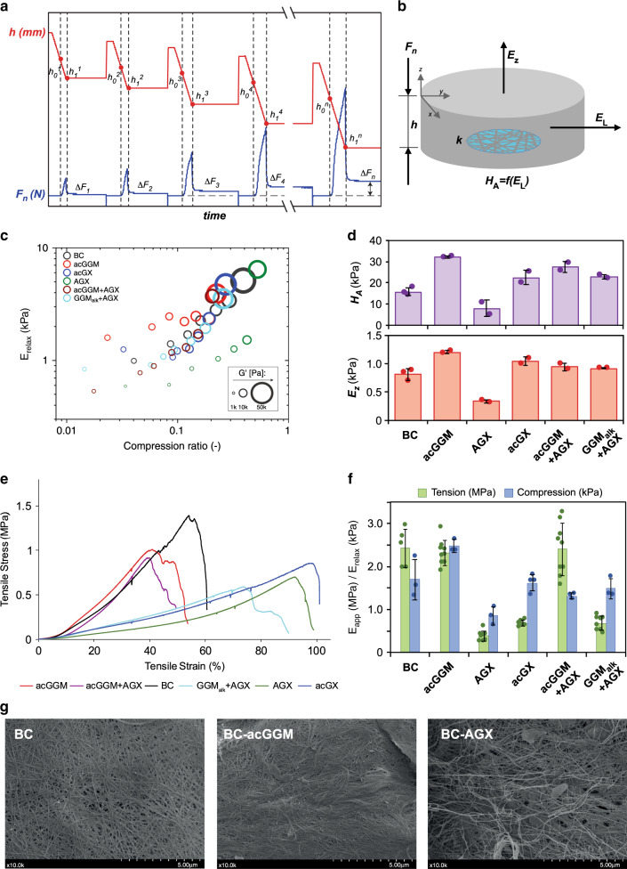
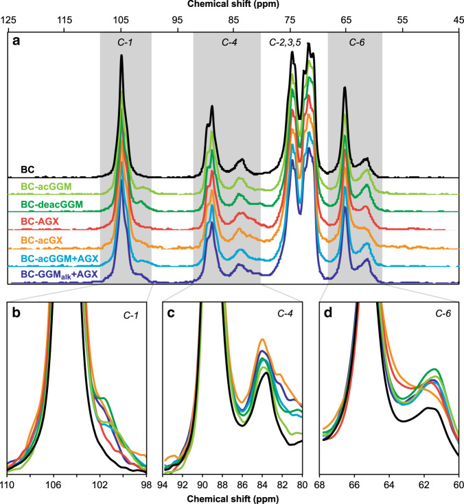
Hemicelluloses, a family of heterogeneous polysaccharides with complex molecular structures, constitute a fundamental component of lignocellulosic biomass. However, the contribution of each hemicellulose type to the mechanical properties of secondary plant cell walls remains elusive. Here we homogeneously incorporate different combinations of extracted and purified hemicelluloses (xylans and glucomannans) from softwood and hardwood species into self-assembled networks during cellulose biosynthesis in a bacterial model, without altering the morphology and the crystallinity of the cellulose bundles. These composite hydrogels can be therefore envisioned as models of secondary plant cell walls prior to lignification. The incorporated hemicelluloses exhibit both a rigid phase having close interactions with cellulose, together with a flexible phase contributing to the multiscale architecture of the bacterial cellulose hydrogels. The wood hemicelluloses exhibit distinct biomechanical contributions, with glucomannans increasing the elastic modulus in compression, and xylans contributing to a dramatic increase of the elongation at break under tension. These diverging effects cannot be explained solely from the nature of their direct interactions with cellulose, but can be related to the distinct molecular structure of wood xylans and mannans, the multiphase architecture of the hydrogels and the aggregative effects amongst hemicellulose-coated fibrils. Our study contributes to understanding the specific roles of wood xylans and glucomannans in the biomechanical integrity of secondary cell walls in tension and compression and has significance for the development of lignocellulosic materials with controlled assembly and tailored mechanical properties.


Conflict of interest statement

The authors declare no competing interests.

Figures

Fig. 1
Fig. 1. Structure of wood hemicelluloses and their incorporation in BC-H model networks.
a Schematic illustration of the polymeric network in a model BC-H system mimicking the secondary cell walls of softwoods and hardwoods prior to lignification. b Average molecular structure of major repeating motifs of wood hemicelluloses. Softwood O-acetyl-galactoglucomannan (acGGM, from spruce) comprises a main chain of β-(1→4)-linked d-mannose (Man) and d-glucose units (Glc) partially substituted by α-(1→6)-linked d-galactose units (Gal) exclusively on the Man units and partial acetylation at the C2 and C3 hydroxyl groups of Man. Hardwood O-acetyl-glucomannan (acGM) has similar structure as acGGM without Gal substitutions. Softwood arabino-4-O-methylglucuronoxylan (AGX, from spruce) consists of a backbone of β-(1→4)-linked d-Xyl with partial substitution by both α-(1→2)-linked mGlcA and α-(1→3)-linked l-arabinofuranose (Ara),. Hardwood O-acetyl-4-O-methylglucuronoxylan (acGX, from birch) consists of a backbone of β-(1→4)-linked d-xylose units (Xyl) with partial substitution of α-(1→2)-linked 4-O-d-methylglucuronic acids (mGlcA) and O-acetylation (OAc) at some of the C2 and C3 units of Xyl,. c Oligomeric mass profiling (OLIMP) by mass spectrometry of the wood hemicelluloses (AGX, acGX, and acGGM) after enzymatic hydrolysis. Note: P refers to a pentose (Xyl or Ara), H refers to a hexose (Man, Gal or Glc), Ac refers to an acetyl group, and mU refers to mGlcA. Source data provided as a Source Data file.
Fig. 2
Fig. 2. Composition and microstructure of the BC-H hydrogels.
The sample names for the BC-H materials correspond to the hemicellulose content. a Monosaccharide composition of BC-H composite pellicles. The y axis ends at 30% to amplify the composition of hemicellulose sugars; however, the glucose (Glc) fraction continues up to 100%. SDs: ±0.0–0.8. Values correspond to the fraction of hemicellulose-related sugars, where the Glc contribution from hemicelluloses as well as uronic acid composition was estimated by assuming the same ratio to mannose (Man) or xylose (Xyl) as in the extracted hemicellulose. Source data are provided as a Source Data file. b Confocal microscopy images of antibody-labeled BC-acGGM+AGX. Xylans are labeled by LM11 (in red) and mannans are labeled by LM21 (in green). The length of the scale bar corresponds to 5 μm. c Scanning electron microscopy (SEM) images of BC-H pellicles (at ×30 magnification). The length of the scale bar corresponds to 1 μm.
Fig. 3
Fig. 3. Mechanical properties of the BC-H hydrogels under compression and tension.
The sample names for the BC-H materials correspond to the incorporated hemicelluloses. a Graphical scheme of the compression–relaxation mechanical analysis, where Fn(N) is the normal force and h is the pellicle thickness. b Parameters for the poroelastic model in compression, where Ez is the out-of-plane modulus; HA is the aggregate modulus, which is a function of the lateral (in-plane) modulus (HA = f(EL) (see Supplementary Methods, Supplementary Eq. 1); and k is the permeability. c Compression–relaxation mechanical analyses. Values are averages from three replicates. Source data are provided as a Source Data file. d Moduli obtained from the poroelastic model (at a compression rate of 0.1). Error bars are based on SDs from two to three measurements. e Representative stress–strain curves from the tensile testing of BC-H hydrogels after compression. f Comparison between the E-modulus in tension (Eapp) and compression (Erelax at a compression rate of 0.1). Error bars correspond to SDs (n =  5–9 for tension and n = 3–4 for compression measurements). g Scanning electron microscopy (SEM) images of selected BC-H hydrogels after compression (at ×10 magnification). The length of the scale bar corresponds to 5 μm.
Fig. 4
Fig. 4. Interactions in BC-H hydrogels by solid-state 13C CP/MAS NMR analysis.
a Full spectra of the hydrated BC-H samples. bd Magnification of the C-1 and C-4 regions of the NMR spectra for the spruce wood analogs, including BC-acGGM, BC-deacGGM (the same pellicle after a pH 10 wash for acetyl group removal), BC-GGMalk + AGX, and BC-acGGM + AGX. Source data are provided as a Source Data file.
Fig. 5
Fig. 5. Proposed organization of the BC-H hydrogels with incorporated wood hemicelluloses.
a Molecular interactions between the cellulose microfibrils and the wood hemicelluloses: birch xylan (acGX), spruce glucomannan (acGGM), and spruce xylan (AGX). Here we depict individual xylan and glucomannan polymers that exhibit both regular molecular motifs interacting with the cellulose surfaces in a rigid phase and non-patterned domains in a flexible phase. b Proposed scheme for the architecture of the BC-H fibrillar networks. In the rigid phases, the hemicelluloses attain extended conformations directly interacting with the cellulose surfaces. In the flexible phases, the hemicelluloses may adopt more coiled conformations where they can interact with each other through bridging adhesion of different intensities (stronger for glucomannans, weaker for xylans).The presence of rigid and flexible phases in BC-H hydrogels has been estimated as 50% for each phase, based on the results from 13C CP/MAS NMR analysis.

References

    1. Carpita NC, Gibeaut DM. Structural models of primary cell walls in flowering plants: consistency of molecular structure with the physical properties of the walls during growth. Plant J. 1993;3:1–30. - PubMed
    1. Burgert I, Keplinger T. Plant micro- and nanomechanics: experimental techniques for plant cell-wall analysis. J. Exp. Bot. 2013;64:4635–4649. - PubMed
    1. Cosgrove D. J. & Jarvis M. C. Comparative structure and biomechanics of plant primary and secondary cell walls. Fron. Plant Sci.3, 204 (2012). - PMC - PubMed
    1. Whitney SEC, et al. Effects of structural variation in xyloglucan polymers on interactions with bacterial cellulose. Am. J. Bot. 2006;93:1402–1414. - PubMed
    1. Kang X, et al. Lignin-polysaccharide interactions in plant secondary cell walls revealed by solid-state NMR. Nat. Commun. 2019;10:347. - PMC - PubMed

Publication types