Multicenter Study
. 2020 Sep 17;10(1):15245.
doi: 10.1038/s41598-020-72270-6.

Diagnosis of thyroid nodules on ultrasonography by a deep convolutional neural network

Affiliations
Multicenter Study

Diagnosis of thyroid nodules on ultrasonography by a deep convolutional neural network

Jieun Koh et al. Sci Rep. .

Abstract

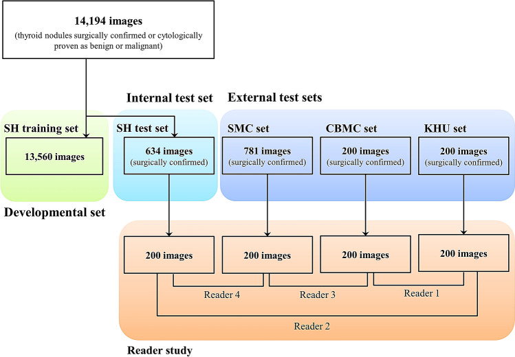
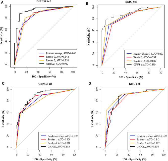
The purpose of this study was to evaluate and compare the diagnostic performances of the deep convolutional neural network (CNN) and expert radiologists for differentiating thyroid nodules on ultrasonography (US), and to validate the results in multicenter data sets. This multicenter retrospective study collected 15,375 US images of thyroid nodules for algorithm development (n = 13,560, Severance Hospital, SH training set), the internal test (n = 634, SH test set), and the external test (n = 781, Samsung Medical Center, SMC set; n = 200, CHA Bundang Medical Center, CBMC set; n = 200, Kyung Hee University Hospital, KUH set). Two individual CNNs and two classification ensembles (CNNE1 and CNNE2) were tested to differentiate malignant and benign thyroid nodules. CNNs demonstrated high area under the curves (AUCs) to diagnose malignant thyroid nodules (0.898-0.937 for the internal test set and 0.821-0.885 for the external test sets). AUC was significantly higher for CNNE2 than radiologists in the SH test set (0.932 vs. 0.840, P < 0.001). AUC was not significantly different between CNNE2 and radiologists in the external test sets (P = 0.113, 0.126, and 0.690). CNN showed diagnostic performances comparable to expert radiologists for differentiating thyroid nodules on US in both the internal and external test sets.


Conflict of interest statement

The authors declare no competing interests.

Figures

Figure 1
Figure 1
Diagram of the study cohort. For the algorithm development, 13,560 images of thyroid nodules were collected from Severance Hospital (SH training set). For the internal test, 634 images of thyroid nodules were additionally obtained from Severance Hospital (SH test set). For the external test, 1,181 images of thyroid nodules were obtained from three different hospitals (Samsung Medical Center, SMC set; CHA Bundang Medical Center, CBMC set; Kyung Hee University Hospital, KUH set). For the four test sets, 200 images were selected and four readers retrospectively reviewed two sets of images to compare diagnostic performance between expert radiologists and CNN.
Figure 2
Figure 2
Image acquisition process. To extract the ROI without unnecessary interference from the color bounding box used to indicate the ROI’s border, location information was harvested and applied to a duplicate image that did not have the ROI box drawn on it.
Figure 3
Figure 3
Structure of CNN with fine-tuning. The last few layers are modified to produce two output results.
Figure 4
Figure 4
Structure of the classification ensemble. When multiple CNNs were selected, the probability results were collected from each CNN as shown in Fig. 3 and these probabilities were averaged to generate a new probability for the final decision.
Figure 5
Figure 5
ROC curves of CNNE2 and expert radiologists for differentiating thyroid nodules. A. AUC of CNNE2 was significantly higher than radiologists in the SH test set (0.932 vs. 0.840, P < 0.001). AUC of CNNE2 was higher than radiologists in the SMC set (B) and CBMC set (C) without statistical significance (0.899 vs. 0.823 and 0.885 vs. 0.830, P = 0.113 and 0.126) D. AUC of radiologists was higher than CNNE2 in the KHU set without statistical significance (0.870 vs. 0.854, P = 0.690). (Black: CNNE2, Blue: readers average, red and orange: individual reader).

References

    1. Guth S, Theune U, Aberle J, Galach A, Bamberger C. Very high prevalence of thyroid nodules detected by high frequency (13 MHz) ultrasound examination. Eur. J. Clin. Investig. 2009;39:699–706. doi: 10.1111/j.1365-2362.2009.02162.x. - DOI - PubMed
    1. Hegedüs L. The thyroid nodule. N. Engl. J. Med. 2004;351:1764–1771. doi: 10.1056/NEJMcp031436. - DOI - PubMed
    1. Bray F, et al. Global cancer statistics 2018: GLOBOCAN estimates of incidence and mortality worldwide for 36 cancers in 185 countries. CA Cancer J. Clin. 2018;68:394–424. doi: 10.3322/caac.21492. - DOI - PubMed
    1. Moon WJ, et al. Benign and malignant thyroid nodules: US differentiation–multicenter retrospective study. Radiology. 2008;247:762–770. doi: 10.1148/radiol.2473070944. - DOI - PubMed
    1. Kim EK, et al. New sonographic criteria for recommending fine-needle aspiration biopsy of nonpalpable solid nodules of the thyroid. AJR Am. J. Roentgenol. 2002;178:687–691. doi: 10.2214/ajr.178.3.1780687. - DOI - PubMed

Publication types