. 2020 Sep 17;10(1):15271.
doi: 10.1038/s41598-020-72227-9.

Nociceptive mechanisms driving pain in a post-traumatic osteoarthritis mouse model

Affiliations

Nociceptive mechanisms driving pain in a post-traumatic osteoarthritis mouse model

C J Alves et al. Sci Rep. .

Abstract

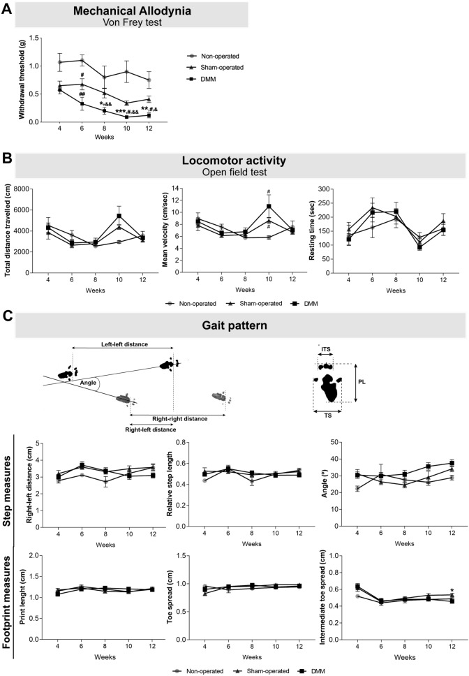
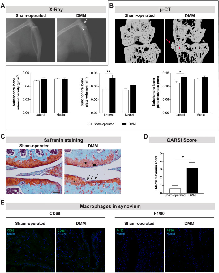
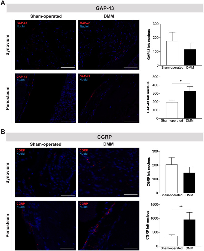
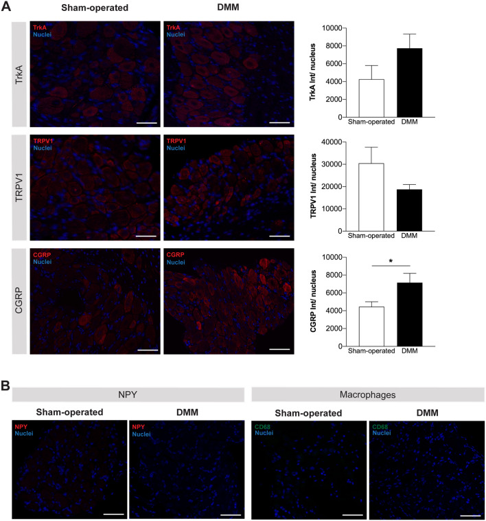
In osteoarthritis (OA), pain is the dominant clinical symptom, yet the therapeutic approaches remain inadequate. The knowledge of the nociceptive mechanisms in OA, which will allow to develop effective therapies for OA pain, is of utmost need. In this study, we investigated the nociceptive mechanisms involved in post-traumatic OA pain, using the destabilization of the medial meniscus (DMM) mouse model. Our results revealed the development of peripheral pain sensitization, reflected by augmented mechanical allodynia. Along with the development of pain behaviour, we observed an increase in the expression of calcitonin gene-related peptide (CGRP) in both the sensory nerve fibers of the periosteum and the dorsal root ganglia. Interestingly, we also observed that other nociceptive mechanisms commonly described in non-traumatic OA phenotypes, such as infiltration of the synovium by immune cells, neuropathic mechanisms and also central sensitization were not present. Overall, our results suggest that CGRP in the sensory nervous system is underlying the peripheral sensitization observed after traumatic knee injury in the DMM model, highlighting the CGRP as a putative therapeutic target to treat pain in post-traumatic OA. Moreover, our findings suggest that the nociceptive mechanisms involved in driving pain in post-traumatic OA are considerably different from those in non-traumatic OA.


Conflict of interest statement

The authors declare no competing interests.

Figures

Figure 1
Figure 1
The OA post-traumatic DMM model was characterized by the presence of mechanical allodynia. Mechanical allodynia (A), and alterations in locomotor activity (B) and gait pattern (C) were assessed 4, 6, 8, 10 and 12 weeks after the knee joint traumatic lesion. The Von Frey data analysis showed that DMM mice presented increased mechanical allodynia when compared with sham-operated, starting at week 8. No effects were observed in the locomotor activity. A decrease in the ITS was observed in the DMM mice. Results are presented as mean ± SEM, n = 5 in the control group and n = 8 in the sham-operated and DMM groups. *Indicates differences between sham-operated and DMM groups; #indicates differences when compared with non-operated group; &indicates differences when compared with week 4; *p < 0.05; **p < 0.01; ***p < 0.001; #p < 0.05; ##p < 0.01; &p < 0.05; &&p < 0.01. ITS intermediate toe spread, PL print length, TS toe spread.
Figure 2
Figure 2
Joint damage was observed 12 weeks after the induction of the OA post-traumatic DMM model. X-ray (A) and μ-CT (B) images were acquired 12 weeks post-surgery in sham-operated and DMM mice. The narrowing of the knee joint space (arrowhead) and the formation of osteophytes (arrow) were observed in both technical approaches. μ-CT quantitative analyses were also performed (B). Increased subchondral bone volume and subchondral bone thickness were observed in the DMM mice. The safranin O staining (C) revealed intense fibrillation and thinning of the cartilage (arrows) and lower intensity in red staining (proteoglycan) of cartilage (highlighted with *) in the DMM mice (scale bar = 100 μm). A higher OARSI maximum score of articular cartilage was calculated for the DMM mice (D). The infiltration of synovium by macrophages was assessed by the analysis of CD68 and F4/80 expression and no macrophages were observed (E) (scale bar = 50 μm). Results are presented as mean ± SEM, n = 5 per group. *p < 0.05, **p < 0.01.
Figure 3
Figure 3
CGRP innervation was increased in the periosteum after the induction of the OA post-traumatic DMM model. The presence of nerve fibers positive for GAP-43 (A) and positive for CGRP (B) was assessed in the knee joint 12 weeks post-surgery. No alterations in the GAP-43 and CGRP innervation patterns were observed in the synovium of DMM mice, however, increased GAP-43 and CGRP innervation was observed in the periosteum of these mice. Scale bar = 50 μm. Results are presented as mean ± SEM, n = 5 per group. *p < 0.05, **p < 0.01.
Figure 4
Figure 4
CGRP expression was increased in the DRG after the induction of the OA post-traumatic DMM model. The expression of TrkA, TRPV1, and CGRP (A) was assessed in the DRG 12 weeks post-surgery; quantitative analyses of the staining intensity were performed. The expression of NPY in DRG and the infiltration of DRG by macrophages were also assessed at the same time point of disease progression (B). Increased expression levels of CGRP in the DRG of DMM mice were observed at 12 weeks after the knee traumatic lesion. No differences were observed in the expression of TrkA and TRPV1 between DMM and sham-operated mice. No expression of NPY was found in the DRG, and no macrophages were infiltrating the DRG 12 weeks after DMM surgery. Results are presented as mean ± SEM, n = 5 per group. *p < 0.05. Scale bar = 50 μm.
Figure 5
Figure 5
Markers of central sensitization were not altered in the OA post-traumatic DMM model. The protein levels of c-Fos (A), p-ERK1/2 (B), GFAP (C) and IBA-1 (D) were quantified in the spinal cord of the DMM mice 12 weeks after surgery by western blot. The p-ERK1/2 expression was normalized to ERK1/2, and the c-Fos, GFAP and IBA-1 expression were normalized to the GAPDH. No differences were observed in the expression levels of both markers between DMM and sham-operated mice. Results are presented as mean ± SEM, n = 5 per group.

Similar articles

Cited by

References

    1. Neogi T. The epidemiology and impact of pain in osteoarthritis. Osteoarthritis Cartil. 2013;21:1145–1153. doi: 10.1016/j.joca.2013.03.018. - DOI - PMC - PubMed
    1. Sutton PM, Holloway ES. The young osteoarthritic knee: Dilemmas in management. BMC Med. 2013;11:14. doi: 10.1186/1741-7015-11-14. - DOI - PMC - PubMed
    1. Amoako AO, Pujalte GG. Osteoarthritis in young, active, and athletic individuals. Clin. Med. Insights Arthritis Musculoskelet. Disord. 2014;7:27–32. doi: 10.4137/CMAMD.S14386. - DOI - PMC - PubMed
    1. Hawker GA, et al. Differences between men and women in the rate of use of hip and knee arthroplasty. N. Engl. J. Med. 2000;342:1016–1022. doi: 10.1056/NEJM200004063421405. - DOI - PubMed
    1. Cope PJ, Ourradi K, Li Y, Sharif M. Models of osteoarthritis: The good, the bad and the promising. Osteoarthritis Cartil. 2019;27:230–239. doi: 10.1016/j.joca.2018.09.016. - DOI - PMC - PubMed

Publication types

MeSH terms

Substances