

MSC-based therapy in female pelvic floor disorders
- PMID: 32944218
- PMCID: PMC7488254
- DOI: 10.1186/s13578-020-00466-4
MSC-based therapy in female pelvic floor disorders
Abstract
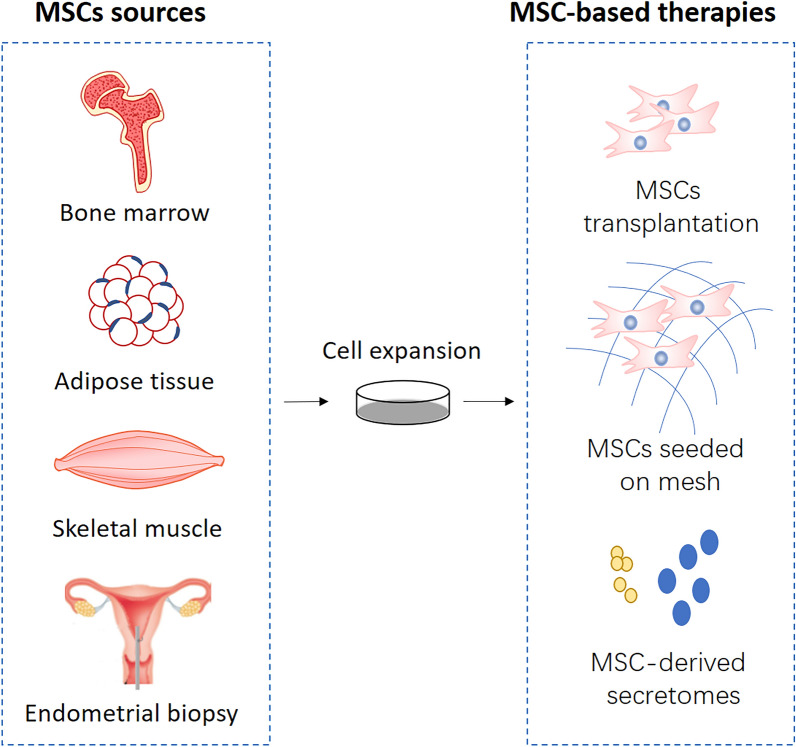
Mesenchymal stem cells (MSCs), also referred to as multipotent stromal cells or mesenchymal stromal cells, are present in multiple tissues and capable of differentiating into diverse cell lineages, holding a great promise in developing cell-based therapy for a wide range of conditions. Pelvic floor disorders (PFDs) is a common degenerative disease in women and may diminish a woman's quality of life at any age. Since the treatments for this disease are limited by the high rates of recurrence and surgical complications, seeking an ideal therapy in the restoration of pelvic floor function is an urgent issue at present. Herein, we summarize the cell sources of MSCs used for PFDs and discuss the potential mechanisms of MSCs in treating PFDs. Specifically, we also provide a comprehensive review of current preclinical and clinical trials dedicated to investigating MSC-based therapy for PFDs. The novel therapy has presented promising therapeutic effects which include relieving the symptoms of urinary or fecal incontinence, improving the biological properties of implanted meshes and promoting the injured tissue repair. Nevertheless, MSC-based therapies for PFDs are still experimental and the unstated issues on their safety and efficacy should be carefully addressed before their clinical applications.
Keywords: Cell- and tissue-based therapy; Mesenchymal stem cells; Mesenchymal stem cells transplantation; Pelvic floor disorders.
© The Author(s) 2020.
Conflict of interest statement
Competing interestsThe authors declare that they have no competing interests.
Figures

Similar articles
-
Exosomes derived from mesenchymal stromal cells: a promising treatment for pelvic floor dysfunction.Hum Cell. 2023 May;36(3):937-949. doi: 10.1007/s13577-023-00887-6. Epub 2023 Mar 20. Hum Cell. 2023. PMID: 36940057 Review.
-
Preliminary study on mesenchymal stem cells in repairing nerve injury in pelvic floor denervation.Front Bioeng Biotechnol. 2023 Jun 22;11:1190068. doi: 10.3389/fbioe.2023.1190068. eCollection 2023. Front Bioeng Biotechnol. 2023. PMID: 37425357 Free PMC article.
-
A Review of Phytoestrogens and Their Association With Pelvic Floor Conditions.Female Pelvic Med Reconstr Surg. 2018 May/Jun;24(3):193-202. doi: 10.1097/SPV.0000000000000559. Female Pelvic Med Reconstr Surg. 2018. PMID: 29432329 Free PMC article. Review.
-
Obesity and Pelvic Floor Dysfunction: Battling the Bulge.Obstet Gynecol Surv. 2016 Feb;71(2):114-25. doi: 10.1097/OGX.0000000000000274. Obstet Gynecol Surv. 2016. PMID: 26894803 Review.
-
Status, challenges, and future prospects of stem cell therapy in pelvic floor disorders.World J Clin Cases. 2020 Apr 26;8(8):1400-1413. doi: 10.12998/wjcc.v8.i8.1400. World J Clin Cases. 2020. PMID: 32368533 Free PMC article. Review.
Cited by
-
Exosomes derived from mesenchymal stromal cells: a promising treatment for pelvic floor dysfunction.Hum Cell. 2023 May;36(3):937-949. doi: 10.1007/s13577-023-00887-6. Epub 2023 Mar 20. Hum Cell. 2023. PMID: 36940057 Review.
-
Mesenchymal Stromal Cells Rapidly Suppress TCR Signaling-Mediated Cytokine Transcription in Activated T Cells Through the ICAM-1/CD43 Interaction.Front Immunol. 2021 Feb 22;12:609544. doi: 10.3389/fimmu.2021.609544. eCollection 2021. Front Immunol. 2021. PMID: 33692786 Free PMC article.
-
Preliminary study on mesenchymal stem cells in repairing nerve injury in pelvic floor denervation.Front Bioeng Biotechnol. 2023 Jun 22;11:1190068. doi: 10.3389/fbioe.2023.1190068. eCollection 2023. Front Bioeng Biotechnol. 2023. PMID: 37425357 Free PMC article.
-
Progress of mesenchymal stem cells affecting extracellular matrix metabolism in the treatment of female stress urinary incontinence.Stem Cell Res Ther. 2025 Feb 25;16(1):95. doi: 10.1186/s13287-025-04220-w. Stem Cell Res Ther. 2025. PMID: 40001265 Free PMC article. Review.
-
Perinatal mesenchymal stromal cells of the human decidua restore continence in rats with stress urinary incontinence induced by simulated birth trauma and regulate senescence of fibroblasts from women with stress urinary incontinence.Front Cell Dev Biol. 2023 Jan 18;10:1033080. doi: 10.3389/fcell.2022.1033080. eCollection 2022. Front Cell Dev Biol. 2023. PMID: 36742196 Free PMC article.
References
-
- Dooley Y, et al. Urinary incontinence prevalence: results from the National Health and Nutrition Examination Survey. J Urol. 2008;179(2):656–661. - PubMed
Publication types
LinkOut - more resources
Full Text Sources
