

Electrocardiographic features of 431 consecutive, critically ill COVID-19 patients: an insight into the mechanisms of cardiac involvement
- PMID: 32944767
- PMCID: PMC7543398
- DOI: 10.1093/europace/euaa258
Electrocardiographic features of 431 consecutive, critically ill COVID-19 patients: an insight into the mechanisms of cardiac involvement
Abstract
Aims: Our aim was to describe the electrocardiographic features of critical COVID-19 patients.
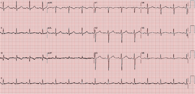
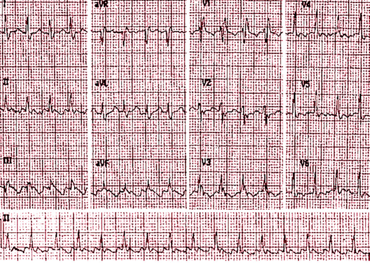
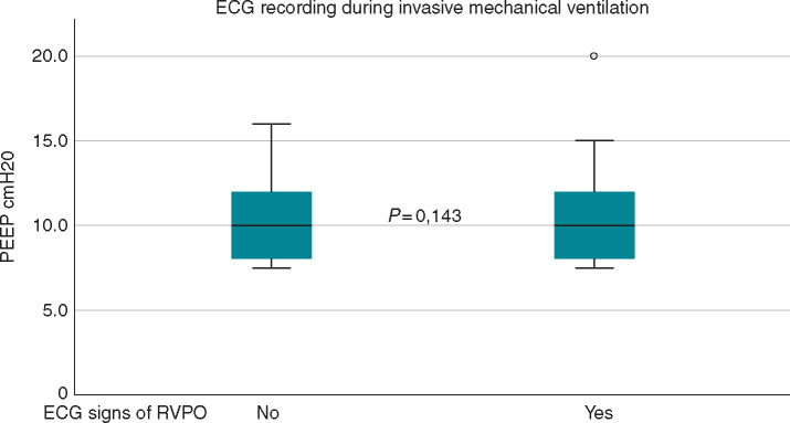
Methods and results: We carried out a multicentric, cross-sectional, retrospective analysis of 431 consecutive COVID-19 patients hospitalized between 10 March and 14 April 2020 who died or were treated with invasive mechanical ventilation. This project is registered on ClinicalTrials.gov (identifier: NCT04367129). Standard ECG was recorded at hospital admission. ECG was abnormal in 93% of the patients. Atrial fibrillation/flutter was detected in 22% of the patients. ECG signs suggesting acute right ventricular pressure overload (RVPO) were detected in 30% of the patients. In particular, 43 (10%) patients had the S1Q3T3 pattern, 38 (9%) had incomplete right bundle branch block (RBBB), and 49 (11%) had complete RBBB. ECG signs of acute RVPO were not statistically different between patients with (n = 104) or without (n=327) invasive mechanical ventilation during ECG recording (36% vs. 28%, P = 0.10). Non-specific repolarization abnormalities and low QRS voltage in peripheral leads were present in 176 (41%) and 23 (5%), respectively. In four patients showing ST-segment elevation, acute myocardial infarction was confirmed with coronary angiography. No ST-T abnormalities suggestive of acute myocarditis were detected. In the subgroup of 110 patients where high-sensitivity troponin I was available, ECG features were not statistically different when stratified for above or below the 5 times upper reference limit value.
Conclusions: The ECG is abnormal in almost all critically ill COVID-19 patients and shows a large spectrum of abnormalities, with signs of acute RVPO in 30% of the patients. Rapid and simple identification of these cases with ECG at hospital admission can facilitate classification of the patients and provide pathophysiological insights.
Keywords: Critically ill COVID-19; ECG; Right ventricular pressure overload.
Published on behalf of the European Society of Cardiology. All rights reserved. © The Author(s) 2020. For permissions, please email: journals.permissions@oup.com.
Figures
References
-
- Surawicz B, Knilans T. Chou’s Electrocardiography in Clinical Practice 6th ed. Saunders; 2008.
Publication types
MeSH terms
Substances
Associated data
LinkOut - more resources
Full Text Sources
Medical
Miscellaneous
