. 2020 Oct;61(10):2301-2312.
doi: 10.1111/epi.16670. Epub 2020 Sep 17.

Endoplasmic reticulum stress increases inflammatory cytokines in an epilepsy mouse model Gabrg2+/Q390X knockin: A link between genetic and acquired epilepsy?

Affiliations

Endoplasmic reticulum stress increases inflammatory cytokines in an epilepsy mouse model Gabrg2+/Q390X knockin: A link between genetic and acquired epilepsy?

Wangzhen Shen et al. Epilepsia. 2020 Oct.

Abstract

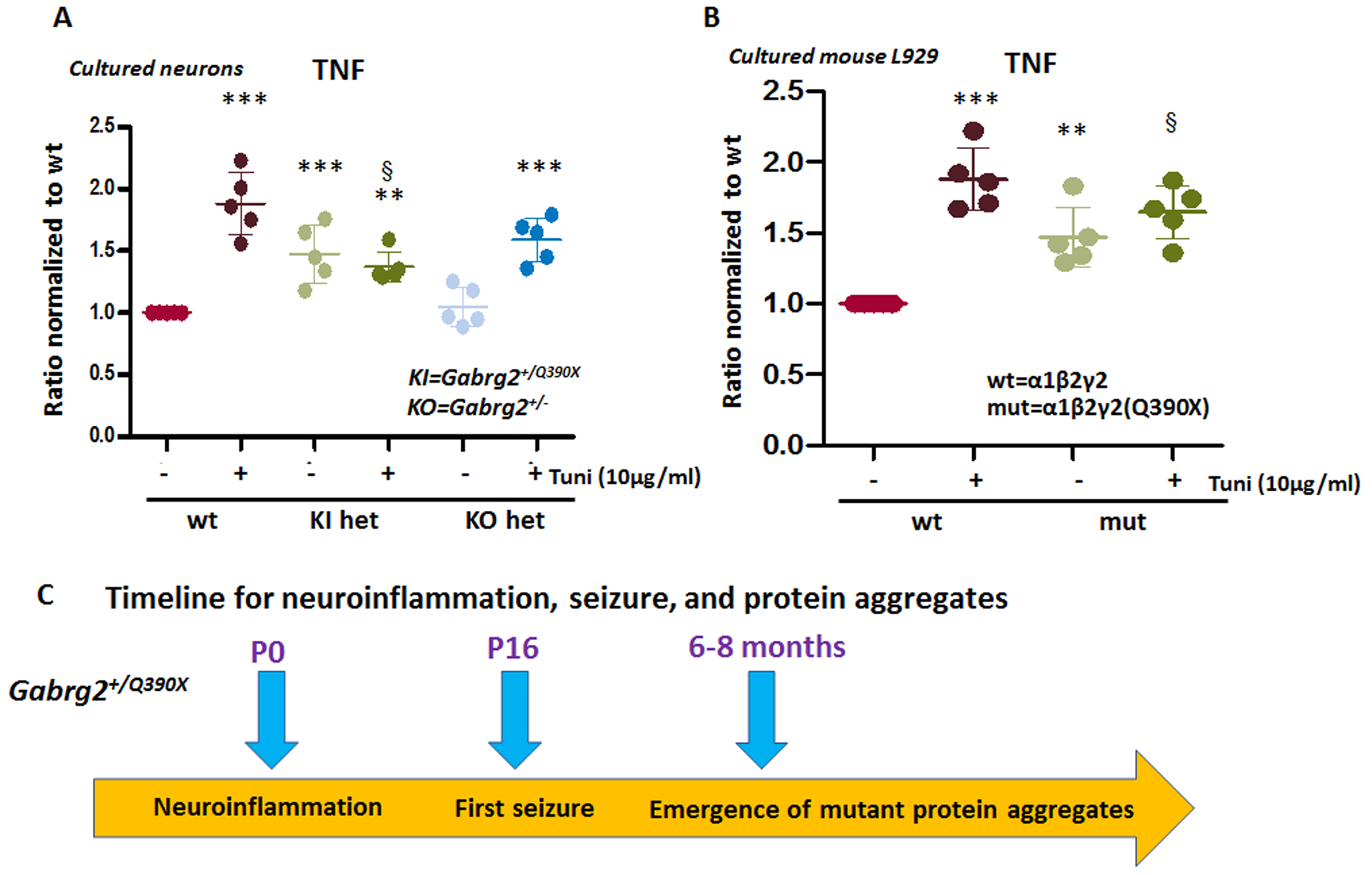
Objective: Neuroinflammation is a major theme in epilepsy, which has been characterized in acquired epilepsy but is poorly understood in genetic epilepsy. γ-Aminobutyric acid type A receptor subunit gene mutations are significant causes of epilepsy, and we have studied the pathophysiology directly resulting from defective receptor channels. Here, we determined the proinflammatory factors in a genetic mouse model, the Gabrg2+/Q390X knockin (KI). We have identified increased cytokines in multiple brain regions of the KI mouse throughout different developmental stages and propose that accumulation of the trafficking-deficient mutant protein may increase neuroinflammation, which would be a novel mechanism for genetic epilepsy.

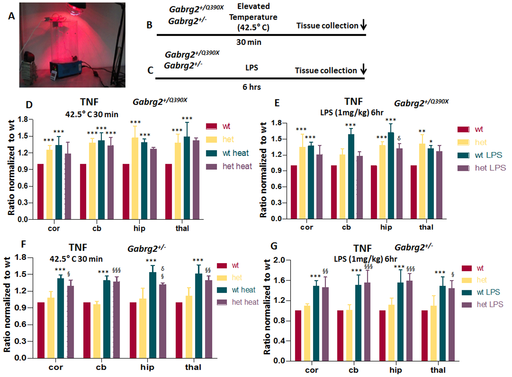
Methods: We used enzyme-linked immunosorbent assay, immunoprecipitation, nuclei purification, immunoblot, immunohistochemistry, and confocal microscopy to characterize increased neuroinflammation and its potential causes in a Gabrg2+/Q390X KI mouse and a Gabrg2+/- knockout (KO) mouse, each associated with a different epilepsy syndrome with different severities.

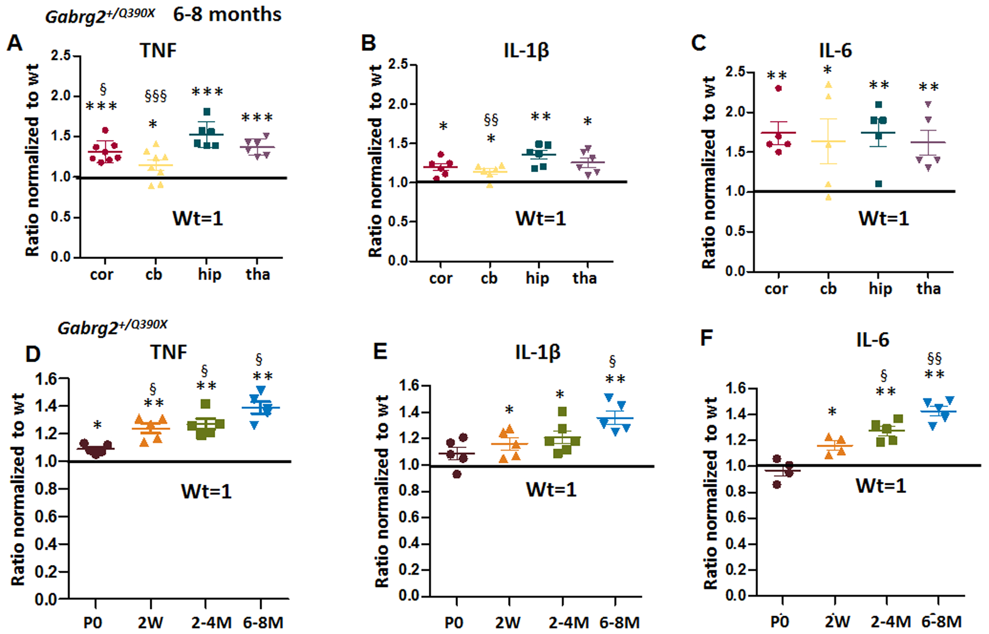
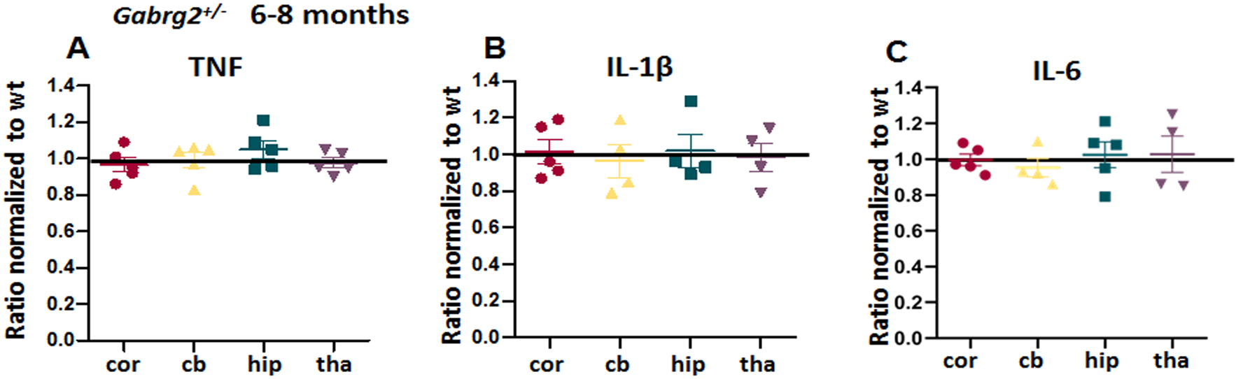
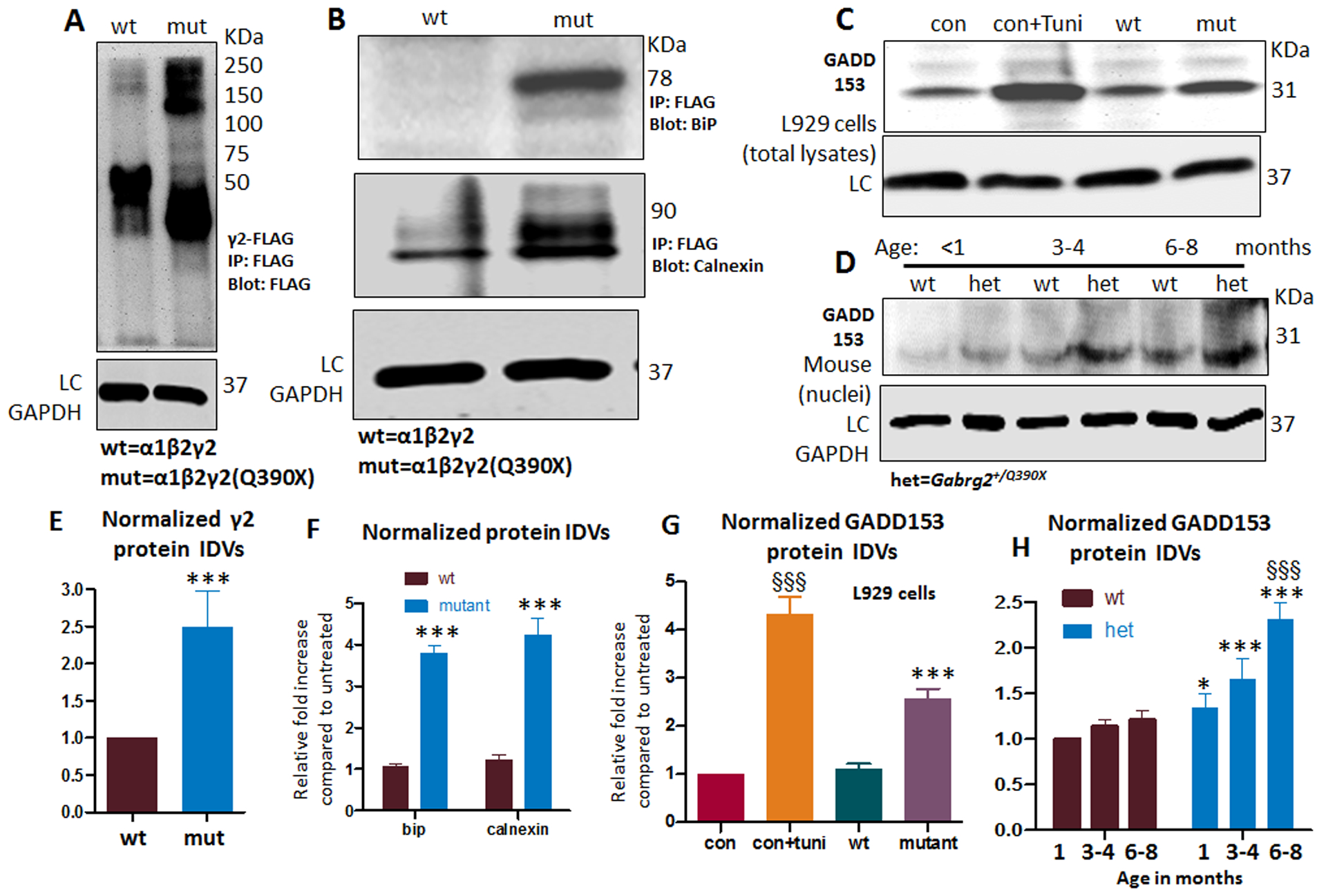
Results: We found that proinflammatory cytokines such as tumor necrosis factor alpha, interleukin 1-beta (IL-1β), and IL-6 were increased in the KI mice but not in the KO mice. A major underlying basis for the discrepancy in cytokine expression between the two mouse models is likely chronic mutant protein accumulation and endoplasmic reticulum (ER) stress. The presence of mutant protein dampened cytokine induction upon further cellular stimulation or external stress such as elevated temperature. Pharmacological induction of ER stress upregulated cytokine expression in the wild-type and KO but not in the KI mice. The increased cytokine expression was independent of seizure occurrence, because it was upregulated in both mice and cultured neurons.

Significance: Together, these data demonstrate a novel pathophysiology for genetic epilepsy, increased neuroinflammation, which is a common mechanism for acquired epilepsy. The findings thus provide the first link of neuroinflammation between genetic epilepsy associated with an ion channel gene mutation and acquired epilepsy.

Keywords: ER stress; GABAA receptors; Gabrg2+/Q390X knockin (KI) mice; epilepsy; neuroinflammation; proinflammatory cytokines.


Conflict of interest statement

Conflict of Interest statement: None of authors declared any conflict of interest.

Figures

Figure 1.
Figure 1.. Increased proinflammatory cytokines tumor-necrosis factor alpha (TNF), interleukin 1β (IL-1β) and interleukin 6 (IL-6) in Gabrg2+/Q390X, a genetic mouse model of epileptic encephalopathy
A-C. The brains from 6–8 months old Gabrg2+/Q390X mice were dissected and processed for measurement of pro-inflammatory cytokines. Equal amounts of protein lysates (30μg) from each brain region were determined for cytokines including tumor-necrosis factor alpha (TNF) (A), interleukin 1β (IL-1β) (B) and interleukin 6 (IL-6) (C) with enzyme-linked immunosorbent assay (ELISA). The measurements in the heterozygotes (het) were normalized to the same brain region of their own wildtype (WT) littermates. D-F. The forebrain cortex from the Gabrg2+/Q390X mice at different ages (P0=postnatal day 0, 2W=2 weeks, 2–4M=2–4 months, 6–8M=6–8 months) were dissected and processed for measurement of TNF (D), IL-1β (E) and IL-6 (F) with ELISA. Equal amounts of protein lysates (30μg) from the mouse cortex of different ages were determined. In D-F, the measurements in the het were normalized to the cortex of their own wildtype littermates. (In A-F, *p < 0.05; ** p < 0.01; *** p < 0.001 vs wt, In A-B, § P< 0.05; §§ P< 0.01 §§§ P< 0.001 vs hip in het; in D-F, § P< 0.05; §§P< 0.01 vs P0). Data were presented as mean ± S.E.M. In A-C, N=5–8 mice. In D-F, N=4–6 mice.
Figure 2.
Figure 2.. There was no increase of proinflammatory cytokines TNF, IL-1β and IL-6 in Gabrg2+/− knockout, a mouse model of infrequent absence epilepsy
A-C. The brains from 6–8 months old Gabrg2+/− mice were dissected and processed for measurement of pro-inflammatory cytokines. Equal amounts of protein lysates (30μg) from each brain region were determined for cytokines including tumor-necrosis factor alpha (TNF) (A), interleukin 1β (IL-1β) (B) and IL-6 (C) with enzyme-linked immunosorbent assay (ELISA). The measurements in the het were normalized to the same brain region of their own wildtype littermates. Data were presented as mean ± S.E.M. N=5 mice for each group.
Figure 3.
Figure 3.. Treatment with elevated temperature or lipopolysaccharide had dampened response in Gabrg2+/Q390X mice
(A) The temperature induction apparatus and heating setup are shown. (B, C) A diagram of stress with temperature elevation (B) or lipopolysaccharide (LPS) treatment (C) procedure in mice is shown. In B, the mice were heated up with a heating lamp and the core temperature was maintained for 30 min followed by tissue harvest. In C, a single dose of LPS (1 mg/kg i.p) was injected to induce neuroinflammation and the brain was harvested 6hr after drug administration. D-G. The brains from 6–8 months old Gabrg2+/Q390X (D, E) or Gabrg2+/− mice (F, G) treated with elevated temperature (D, F) or LPS (E, G) were dissected and processed for measurement of pro-inflammatory cytokines. Equal amounts of protein lysates (30μg) from each brain region were determined for TNF with enzyme-linked immunosorbent assay (ELISA). The measurements in the het was normalized to the same brain region of the wt without temperature elevation (D, F) or LPS treatment (E, G). Data were presented as mean ± S.E.M (*p < 0.05, **p < 0.01, ***p < 0.001 vs wt, δ P< 0.05 vs wt heat, § p<0.05, §§ P<0.01, §§§ P<0.001 vs het heat). In D, N=4–5 mice for each group. In E, F, G, N=4 mice for each group.
Figure 4.
Figure 4.. The Gabrg2+/Q390X mice, but not the Gabrg2+/− mice, had toxic accumulation of the trafficking deficient γ2 subunits.
A, B. Brains from Gabrg2+/Q390X knockin (KI) (A) or Gabrg2+/− knockout mice (KO) (B) were short fixed in 4% PFA for 30 min and then processed for immunostaining. Images from cortical layers 5–6 in the somatosensory region of brain section were stained with rabbit anti-γ2 subunit (green) antibody and the nuclei marker TO-PRO-3. C. The arrow pointed regions in A and B were enlarged for a better visualization. D. The γ2 subunit fluorescence intensities were analyzed by ImageJ. The γ2 subunit fluorescence signals in somatic region of each individual neurons in the whole sampled region were measured by subtracting the background value in the nuclei region from the total raw. Data were presented as mean ± S.E.M (***p < 0.05; ***p < 0.001 vs wt; §§§ P< 0.05 vs het Gabrg2+/Q390X, In Gabrg2+/Q390X , N=12 sections for wt and het. In Gabrg2+/− , N=14 sections for wt and 11 for het. Brain sections were obtained from 4 mice in each group.
Figure 5.
Figure 5.. The mutant γ2(Q390X) subunit protein increased conjugation with ER chaperones and caused ER stress.
(A-B) Total lysates from HEK293T cells expressing γ2FLAG subunits (cDNA 3μg) were immunoprecipitated with FLAG beads and immunoblotted with anti-FLAG antibody (A) or GRP78 (B, upper panel) or calnexin (B, lower panel) antibody. The γ2FLAG subunits, GRP78, and calnexin were normalized to the wildtype condition. In A, the γ2FLAG subunits were measured from about 75 KDa to 250 KDa. Individual wildtype γ2FLAG subunits were predicted to be 55 KDa and migrated at about 50 KDa, and individual FLAG-tagged truncated mutant γ2(Q390X) subunits migrated at lower molecular masses predicted to be about 40 KDa. (C). Total lysates from mouse L929 cells expressing wildtype or mutant γ2 subunits in combination with the wildtype partnering α1 and β2 subunits were analyzed by SDS-PAGE and immunoblotted with mouse monoclonal anti-GADD153 antibody. In A-D, LC stands for loading control GAPDH. (D). The nuclei portion of the total forebrain of Gabrg2+/Q390X knockin mice at different ages was purified and lysed. Equal amounts of the protein were analyzed by SDS-PAGE and immunoblotted with GADD153. (E-F). Total mutant subunit band IDVs of γ2FLAG subunits (E) or the conjugated chaperone GRP78 and calnexin (F) were normalized to the wildtype conditions. (G) The total amounts of endogenous GADD153 were normalized to untreated controls (con). (H) The endogenous GADD153 in the nuclei of forebrain in the Gabrg2+/Q390X knockin mice at different ages were normalized to wt in the youngest group (<1 month). Seizure onset is around P19 while mutant protein aggregates become detectable around 6mo. (In E, F and G, *p < 0.05, **p < 0.01, ***p<0.001 vs wt. In G, §§§ P< 0.001 vs con, †† vs P< 0.01 vs con+Tuni, N=5 batches of cells. In H, §§§ P< 0.001 vs 1 month old. In E to G, n=5 batch of cells, in H, N=4 mice for each group.)
Figure 6.
Figure 6.. ER stress increased TNF in cultured cells but the cells expressing the mutant γ2(Q390X) had dampened response.
A. The forebrain cortex of postnatal day 0 old pups of Gabrg2+/Q390X and Gabrg2+/− mouse lines were dissected, disassociated and the neurons were cultured for 15 days in dish before harvest. Equal amounts of cell protein lysates (30μg) from the wildtype or the heterozygous mice were determined for TNF with enzyme-linked immunosorbent assay (ELISA). The measurements in the het were normalized to the wildtype sister cultures. B. Mouse L929 cells were transfected with the wildtype or the mutant γ2(Q390X) subunits in combination with the wildtype α1 and β2 subunits for 48 hrs. Sister cultures were treated with or without tunicamycin (10μg/ml) for 16 hrs before harvest. Equal amounts of cell protein lysates (30μg) from each group were determined for TNF with ELISA. (**p < 0.01 vs wt, ***p < 0.001 vs wt, § p<0.05 vs Wt+Tuni. n = 4–5 batches of cultures. Data were presented as mean ± S.E.M).

Similar articles

Cited by

References

    1. McElroy PB, Liang LP, Day BJ, Patel M. Scavenging reactive oxygen species inhibits status epilepticus-induced neuroinflammation. Exp Neurol 2017;298:13–22. - PMC - PubMed
    1. Barker-Haliski ML, Loscher W, White HS, Galanopoulou AS. Neuroinflammation in epileptogenesis: Insights and translational perspectives from new models of epilepsy. Epilepsia 2017;58 Suppl 3:39–47. - PMC - PubMed
    1. Vezzani A, Balosso S, Ravizza T. Neuroinflammatory pathways as treatment targets and biomarkers in epilepsy. Nat Rev Neurol 2019;15:459–72. - PubMed
    1. Aronica E, Bauer S, Bozzi Y et al. Neuroinflammatory targets and treatments for epilepsy validated in experimental models. Epilepsia 2017;58 Suppl 3:27–38. - PMC - PubMed
    1. Kaufer C, Chhatbar C, Broer S et al. Chemokine receptors CCR2 and CX3CR1 regulate viral encephalitis-induced hippocampal damage but not seizures. Proc Natl Acad Sci U S A 2018;115:E8929–E8938. - PMC - PubMed

Publication types