Case Reports
. 2020 Oct;61(10):e135-e139.
doi: 10.1111/epi.16683. Epub 2020 Sep 18.

Post-COVID-19 inflammatory syndrome manifesting as refractory status epilepticus

Affiliations
Case Reports

Post-COVID-19 inflammatory syndrome manifesting as refractory status epilepticus

Elizabeth Carroll et al. Epilepsia. 2020 Oct.

Abstract

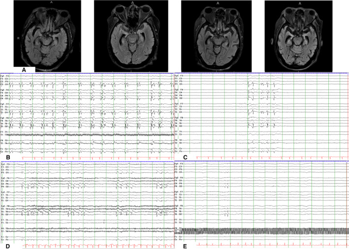
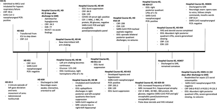
There have been multiple descriptions of seizures during the acute infectious period in patients with COVID-19. However, there have been no reports of status epilepticus after recovery from COVID-19 infection. Herein, we discuss a patient with refractory status epilepticus 6 weeks after initial infection with COVID-19. Extensive workup demonstrated elevated inflammatory markers, recurrence of a positive nasopharyngeal SARS-CoV-2 polymerase chain reaction, and hippocampal atrophy. Postinfectious inflammation may have triggered refractory status epilepticus in a manner similar to the multisystemic inflammatory syndrome observed in children after COVID-19.

Keywords: COVID-19; SARS-CoV-2; inflammatory response; postinfectious; refractory status epilepticus; seizures.


Conflict of interest statement

None of the authors has any conflict of interest to disclose.

Figures

FIGURE 1
FIGURE 1
A, Magnetic resonance imaging brain axial fluid‐attenuated inversion recovery sequences, from left to right, obtained 3 months prior to diagnosis of COVID‐19, on hospital day (HD) #8, HD #15, and HD #29 of hospital course #2, demonstrating global cerebral atrophy and progressive right > left hippocampal atrophy. B, Electroencephalogram (EEG) from HD #8 of hospital course #2, demonstrating bilateral synchronous periodic discharges maximal over the right posterior quadrant and the left occipital pole. C, EEG from HD #9 of hospital course #2, demonstrating a burst suppression pattern with 20% of the page showing bursts, following initiation of propofol, midazolam, and ketamine drips. Bursts consist of synchronous polyspike and wave discharges. D, EEG from HD #29 of hospital course #2, demonstrating increased burden of brief runs of rhythmic discharges that are again maximal biposteriorly. E, EEG from HD #11 of hospital course #3 demonstrating right posterior quadrant discharges with moderate to severe generalized background slowing. All EEG studies in Figure 2 are shown in time base of 30 mm/s, read at 7 µV/mm, with notch filter of 60 Hz applied, in double banana montage with subtemporal leads
FIGURE 2
FIGURE 2
Timeline of the 114 days from admission for COVID‐19 to discharge from her third hospitalization. BIRD, brief potentially ictal rhythmic discharge; CRP, C‐reactive protein; CSF, cerebrospinal fluid; EEG, electroencephalography; ESR, erythrocyte sedimentation rate; HD, hospital day; ICU, intensive care unit; IVIG, intravenous immunoglobulin; L, left; LPD, lateralized periodic discharge; MICU, medical ICU; MRI, magnetic resonance imaging; NCHCT, noncontrast head computed tomography; NYU, New York University Hospital; PCR, polymerase chain reaction; R, right; RBC, red blood cells; SAR, subacute rehabilitation; SARS‐CoV‐2, severe acute respiratory syndrome coronavirus 2; WBC, white blood cells.

Similar articles

Cited by

References

    1. Sohal S, Mansur M.COVID‐19 presenting with seizures. IDCases. 2020;20:e00782. - PMC - PubMed
    1. Somani S, Pati S, Gaston T, Chitlangia A, Agnihotri S. De novo status epilepticus in patients with COVID‐19. Ann Clin Transl Neurol. 2020;7(7):1240–4. - PMC - PubMed
    1. Balloy G, Mahe P‐J, Leclair‐Visonneau L, Pereon Y, Derkinderen P, Magot A, et al. Non‐lesional status epilepticus in a patient with coronavirus disease 2019. Clin Neurophysiol. 2020;131(8):2059–61. - PMC - PubMed
    1. Lu L, Xiong W, Liu D, Liu J, Yang D, Li N, et al. New onset acute symptomatic seizure and risk factors in coronavirus disease 2019: a retrospective multicenter study. Epilepsia. 2020;61(6):e49–53. - PMC - PubMed
    1. Achiriloaie A, Michelson D, Lei L, Denham L, Oberg K, Raghavan R. Acute postviral encephalopathy. Child Neurol Open. 2016;3:2329048X1665884. - PMC - PubMed

Publication types