. 2020 Dec;598(24):5639-5659.
doi: 10.1113/JP279909. Epub 2020 Oct 6.

TRPV1 expressed throughout the arterial circulation regulates vasoconstriction and blood pressure

Affiliations

TRPV1 expressed throughout the arterial circulation regulates vasoconstriction and blood pressure

Thieu X Phan et al. J Physiol. 2020 Dec.

Abstract

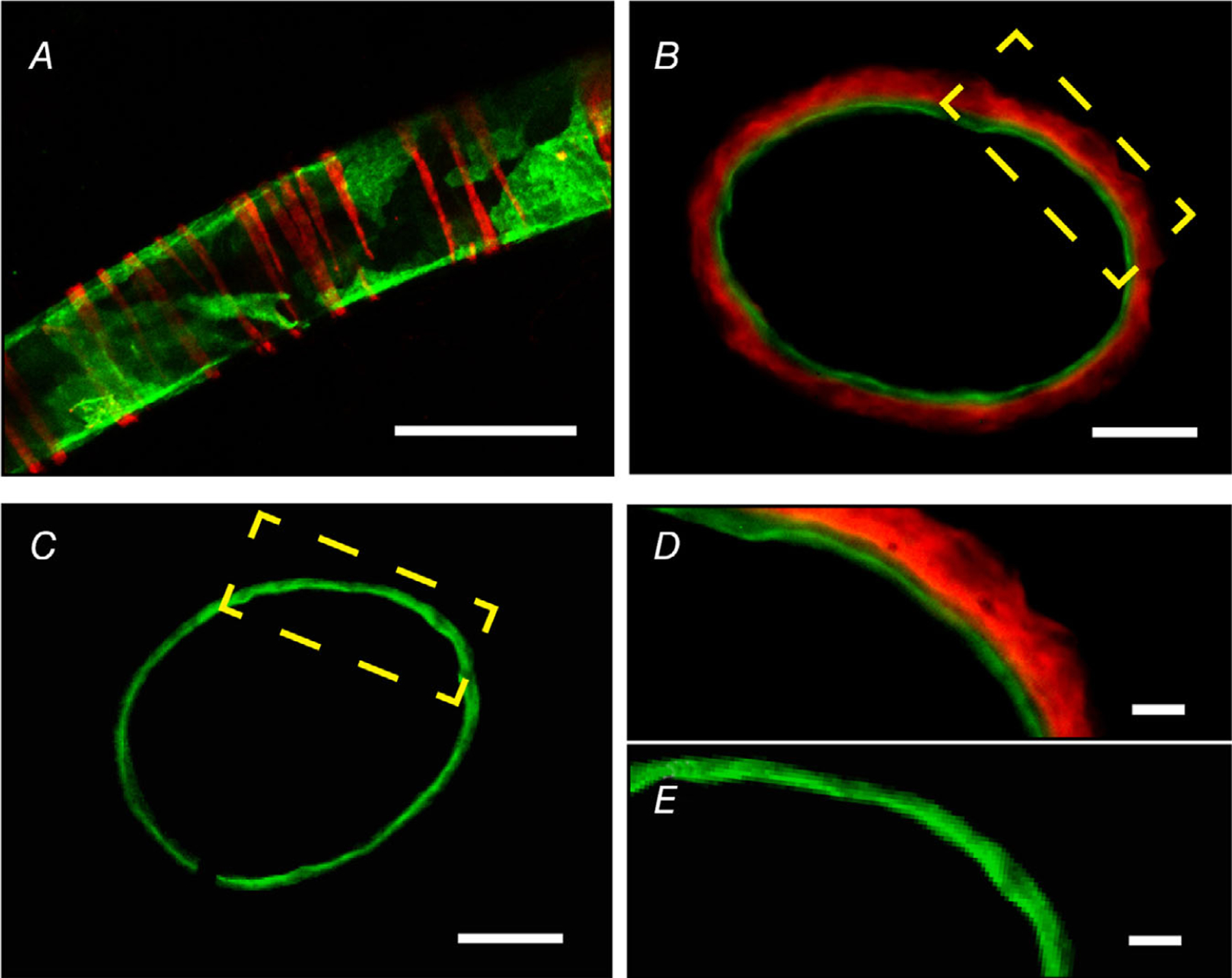
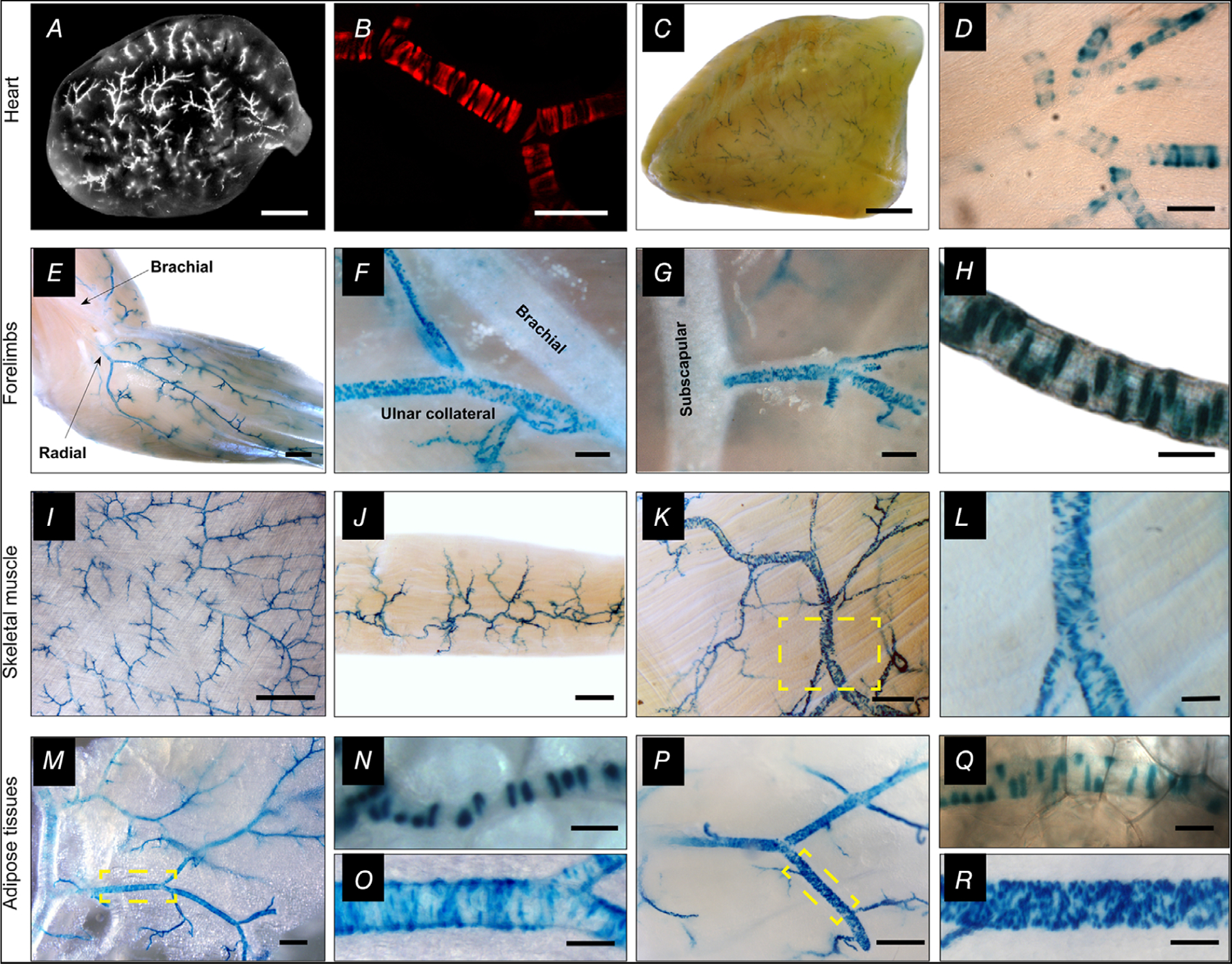
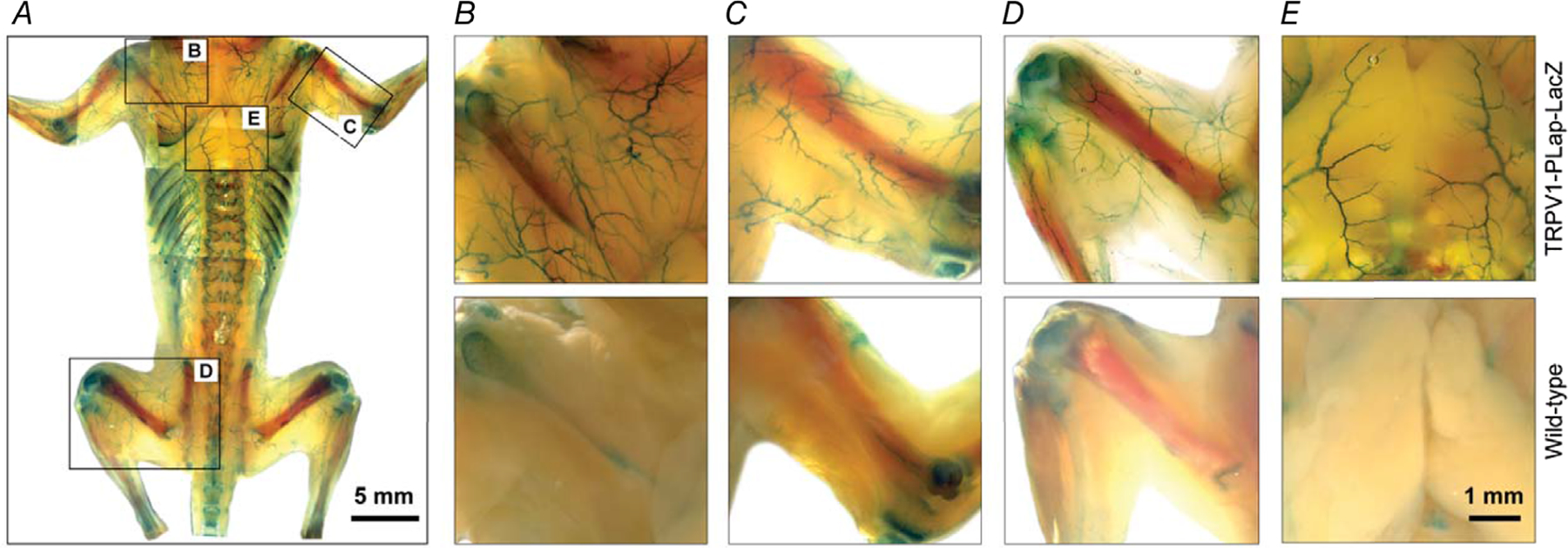
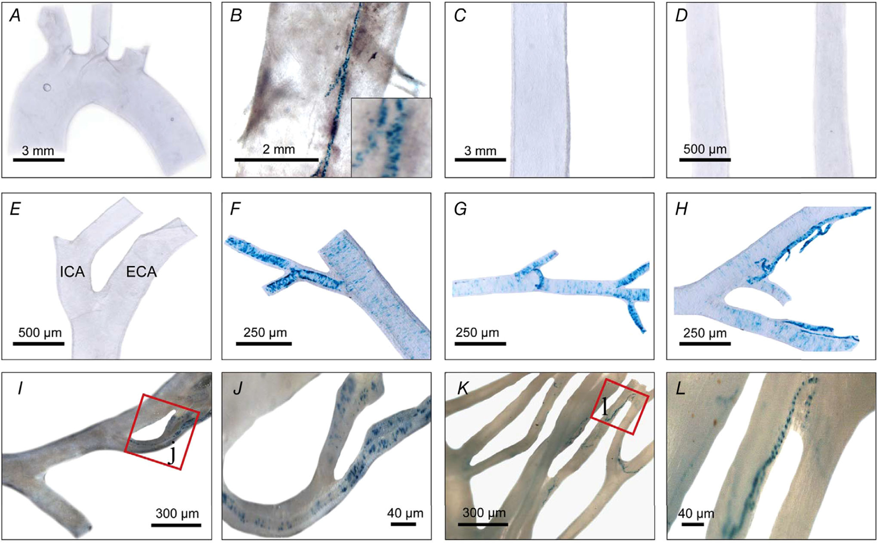
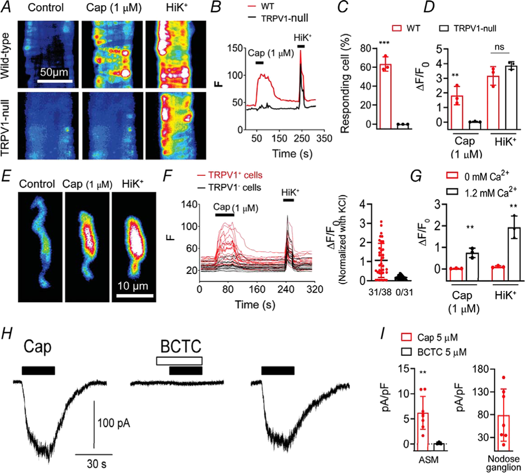
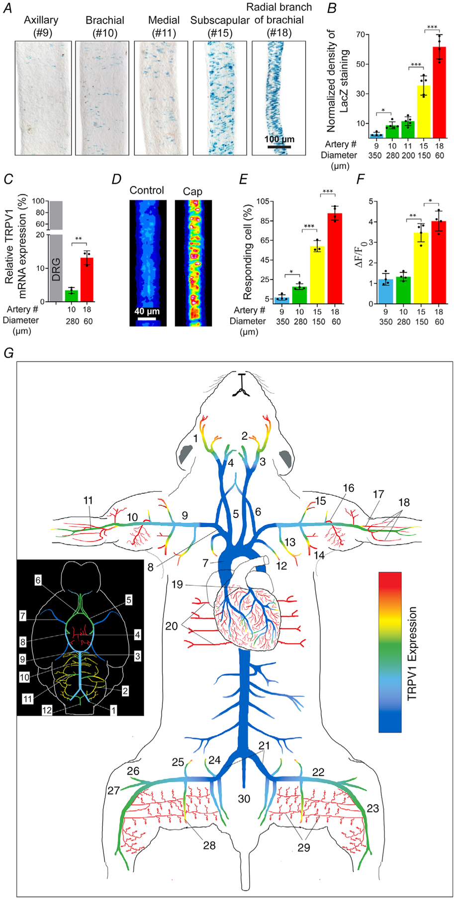
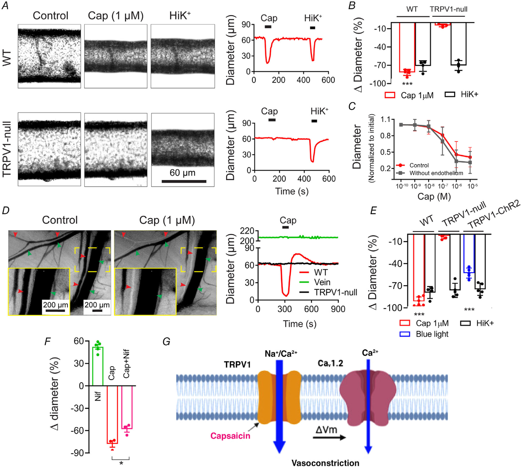
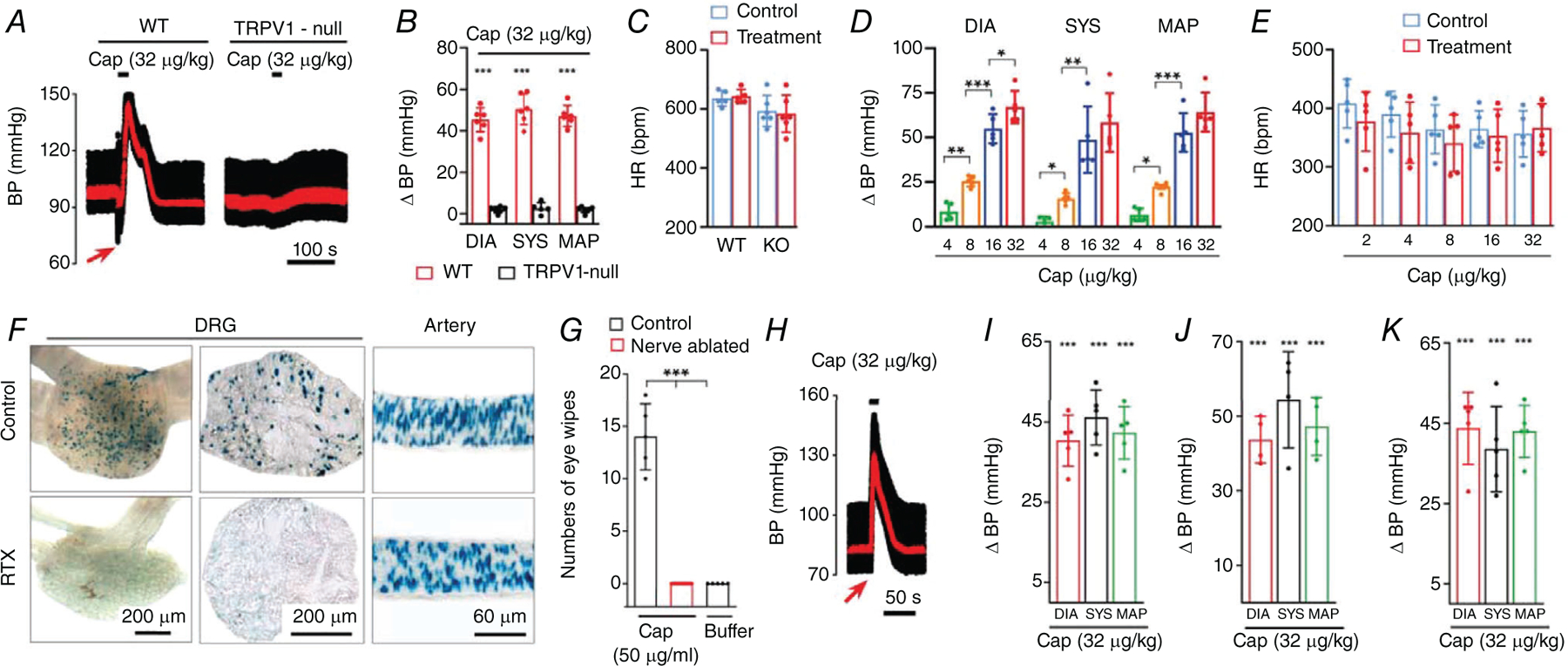
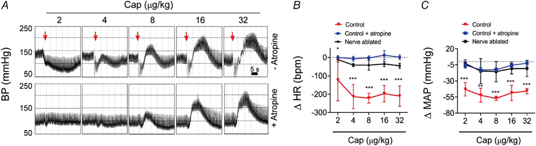
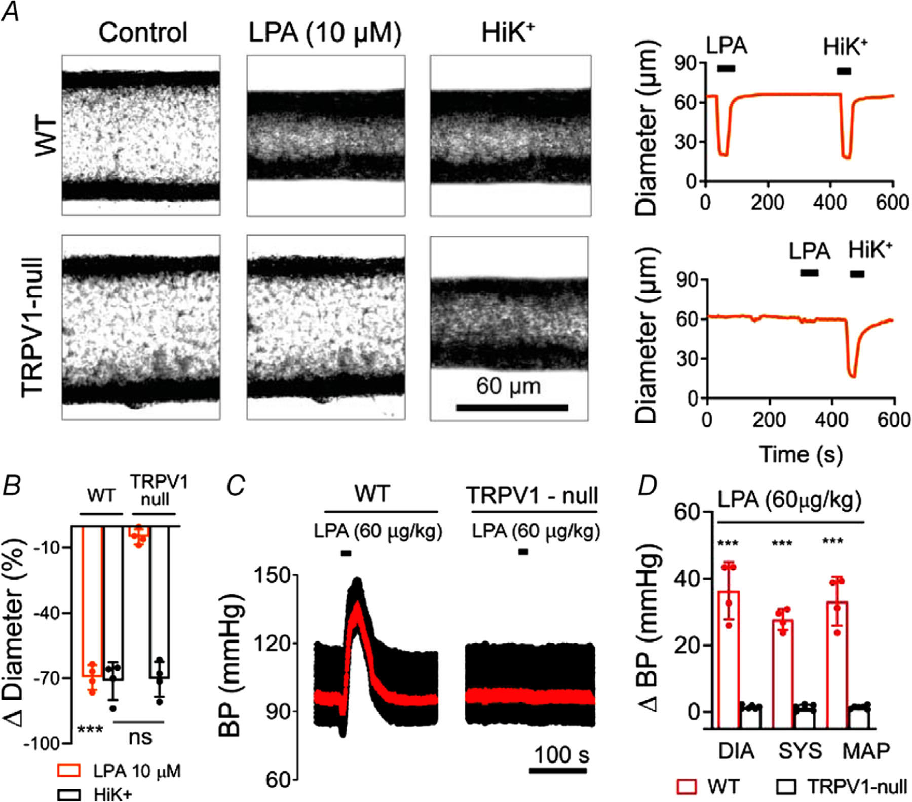
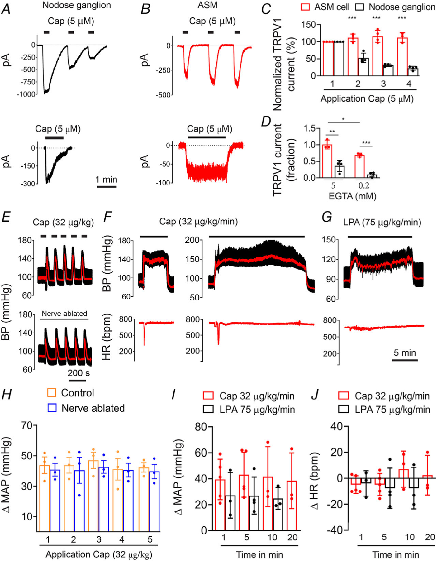
Key points: The functional roles of the capsaicin receptor, TRPV1, outside of sensory nerves are unclear. We mapped TRPV1 in the mouse circulation, revealing extensive expression in the smooth muscle of resistance arterioles supplying skeletal muscle, heart and adipose tissue. Activation of TRPV1 in vascular myocytes constricted arteries, reduced coronary flow in isolated hearts and increased systemic blood pressure. These functional effects were retained after sensory nerve ablation, indicating specific signalling by arterial TRPV1. TRPV1 mediated the vasoconstrictive and blood pressure responses to the endogenous inflammatory lipid lysophosphatidic acid. These results show that TRPV1 in arteriolar myocytes modulates regional blood flow and systemic blood pressure, and suggest that TRPV1 may be a target of vasoactive inflammatory mediators.

Abstract: The capsaicin receptor, TRPV1, is a key ion channel involved in inflammatory pain signalling. Although mainly studied in sensory nerves, there are reports of TRPV1 expression in isolated segments of the vasculature, but whether the channel localizes to vascular endothelium or smooth muscle is controversial and the distribution and functional roles of TRPV1 in arteries remain unknown. We mapped functional TRPV1 expression throughout the mouse arterial circulation. Analysis of reporter mouse lines TRPV1PLAP-nlacZ and TRPV1-Cre:tdTomato combined with Ca2+ imaging revealed specific localization of TRPV1 to smooth muscle of terminal arterioles in the heart, adipose tissue and skeletal muscle. Capsaicin evoked inward currents (current density ∼10% of sensory neurons) and raised intracellular Ca2+ levels in arterial smooth muscle cells, constricted arterioles ex vivo and in vivo and increased systemic blood pressure in mice and rats. Further, capsaicin markedly and dose-dependently reduced coronary flow. Pharmacological and/or genetic disruption of TRPV1 abolished all these effects of capsaicin as well as vasoconstriction triggered by lysophosphatidic acid, a bioactive lipid generated by platelets and atherogenic plaques. Notably, ablation of sensory nerves did not affect the responses to capsaicin revealing a vascular smooth muscle-restricted signalling mechanism. Moreover, unlike in sensory nerves, TRPV1 function in arteries was resistant to activity-induced desensitization. Thus, TRPV1 activation in vascular myocytes enables a persistent depolarizing current, leading to constriction of coronary, skeletal muscle and adipose arterioles and a sustained increase in systemic blood pressure.

Keywords: TRPV1; blood pressure; capsaicin; lysophosphatidic acid; vascular smooth muscle.


Conflict of interest statement

Competing interests

T.P. and G.A. are co-inventors of a provisional patent application related to technology presented in this article.

Figures

Figure 1.
Figure 1.. TRPV1 expression in arteries is restricted to vascular smooth muscle
A, TdTomato fluorescence in a muscle artery from a TRPV1-Cre:Tomato mouse. The endothelium is stained with DioC18 (green). Data were obtained from >4 arteries from 5 mice. BE, LacZ and CD31 immunostaining in artery cross-sections from a TRPV1PLAP-nlacZ (B and D) and WT (C and E) mouse. Scale bar: 100 μm (A), 20 μm (B and C), 10 μm (D and E). Data were obtained from >3 arterial sections from 3 mice.
Figure 2.
Figure 2.. TRPV1 expression in arteriolar smooth muscle of the myocardium, skeletal muscle and adipose
A–D, analysis of whole hearts and transverse heart sections from TRPV1-Cre:tdTomato or TRPV1PLAP-nlacZ mice reveals TRPV1 expression in small arterioles of the ventricular myocardium. ER, nuclear LacZ staining in forelimb arteries (E–H), arteries in latissimus dorsi, gracilis and trapezius skeletal muscles (I–L), and arteries supplying white (M–O) and brown (P–R) adipose tissues. Insets (yellow boxes) in K, M and P are expanded in L, O and R, respectively. Scale bar: 1 mm (A, C, E, I), 300 μm (F, J, M, P), 100 μm (B, D, G, L, O, R), 20 μm (H, N, Q). These representative images were compiled from a total of 30 male mice and six female mice and no apparent sex differences were noted.
Figure 3.
Figure 3.. Arterial TRPV1 expression in a whole mouse preparation
Representative whole-animal nLacZ staining in a 2-week-old TRPV1PLAP-nlacZ mouse (skin removed) versus a control (wild-type) mouse shows extensive arterial TRPV1 expression in skeletal muscles (A–D) and interscapular brown adipose tissue (E). Note the non-specific staining in bone tissues. Data are representative of five TRPV1PLAP-nlacZ and two wild-type mice.
Figure 4.
Figure 4.. Limited TRPV1 expression in major mouse arteries
A and B, nLacZ staining in the aortic arch (A) and descending aorta (B) showing TRPV1 expression is restricted to small feeding arteries ‘vasa vasorum’ (see inset). CL, abdominal aorta (C), common carotid (D) and external (ECA) and internal carotid (ICA) arteries (E), facial artery (F), maxillary artery (G), superficial temporal artery (H) and mesenteric arteries (I–L) (note restricted expression to small diameter branches). Data were compiled from 10 mice.
Figure 5.
Figure 5.. TRPV1 functionality in arterial smooth muscle cells
A–D, Ca2+ transients evoked by capsaicin (1 μm) and KCl (50 mm) in isolated cerebellar arteries from wild-type and TRPV1-null mice (n = 40–70 cells in the groups from three independent experiments, unpaired t test, ***P < 0.001; **P = 0.0079 and ns, P = 0.15). E and F, Capsaicin-evoked Ca2+ signalling in dissociated ASM cells from TRPV1-Cre:tdTomato mice is restricted to TRPV1+ cells (31/38 TRPV1+ and 0/31 TRPV1−/− cells). G, capsaicin- and K+-evoked responses require extracellular Ca2+ (0 mm Ca2+, n = 20, 1.2 mm Ca2+, n = 30; unpaired t test, **P < 0.01. H, representative current traces in a voltage-clamped (−50 mv) ASM cell (10 pF) in response to capsaicin (filled bars, 5 μm) with or without the TRPV1 antagonist BCTC (open bars, 5 μM), and recovery after washout. I, mean current density in ASM cells in response to capsaicin (n = 9) and capsaicin + BCTC (n = 3, unpaired t test, **P = 0.0013) and in nodose ganglion neurons (n = 7).
Figure 6.
Figure 6.. An arterial map of functional TRPV1 expression
A and B, TRPV1 expression (nuclear LacZ staining) in forelimb arteries and muscle branches versus vessel diameter (n = 4–6 arteries from five mice per group, one-way ANOVA, *P < 0.05, ***P < 0.001). C, TRPV1 mRNA measured by qRT-PCR in small and large-diameter arteries relative to DRG (n = 3–4 mice, unpaired t test, **P = 0.0017. D–F, capsaicin (1 μm)-evoked Ca2+ responses in forelimb arteries of different diameter (no. 9, n = 85; no. 10, n = 92; no. 15, n = 136; no. 18, n = 150 from three independent experiments, one-way ANOVA, *P < 0.05, **P = 0.0029, ***P < 0.001). G, heat-map of TRPV1 expression in arteries based on TRPV1PLAP-nlacZ mice (n = 15 mice) and confirmed by functional imaging. The inset shows the density of TRPV1 expression in cerebral arteries. Arterial colour-coding is similarly applied to B–F. Artery nomenclature is included in Table 1.
Figure 7.
Figure 7.. TRPV1 agonists constrict skeletal muscle arterioles
A and B, capsaicin (1 μm) constricts isolated, pressurized (60 mmHg) skeletal muscle arteries from wild-type but not TRPV1-null mice (WT, n = 5; TRPV1-null, n = 4; unpaired t test, ***P < 0.001). C, capsaicin constricts intact and endothelium-denuded gracilis arteries from rats with similar potency (n = 6). D and E, intravital imaging shows that capsaicin constricts radial branch arteries (red arrowheads) without affecting veins (green arrowheads). The insets (continuous yellow boxes) show expanded views of the designated area (dashed yellow boxes). Arteries from TRPV1-null mice are unresponsive to capsaicin and blue-light constricts arteries from TRPV1-Cre:ChR2 mice (WT, n = 7; KO, n = 5; TRPV1-ChR2, n = 5 arteries obtained from 3 (KO and TRPV1-ChR2) and 5 (WT) mice, one-way ANOVA, ***P < 0.001). F, in vivo arteriole diameter following treatment with nifidepine (3 μm, n = 5), capsaicin (1 μm, n = 3), and nifedipine–capsaicin (n = 3, from three mice, unpaired t test, *P = 0.027). G, proposed model for Ca2+-entry pathways underlying capsaicin-induced vasoconstriction.
Figure 8.
Figure 8.. Capsaicin constricts coronary arterioles and reduces coronary perfusion
A and B, sagittal sections (150 μm) of hearts from TRPV1-Cre:tdTomato and TRPV1-nLacZ mice reveal TRPV1 expression in small arteriole branches (yellow arrowheads denote the left coronary artery). C–E, capsaicin preferentially constricts small arterioles in situ in slice preparations from the hearts of TRPV1-Cre:tdTomato mice. Data are normalized to KCl (40 mm) (n = 5, one-way ANOVA, *P < 0.05, **P < 0.01). F–H, capsaicin dose-dependently decreases coronary perfusion in isolated rat hearts (n = 7) without affecting the heart rate. BCTC (n = 3) abolishes the effect of capsaicin (one-way ANOVA, *P < 0.05, ***P < 0.001).
Figure 9.
Figure 9.. Arterial TRPV1 regulates systemic blood pressure
AC, blood pressure (BP) and heart rate (HR) changes in wild-type and TRPV1-null mice in response to intravenous (I.V.) infusion (20 s) of capsaicin (n = 4–7, unpaired t test, ***P < 0.001). Mean arterial pressure (MAP) is shown in red. D and E, mean changes in systolic BP, diastolic BP, MAP and HR in rats during bolus i.v. capsaicin (n = 6, one-way ANOVA, *P=0.0475,**P < 0.01, ***P < 0.001). F, nuclear LacZ staining in L5 dorsal root ganglion (section and whole ganglion) and arteries from mice with or without neonatal RTX treatment. G, mean eye-wipes in response to buffer (n = 5), or capsaicin in control (n = 5) and nerve-ablated rats (n = 14, one-way ANOVA, ***P < 0.001). H and I, BP responses to i.v. capsaicin in sensory nerve ablated mice (n = 5, unpaired t test, ***P < 0.001) and J, rats (n = 4, unpaired t test, ***P < 0.001). K, change in BP in conscious mice (nerve ablated) in response to i.v. administration (20 s) of capsaicin (n = 5, unpaired t test, ***P < 0.001).
Figure 10.
Figure 10.. TRPV1 in sensory nerves mediates the Bezold-Jarisch reflex
A, BP recordings in a rat in response to escalating bolus i.v. doses of capsaicin with or without atropine pre-treatment (note: atropine abolishes the rapid decrease in BP reflecting a Bezold–Jarisch reflex). B and C, mean changes in HR and MAP measured immediately after bolus i.v. capsaicin in control, atropine-treated or sensory nerve ablated rats (n = 6, one-way ANOVA, *P < 0.05, **P < 0.01, ***P < 0.001).
Figure 11.
Figure 11.. TRPV1 is critical for lysophosphatidic acid-induced vasoconstriction
A and B, LPA (C18:1, 10 μm) constricts isolated, pressurized (60 mmHg) skeletal muscle arteries from wild-type but not TRPV1-null mice (n = 4, unpaired t test, ***P < 0.001). C and D, BP changes in wild-type (n = 4) and TRPV1-null (n = 6) mice in response to i.v. infusion (20 s)=of LPA (C18:1; unpaired t test, ***P < 0.001).
Figure 12.
Figure 12.. TRPV1 mediates persistent vasoconstriction
A and B, representative current traces in nodose ganglion neurons and ASM cells in response to repetitive or sustained application of capsaicin (holding potential, −50 mV). C, mean normalized peak current evoked by repeated capsaicin treatment in ASM cells (n = 5) or neurons (n = 4) (unpaired t test, ***P < 0.001, neurons versus ASM cells). D, mean current (fraction of initial current) after 90 s application of capsaicin in ASM cells (n = 3) and nodose neurons (n = 4) measured with either low (0.2 mm) or high (5 mm) cytoplasmic EGTA (unpaired t test, ***P = 7.19 × 10⁻⁵, **P = 0.0028,*P = 0.0188). E–G, representative BP traces in response to bolus or continuous infusion of capsaicin or LPA. H–J, mean changes in BP and heart rate (HR) in response to repeated or continuous administration of capsaicin (n = 5) or LPA (n = 3).

Similar articles

Cited by

References

    1. Baluk P (1997). Neurogenic inflammation in skin and airways. J Investig Dermatol Symp Proc 2, 76–81. - PubMed
    1. Basbaum AI, Bautista DM, Scherrer G & Julius D (2009). Cellular and molecular mechanisms of pain. Cell 139, 267–284. - PMC - PubMed
    1. Bautista DM, Wilson SR & Hoon MA (2014). Why we scratch an itch: the molecules, cells and circuits of itch. Nat Neurosci 17, 175–182. - PMC - PubMed
    1. Bohlen CJ, Priel A, Zhou S, King D, Siemens J & Julius D (2010). A bivalent tarantula toxin activates the capsaicin receptor, TRPV1, by targeting the outer pore domain. Cell 141, 834–845. - PMC - PubMed
    1. Bratz IN, Dick GM, Tune JD, Edwards JM, Neeb ZP, Dincer UD & Sturek M (2008). Impaired capsaicin-induced relaxation of coronary arteries in a porcine model of the metabolic syndrome. Am J Physiol Heart Circ Physiol 294, H2489–H2496. - PubMed

Publication types