Review
. 2020 Nov 12;100(18):adv00317.
doi: 10.2340/00015555-3639.

Non-contrast-enhanced 3-Tesla Magnetic Resonance Imaging Using Surface-coil and Sonography for Assessment of Hidradenitis Suppurativa Lesions

Affiliations
Review

Non-contrast-enhanced 3-Tesla Magnetic Resonance Imaging Using Surface-coil and Sonography for Assessment of Hidradenitis Suppurativa Lesions

Fatemeh Jabbary Lak et al. Acta Derm Venereol. .

Abstract

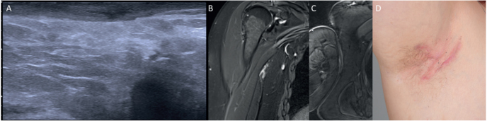
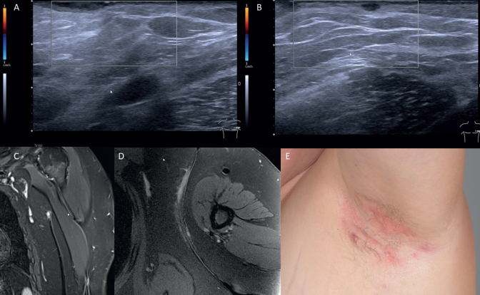
Hidradenitis suppurativa has a substantial negative effect on quality of life of affected persons. Diagnosis is based mainly on clinical examination. However, physi-cal examination alone might underestimate disease severity compared with imaging modalities. We report here the application of non-contrast-enhanced 3-Tesla magnetic resonance imaging using surface-coil and sonography for assessment of hidradenitis suppurativa lesions based on topographic assessment of skin lesions. In addition, we review the literature regarding the application of ultrasound and magnetic resonance imaging in hidradenitis suppurativa.

Keywords: MRI; hidradenitis suppurativa; sonography; surface coil; treatment; 3-Tesla.


Conflict of interest statement

The authors have no conflicts of interest to declare.

Figures

Fig. 1
Fig. 1
Right shoulder. (A) Sonography: dermal thickening without fluid collection or fistula formation. (B, C) Magnetic resonance imaging: Both pictures show dermal thickening in the coronal and axial sequences, with suspicion of beginning of tiny sinus tract formation on the coronal acquisition. (B) TIRM (Turbo-Inversion Recovery-Magnitude) blade coronal right; (C) T2 Blade FS (Fat Suppressed) axial. (D) Clinical image.
Fig. 2
Fig. 2
Left shoulder. (A, B) Sonography: in addition to routine ultrasound (US) imaging, power Doppler and colour Doppler ultrasound modalities can be utilized to visualize hidradenitis suppurativa lesions and differentiate nearby vasculature. Ultrasound shows dermal thickening with some small dermal fluid collection up to 10 mm without sign of fistula formation. (C, D) Magnetic resonance imaging: both pictures show diffuse infiltrating dermal thickening in the axial sequences, with small fluid formation on the coronal acquisition. No fistula formation. (C) TIRM (Turbo-Inversion Recovery-Magnitude) blade coronal. (D) PD (Proton Density) TSE (Turbo Spin Echo) FS (Fat Suppressed) axial. (E) Clinical image.
Fig. 3
Fig. 3
Flow of information during the review.

Similar articles

Cited by

References

    1. Seyed Jafari SM, Knusel E, Cazzaniga S, Hunger RE. A retrospective cohort study on patients with hidradenitis suppurativa. Dermatology 2018; 234: 71–78. - PubMed
    1. Houriet C, Seyed Jafari SM, Thomi R, Schlapbach C, Borradori L, Yawalkar N, et al. . Canakinumab for severe hidradenitis suppurativa: preliminary experience in 2 cases. JAMA Dermatol 2017; 153: 1195–1197. - PubMed
    1. Seyed Jafari SM, Hunger RE, Schlapbach C. Hidradenitis suppurativa: current understanding of pathogenic mechanisms and suggestion for treatment algorithm. Front Med (Lausanne) 2020; 7: 68. - PMC - PubMed
    1. Jemec GB, Heidenheim M, Nielsen NH. Hidradenitis suppurativa – characteristics and consequences. Clin Exp Dermatol 1996; 21: 419–423. - PubMed
    1. Poh F, Wong SK. Imaging of hidradenitis suppurativa and its complications. Case Rep Radiol 2014; 2014: 294753. - PMC - PubMed