. 2020 Oct;46(4):1289-1300.
doi: 10.3892/ijmm.2020.4675. Epub 2020 Jul 15.

MicroRNA‑145 promotes the apoptosis of leukemic stem cells and enhances drug‑resistant K562/ADM cell sensitivity to adriamycin via the regulation of ABCE1

Affiliations

MicroRNA‑145 promotes the apoptosis of leukemic stem cells and enhances drug‑resistant K562/ADM cell sensitivity to adriamycin via the regulation of ABCE1

Zhijun Wuxiao et al. Int J Mol Med. 2020 Oct.

Abstract

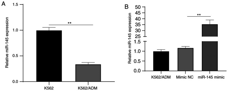
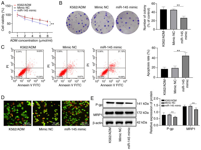
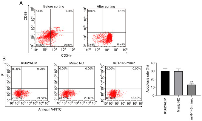
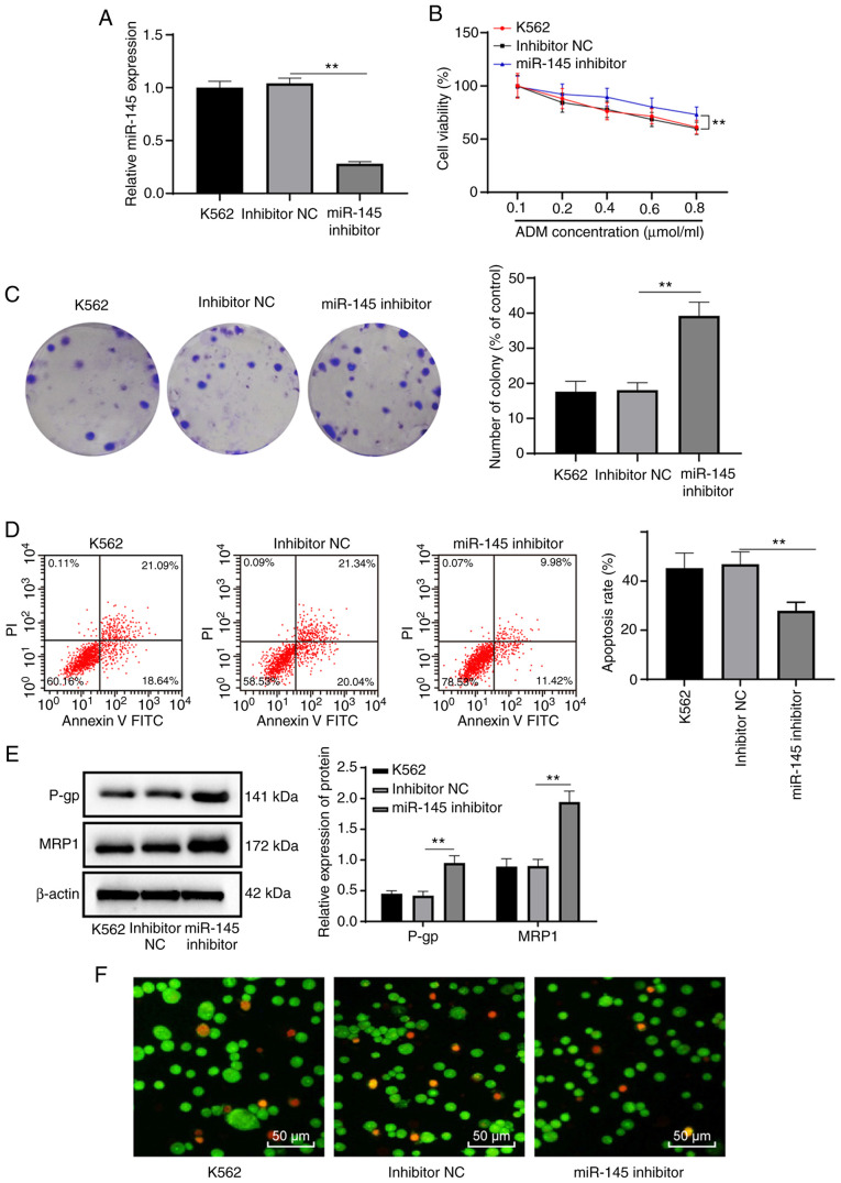
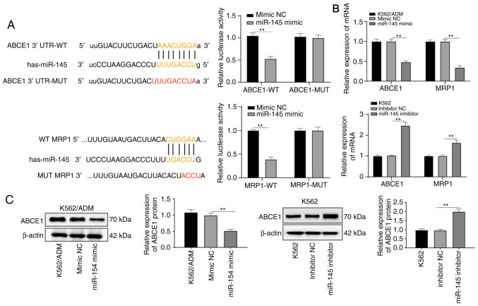
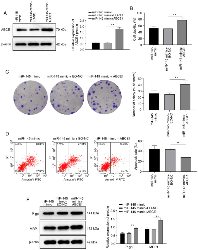
Leukemia is a type of cancer which originates in blood‑forming tissues. MicroRNAs (miRNAs or miRs) have been shown to be involved leukemogenesis. In the present study, following the gain‑ and loss‑function of miR‑145 and ATP‑binding cassette sub‑family E member 1 (ABCE1) in K562 cells and K562 adriamycin‑resistant cells (K562/ADM cells), the levels of multidrug resistance protein 1 (MRP1) and P‑glycoprotein (P‑gp) were measured. The viability of the K562 cells and K562/ADM cells treated with various concentrations of ADM, and cell sensitivity to ADM were measured. The apoptosis of stem cells was detected. K562/ADM cells were transfected with miR‑145 mimic or with miR‑145 mimic together with ABCE1 overexpression plasmid to examine the effects of ABCE1 on the sensitivity of K562/ADM cells to ADM. The association between miR‑145 and ABCE1/MRP1 was then verified. The dose‑ and time‑dependent effects of ADM on the K562 cells and K562/ADM cells were examined. The K562/ADM cells exhibited a greater resistance to ADM, higher levels of MRP1 and P‑gp, and a lower miR‑145 expression. The K562/ADM cells and stem cells in which miR‑145 was overexpressed exhibited a suppressed cell proliferation, decreased MRP1 and P‑gp levels, and an increased apoptotic rate. However, K562 cells with a low expression of miR‑145 exhibited an increased cell proliferation, increased levels of MRP1 and P‑gp, and a suppressed apoptotic rate. Compared with the overexpression of miR‑145, the combination of miR‑145 and ABCE1 decreased the sensitivity of drug‑resistant K562/ADM cells to ADM. The above‑mentioned effects of miR‑145 were achieved by targeting ABCE1. Taken together, the findings of the present study demonstrate that the overexpression of miR‑145 promotes leukemic stem cell apoptosis and enhances the sensitivity of K562/ADM cells to ADM by inhibiting ABCE1.


Figures

Figure 1
Figure 1
K562/ADM cells exhibit potent drug resistance. (A) Inhibition ratio of K562 cells and K562/ADM cells with various concentrations of ADM at 12, 24 and 48 h detected by MTT assay. (B) Maps of cell colonies of K562 and K562/ADM cells treated with various concentrations of ADM detected by colony formation assay. (C) Levels of MRP1 and P-gp in K562 cells and K562/ADM cells detected by western blot analysis; n=3. **P<0.01. Data in (A and C) were analyzed by two-way ANOVA, while data in (B) were analyzed with one-way ANOVA. Tukey's multiple comparisons test was applied as a post hoc test. ADM, adriamycin; MTT, 3-(4,5-dimethylthiazol-2-yl)-2,5-diphenyltetrazolium bromide; MRP1, multidrug resistance protein 1; P-gp, P-glycoprotein; ANOVA, analysis of variance.
Figure 2
Figure 2
miR-145 expression is downregulated in K562/ADM cells. (A) Relative miR-145 expression in K562/ADM cells and K562 cells detected by RT-qPCR. (B) Relative miR-145 expression following transfection detected by RT-qPCR. **P<0.01; n=3. Data in (A) were analyzed by an independent t-test, while data in (B) were analyzed with one-way ANOVA. Tukey's multiple comparisons test was used as a post hoc test. ADM, adriamycin; miR-145, microRNA-145; RT-qPCR, reverse transcription quantitative polymerase chain reaction; ANOVA, analysis of variance.
Figure 3
Figure 3
Overexpression of miR-145 enhances the sensitivity of K562/ADM cells to ADM. (A) Relative cell viability in each group treated with various concentrations of ADM detected by MTT assay. (B) Relative cell colonies in K562/ADM cells with 6 µmol/l ADM measured using colony formation assay. (C) Relative apoptosis of K562/ADM cells treated with 6 µmol/l ADM measured by flow cytometry. (D) The morphology and apoptosis of K562/ADM cells treated with 6 µmol/l ADM observed under a fluorescence microscope. (E) Levels of MRP1 and P-gp of K562/ADM cells treated with 6 µmol/l ADM measured by western blot analysis. **P<0.01; n=3. Data in (A and E) were analyzed by two-way ANOVA, while data in (B and C) were analyzed by one-way ANOVA. Tukey's multiple comparisons test was used as a post hoc test. ADM, adriamycin; miR-145, microRNA-145; MTT, 3-(4,5-dimethylthiazol-2-yl)-2, 5-diphenyltetrazolium bromide; MRP1, multidrug resistance protein 1; P-gp, P-glycoprotein; ANOVA, analysis of variance.
Figure 4
Figure 4
Overexpression of miR-145 promotes the apoptosis of stem cells. (A) CD34+CD38K562/ADM stem cells were sorted from K562/ADM stem cells using magnetic bead sorting methods. (B) Cell apoptosis was detected by flow cytometry. **P<0.01; n=3. Data were analyzed by one-way ANOVA. Tukey's multiple comparisons test was used as a post hoc test. ADM, adriamycin; miR-145, microRNA-145.
Figure 5
Figure 5
Low expression of miR-145 reduces the sensitivity of K562 cells to ADM. (A) Relative miR-145 expression in K562 cells detected by RT-qPCR. (B) Relative viability of K562 cells treated with various concentrations of ADM measured by MTT assay. (C) Relative cell colonies of K562 cells treated with 0.6 µmol/l ADM measured by colony formation assay. (D) Relative cell apoptosis of K562 cells treated with 0.6 µmol/l ADM measured by flow cytometry. (E) Levels of MRP1 and P-gp of K562 cells treated with 0.6 µmol/l ADM measured by western blot analysis. (F) The morphology and apoptosis of K562 cells treated with 0.6 µmol/l ADM were observed under a fluorescence microscope. **P<0.01; n=3. Data in (A, C and D) were analyzed by one-way ANOVA, while data in (B and E) were analyzed by two-way ANOVA. Tukey's multiple comparisons test was used as a post hoc test. ADM, adriamycin; miR-145, microRNA-145; RT-qPCR, reverse transcription quantitative polymerase chain reaction; MTT, 3-(4,5-dimethylthiazol-2-yl)-2,5-diphenyltetrazolium bromide; MRP1, multidrug resistance protein 1; P-gp, P-glycoprotein; ANOVA, analysis of variance.
Figure 6
Figure 6
miR-145 targets MRP1 and ABCE1. (A) Binding sites between miR-145 and ABCE1, and between miR-145 and MRP1 predicted by bioinformatics software and website, and relative ABCE1 and MRP1 luciferase activity measured by dual-luciferase reporter gene assay. (B) Relative mRNA expression of ABCE1 and MRP1 detected by RT-qPCR. (C) Relative protein expression of ABCE1 detected by western blot analysis. **P<0.01; n=3. Data in (A and B) were analyzed by two-way ANOVA, and data in (C) were analyzed by one-way ANOVA. Tukey's multiple comparisons test was used as a post hoc test. ADM, adriamycin; miR-145, microRNA-145; MRP1, multidrug resistance protein 1; ABCE1, adenosine triphosphate (ATP)-binding cassette (ABC) transporter E1.
Figure 7
Figure 7
ABCE1 activation reverses the promoting effects of miR-145 overexpression on K562/ADM cell sensitivity to ADM. (A) Relative protein expression of ABCE1 detected by western blot analysis. (B) Relative cell viability in each group detected by MTT assay. (C) Relative cell colonies in K562/ADM cells treated with 6 µmol/l ADM measured by colony formation assay. (D) Relative apoptosis of K562/ADM cells treated with 6 µmol/l ADM measured by flow cytometry. (E) Levels of MRP1 and P-gp of K562/ADM cells with 6 µmol/l ADM measured by western blot analysis. **P<0.01; n=3. Data in (A-D) were analyzed by one-way ANOVA, and data in (E) were analyzed by two-way ANOVA. Tukey's multiple comparisons test was used as a post hoc test. ADM, adriamycin; miR-145, microRNA-145; MTT, 3-(4,5-dimethylthiazol-2-yl)-2,5-diphenyltetrazolium bromide; MRP1, multidrug resistance protein 1; P-gp, P-glycoprotein.

Similar articles

Cited by

References

    1. Juliusson G, Hough R. Leukemia. Prog Tumor Res. 2016;43:87–100. doi: 10.1159/000447076. - DOI - PubMed
    1. Yamashita Y, Yuan J, Suetake I, Suzuki H, Ishikawa Y, Choi YL, Ueno T, Soda M, Hamada T, Haruta H, et al. Array-based genomic resequencing of human leukemia. Oncogene. 2010;29:3723–3731. doi: 10.1038/onc.2010.117. - DOI - PubMed
    1. Steinmaus C, Smith MT. Steinmaus and Smith respond to 'proximity to gasoline stations and childhood leukemia'. Am J Epidemiol. 2017;185:5–7. doi: 10.1093/aje/kww133. - DOI - PMC - PubMed
    1. Kabat GC, Wu JW, Moore SC, Morton LM, Park Y, Hollenbeck AR, Rohan TE. Lifestyle and dietary factors in relation to risk of chronic myeloid leukemia in the NIH-AARP Diet and Health Study. Cancer Epidemiol Biomarkers Prev. 2013;22:848–854. doi: 10.1158/1055-9965.EPI-13-0093. - DOI - PMC - PubMed
    1. Kosmider O, Gelsi-Boyer V, Slama L, Dreyfus F, Beyne-Rauzy O, Quesnel B, Hunault-Berger M, Slama B, Vey N, Lacombe C, et al. Mutations of IDH1 and IDH2 genes in early and accelerated phases of myelodysplastic syndromes and MDS/myeloproliferative neoplasms. Leukemia. 2010;24:1094–1096. doi: 10.1038/leu.2010.52. - DOI - PubMed

MeSH terms

Substances