. 2021 May;34(5):e4411.
doi: 10.1002/nbm.4411. Epub 2020 Sep 18.

Spectral editing in 1 H magnetic resonance spectroscopy: Experts' consensus recommendations

Affiliations

Spectral editing in 1 H magnetic resonance spectroscopy: Experts' consensus recommendations

In-Young Choi et al. NMR Biomed. 2021 May.

Abstract

Spectral editing in in vivo 1 H-MRS provides an effective means to measure low-concentration metabolite signals that cannot be reliably measured by conventional MRS techniques due to signal overlap, for example, γ-aminobutyric acid, glutathione and D-2-hydroxyglutarate. Spectral editing strategies utilize known J-coupling relationships within the metabolite of interest to discriminate their resonances from overlying signals. This consensus recommendation paper provides a brief overview of commonly used homonuclear editing techniques and considerations for data acquisition, processing and quantification. Also, we have listed the experts' recommendations for minimum requirements to achieve adequate spectral editing and reliable quantification. These include selecting the right editing sequence, dealing with frequency drift, handling unwanted coedited resonances, spectral fitting of edited spectra, setting up multicenter clinical trials and recommending sequence parameters to be reported in publications.

Keywords: J-difference editing; consensus recommendations; multiple quantum filtering; spectral editing; γ-aminobutyric acid, glutathione.


Figures

Figure 1.
Figure 1.. Effect of CSDE on editing efficiency for PRESS editing of lactate (TE = 144 ms) and GABA (TE = 70 ms).
(A) Numerical simulations of the 2D signal distribution of 1.3 ppm lactate resonance (a coupled spin at 4.1 ppm 2D distribution is depicted by the white dashed box). The 2D plane represents two spatial dimensions selected by two 180° localization RF pulses (1 kHz refocusing bandwidth). While severe spatial misregistration between two coupled spins is evident during the EDIT ON scan, the overall signal intensity is relatively uniform. This is due to the editing RF pulses refocusing voxel compartments that did not experience the effect of the 180° localization pulses. However, for the EDIT OFF case, one can observe distinct compartments where the spins of interest exhibit phases opposite that of the theoretical non-localized dual spin-echo experiment. (B) Numerical simulations of the 2D signal distribution of 3.0 ppm GABA resonance (coupled spins at 1.9 ppm 2D distribution is depicted by the white dashed box). The same dynamics are observed for GABA editing, albeit to the less extent due to smaller frequency difference between GABA coupled spins compared to lactate. Image intensity represents the integral of doublets of lactate or two outer peaks of GABA signals and color bars indicate respective intensity ranges. The significant reduction of the signal intensity in the final DIFFERENCE spectra (e.g., ~50% for lactate, ~20% for GABA) due to CSDE is observed at 3T.
Figure 2.
Figure 2.
(A) Representative single voxel MR spectra of GSH from the motor cortex (3.5 × 2.5 × 2.3 cm3) of a subject with ALS using MEGA-PRESS (TE = 68 ms, TR = 2 s, 512 averages, editing pulse at 4.56 ppm for edit on and at 7.5 ppm for edit off) at 3 T. The bottom spectrum is shown without processing. Slight frequency drifts over time lead to small subtraction artifacts as can be seen for NAA (red arrow). The top spectrum shows the same spectrum as below after the individual transients are frequency aligned leading to a clean, flat baseline. Both spectra are shown with 1 Hz exponential line broadening. (B) GSH MRSI measured from the fronto-parietal region of the human brain using the doubly selective MQF editing sequence (TE = 115 ms, TR = 1.5 s, matrix size = 8 × 8, nominal voxel size = 1.25 × 1.25 × 3.0 cm3, field of view = 20 cm, and scan time = 16 min) at 3 T. All spectra were processed with 2 Hz exponential line broadening and are shown in the range from 3.6 to 2.2 ppm (adapted from Reference).
Figure 3.
Figure 3.
Optimized in vivo MRS detection of 2HG from mutant-IDH glioma patients using (A) J-difference editing 3D MEGA-LASER sequence (TR/TE = 1600/68 ms, matrix 10 × 10 × 10, FOV=200 × 200 × 200 mm3, and acquisition time = 9.5 min; reproduced with permission from Reference); (B) long echo 2D PRESS (TR/TE = 1300/97 ms, matrix 16 × 16, FOV = 160 × 160 mm2, slice 15 mm, and acquisition time = 10 min; reproduced with permission from Reference); and (C) 2D COSY-LASER (TR/TE = 1500/30 ms, voxel 3.0 × 3.0 × 3.0 cm3, 64 t1 increments, acquisition time = 11.5 min; reproduced with permission from Reference) at 3 T.
Figure 4.
Figure 4.
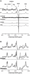
(A) A representative GABA spectrum from the human occipital lobe (4.0 × 2.3 × 3.0 cm3) using MEGA-PRESS (TR/TE = 3000/68 ms, 256 averages) at 3 T. Sub-spectra show traces of best fit, residuals, baseline, GABA and MM. The MM spectrum is an average metabolite-nulled GABA-edited spectrum from 13 subjects. Both metabolite and metabolite-nulled spectra were acquired with editing pulse at 1.9 ppm with resolved averages to monitor motion and frequency drifts and resetting the frequency at 64 scan blocks. (B) GABA+ spectra from the medial parietal region randomly selected from the Big GABA dataset show typical data quality for GE (B, top), Philips (B, middle), Siemens (B, bottom) using MEGA-PRESS (TR/TE = 2000/68 ms, voxel size = 3.0 × 3.0 × 3.0 cm3, 320 averages). See details in Reference.

Similar articles

Cited by

References

    1. Mescher M, Merkle H, Kirsch J, Garwood M, Gruetter R. Simultaneous in vivo spectral editing and water suppression. NMR Biomed. October 1998;11(6):266–272. - PubMed
    1. Star-Lack J, Spielman D, Adalsteinsson E, Kurhanewicz J, Terris DJ, Vigneron DB. In vivo lactate editing with simultaneous detection of choline, creatine, NAA, and lipid singlets at 1.5 T using PRESS excitation with applications to the study of brain and head and neck tumors. J Magn Reson. August 1998;133(2):243–254. - PubMed
    1. Andreychenko A, Boer VO, Arteaga de Castro CS, Luijten PR, Klomp DW. Efficient spectral editing at 7 T: GABA detection with MEGA-sLASER. Magn Reson Med. October 2012;68(4):1018–1025. - PubMed
    1. Bogner W, Gagoski B, Hess AT, et al. 3D GABA imaging with real-time motion correction, shim update and reacquisition of adiabatic spiral MRSI. Neuroimage. December 2014;103:290–302. - PMC - PubMed
    1. Near J, Simpson R, Cowen P, Jezzard P. Efficient gamma-aminobutyric acid editing at 3T without macromolecule contamination: MEGA-SPECIAL. NMR Biomed. December 2011;24(10):1277–1285. - PubMed

Publication types

Substances

LinkOut - more resources