. 2020 Nov 13;6(11):2913-2925.
doi: 10.1021/acsinfecdis.0c00319. Epub 2020 Oct 1.

Attenuating the Selection of Vancomycin Resistance Among Enterococci through the Development of Peptide-Based Vancomycin Antagonists

Affiliations

Attenuating the Selection of Vancomycin Resistance Among Enterococci through the Development of Peptide-Based Vancomycin Antagonists

Ryan W Mull et al. ACS Infect Dis. .

Abstract

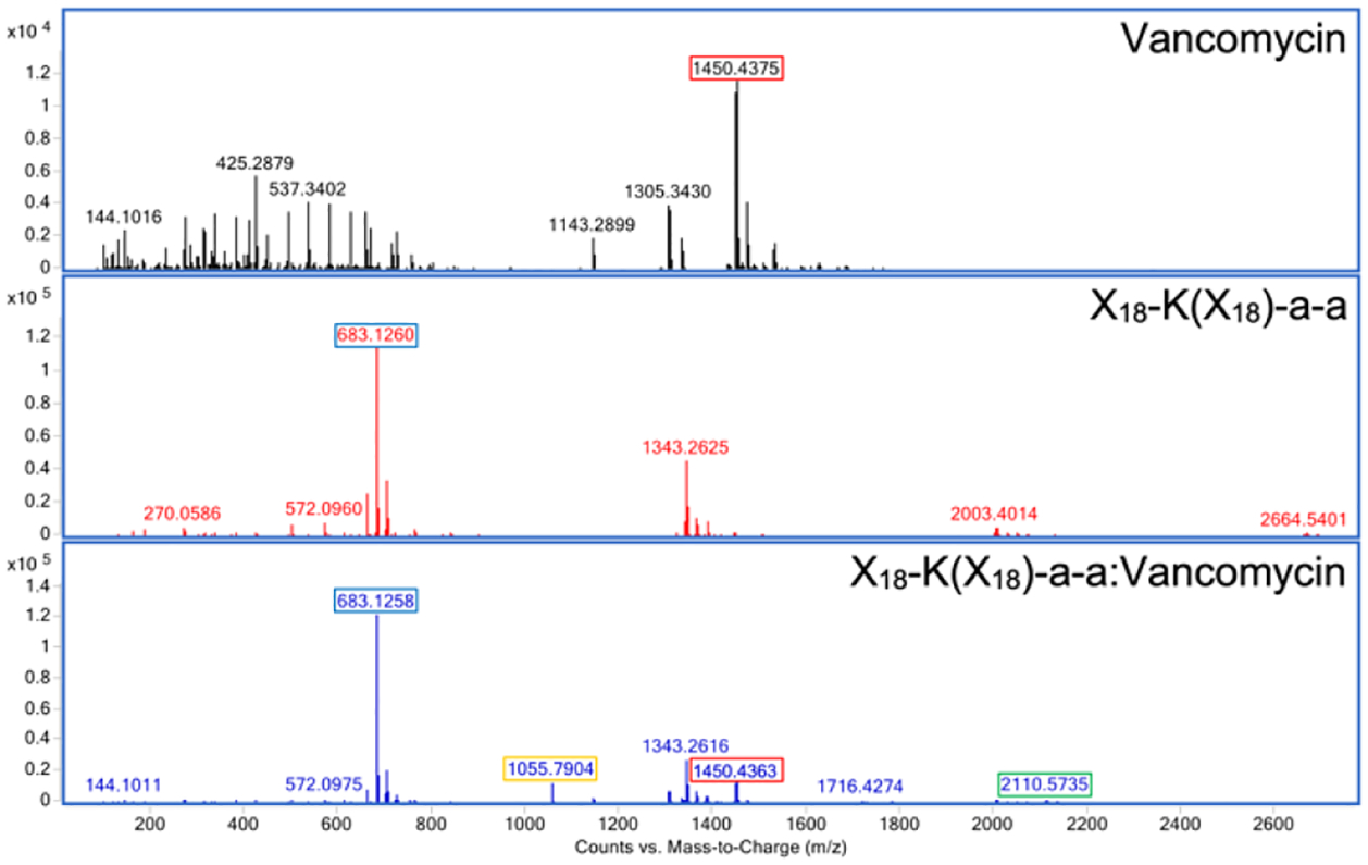
The emergence and spread of multidrug resistant (MDR) pathogens with acquired resistance to almost all available antimicrobial agents has severely threatened the international healthcare community over the last two decades. The last resort antibiotic vancomycin is critical for treatment of several of these pathogens; howeverc vancomycin resistance is spreading due to the undesired accumulation of IV vancomycin in the colon post-treatment. This accumulation exerts selective pressure upon members of the colonic microflora, including Enterococci, which possess vancomycin resistance genes. To ensure the continual effectiveness of vancomycin in the clinical setting by preventing the spread of antibiotic resistance, it is crucial to develop strategies that reduce selective pressure on the colonic microflora while allowing vancomycin to maintain its desired activity at the site of infection. Herein we report that modification of the native l-Lys-d-Ala-d-Ala vancomycin binding site can be used to produce peptides with the ability to competitively bind vancomycin, reducing its activity against susceptible Enterococci. Moreover, several modifications to the N-termini of the native tripeptide have produced compounds with enhanced vancomycin binding activity, including several analogs that were designed to covalently bind vancomycin, thereby acting as suicide inhibitors. Finally, in a mixed culture of susceptible and resistant bacteria, a single lead compound was found to protect high ratios of susceptible bacteria from vancomycin over the course of a week-long period, preventing the selection for vancomycin-resistant Enterococci. These findings demonstrate the ability of these peptides as potential therapeutic adjuvants for counteracting the undesired accumulation of colonic vancomycin, allowing for protection of the colonic microflora.

Keywords: antibiotic resistance; vancomycin; vancomycin-resistant Enterococcus; viability qPCR.


Figures

Figure 1.
Figure 1.
Crystal structure of the complex formed between vancomycin (silver) and the native Acetyl-L-Lys(Acetyl)-D-Ala-D-Ala peptide (yellow). PDB ID: 1FVM.
Figure 2.
Figure 2.
Vancomycin binding peptide Library 1. X1, acetyl; X2, propionyl; X3, butyryl; X4, valeryl; X5, hexanoyl; X6, heptanoyl; X17, 4-(fluorosulfonyl)benzoyl.
Figure 3.
Figure 3.
Vancomycin antagonism by the native vancomycin binding peptide. A. Structure of the native vancomycin binding peptide. B. Antagonism of low (8 μg/mL) and high (32 μg/mL) concentrations of vancomycin at low (5:1) and high (20:1) peptide:vancomycin molar ratios. Inhibition of vancomycin was quantified by observing growth of E. faecium (TX2016), following 24 hr incubation. Data presented is the average of three trials performed in triplicate. See the SI for full experimental data for each peptide.
Figure 4.
Figure 4.
Vancomycin antagonism by X1–6,17-a-a peptides. Antagonism of vancomycin by X1–6,17-a-a peptides was assessed in two different peptide:vancomycin molar ratios (20:1 or 5:1), against two different concentrations of vancomycin (32 μg/mL or 8 μg/mL). Inhibition of vancomycin was quantified by observing growth of E. faecium (TX2016), following 24 hr incubation. Data presented is the average of three trials performed in triplicate. See the SI for full experimental data for each peptide.
Figure 5.
Figure 5.
Vancomycin binding peptide Library 2. Library 2A: X1, acetyl; X2, propionyl; X3, butyryl; X4, valeryl; X5, hexanoyl; X6, heptanoyl. Library 2B: X7, cyclohexanoyl; X8, 4-methylcyclohexanoyl; X9, benzoyl; X10, p-tolyl. Library 2C: X11, 4-(dimethylamino)benzoyl; X12, 4-(trifluoromethyl)benzoyl; X13, 2,3,5,6-(tetrafluoro)p-tolyl; X14, 4-(2-chloroacetamido)benzoyl; X15, 4-(methylsulfonyl)benzoyl; X16, 4-(sulfonyl)benzoyl; X17, 4-(fluorosulfonyl)benzoyl; X18, 3-(fluorosulfonyl)benzoyl.
Figure 6.
Figure 6.
Vancomycin antagonism by X2–18-K(X1)-a-a and X1-K(X2–18)-a-a peptides. Antagonism of vancomycin by peptides was assessed in two different peptide:vancomycin molar ratios (20:1 or 5:1), against two different concentrations of vancomycin (32 μg/mL or 8 μg/mL). Inhibition of vancomycin was quantified by observing growth of E. faecium (TX2016), following 24 hr incubation. Data presented is the average of three trials performed in triplicate. See the SI for full experimental data for each peptide.
Figure 7.
Figure 7.
Vancomycin binding peptide Library 3. Library 3A: X1, acetyl; X2, propionyl; X3, butyryl; X4, valeryl; X5, hexanoyl; X6, heptanoyl. Library 3B: X7, cyclohexanoyl; X8, 4-methylcyclohexanoyl; X9, benzoyl; X10, p-tolyl. Library 3C: X11, 4-(dimethylamino)benzoyl; X12, 4-(trifluoromethyl)benzoyl; X13, 2,3,5,6-(tetrafluoro)p-tolyl; X14, 4-(2-chloroacetamido)benzoyl; X15, 4-(methylsulfonyl)benzoyl; X16, 4-(sulfonyl)benzoyl; X17, 4-(fluorosulfonyl)benzoyl; X18, 3-(fluorosulfonyl)benzoyl.
Figure 8.
Figure 8.
Antagonism of vancomycin by X2–18-K(X2–18)-a-a peptides was assessed in two different peptide:vancomycin molar ratios (20:1 or 5:1), against two different concentrations of vancomycin (32 μg/mL or 8 μg/mL). Inhibition of vancomycin was quantified by observing percent growth of E. faecium (TX2016), following 24 hr incubation. Several peptides (black) were insoluble at the tested concentration, so no data was collected. Data presented is the average of three trials performed in triplicate. See the SI for full experimental data for each peptide.
Figure 9.
Figure 9.
Comparison of vancomycin inhibition between the lead X17-K(X17)-a-a peptide and three lead branched peptides previously reported, as determined by a vancomycin antagonism assay. Inhibition of vancomycin was quantified by observing percent growth of E. faecium (TX2016), following 24 hr incubation. Data presented is the average of three trials performed in triplicate.
Figure 10.
Figure 10.
Assessing the effect of peptide:vancomycin pre-incubation on vancomycin antagonism. Inhibition of vancomycin by the native peptide (A), X14-K(X14)-a-a peptide (B), X17-K(X17)-a-a peptide (C), and X18-K(X18)-a-a peptide (D) was compared following no-incubation (0 h), or standard pre-incubation (1 h) using a vancomycin antagonism assay. Inhibition of vancomycin was quantified by observing percent growth of E. faecium (TX2016), following 24 hr incubation. Data presented is the average of three trials performed in triplicate.
Figure 11.
Figure 11.
Detection of native peptide:vancomycin covalent adducts by ESI-TOF MS. Expected mass of vancomycin (red) [M+H]+ 1450.3. Expected mass of native peptide (blue) [M+Na]+ 395.2.
Figure 12.
Figure 12.
Detection of X14-K(X14)-a-a:vancomycin covalent adducts by ESI-TOF MS. Expected mass of vancomycin [M+H]+ 1450.3 (red). Expected mass of X14-K(X14)-a-a [M+Na]+ 701.2 (blue). Expected mass of X14-K(X14)-a-a:vancomycin [M+Na]+ 2112.6. Expected mass of X14-K(X14)-a-a:vancomycin [M+H+Na]2+ 1056.8.
Figure 13.
Figure 13.
Detection of X17-K(X17)-a-a:vancomycin covalent adducts by ESI-TOF MS. Expected mass of vancomycin [M+H]+ 1450.3 (red). Expected mass of X17-K(X17)-a-a [M+Na]+ 683.1 (blue). Expected mass of X17-K(X17)-a-a:vancomycin [M+Na]+ 2110.6 (green). Expected mass of X17-K(X17)-a-a:vancomycin [M+H+Na]2+ 1055.8 (gold).
Figure 14.
Figure 14.
Detection of X18-K(X18)-a-a:vancomycin covalent adducts by ESI-TOF MS. Expected mass of vancomycin [M+H]+ 1450.3 (red). Expected mass of X18-K(X18)-a-a [M+Na]+ 683.1 (blue). Expected mass of X18-K(X18)-a-a:vancomycin [M+Na]+ 2110.6 (green). Expected mass of X18-K(X18)-a-a:vancomycin [M+H+Na]2+ 1055.8 (gold).
Figure 15.
Figure 15.
Detection of X16-K(X16)-a-a:vancomycin covalent adducts by ESI-TOF MS. Expected mass of vancomycin [M+H]+ 1450.3 (red). Expected mass of X16-K(X16)-a-a [M+H]+ 657.2 (blue). Expected mass of X16-K(X16)-a-a:vancomycin [M+Na]+ 2108.6. Expected mass of X16-K(X16)-a-a:vancomycin [M+H+Na]2+ 1054.8.
Figure 16.
Figure 16.
Detection of X17-K(X1)-a-a:vancomycin covalent adducts by ESI-TOF MS. Expected mass of vancomycin [M+H]+ 1450.3 (red). Expected mass of X17-K(X1)-a-a [M+Na]+ 539.2 (blue). Expected mass of X17-K(X1)-a-a:vancomycin [M+Na]+ 1966.6 (green). Expected mass of X17-K(X1)-a-a:vancomycin [M+H+Na]2+ 983.8 (gold).
Figure 17.
Figure 17.
Detection of X1-K(X17)-a-a:vancomycin covalent adducts by ESI-TOF MS. Expected mass of vancomycin [M+H]+ 1450.3 (red). Expected mass of X1-K(X17)-a-a [M+Na]+ 539.2 (blue). Expected mass of X1-K(X17)-a-a:vancomycin [M+Na]+ 1966.6 (green). Expected mass of X1-K(X17)-a-a:vancomycin [M+H+Na]2+ 983.8 (gold).
Figure 18.
Figure 18.
Peptides containing either the single Nα or Nε X14 or X17 substitution were assessed for their ability to inhibit vancomycin in a 10:1 peptide:vancomycin molar ratio using a vancomycin antagonism assay. Inhibition of vancomycin was quantified by observing percent growth of E. faecium (TX2016), following 24 hr incubation. Data presented is the average of three trials performed in triplicate.
Figure 19.
Figure 19.
The apparent permeability of peptides X14-K(X14)-a-a and X(17)-K(X17)-a-a on Caco-2 cell monolayer. Peptides and controls were tested at a concentration of 10 μM. Data are arbitrary units and represent the mean +/− S.D. for three replicate trials. A; apical, B; basal. Assay performed by Charles River Laboratories.
Figure 20.
Figure 20.
Assessing the changes in population of susceptible (E. faecium TX2016) and resistant (E. faecalis S613) Enterococci present in mixed cultures of susceptible:resistant Enterococci following addition of 32 μg/mL vancomycin and lead peptides in a 5:1 peptide:vancomycin molar ratio over a weeklong period. Populations of bacterial cells in samples containing solely bacteria (solid black line), bacteria with vancomycin (dashed black line), bacteria with native peptide and vancomycin (red), bacteria with peptide X14-K(X14)-a-a and vancomycin (green), and bacteria with peptide X17-K(X17)-a-a and vancomycin (blue), were directly monitored over a week long period using viability qPCR to ensure the amplification of solely viable DNA. Red line is the detection threshold of 2.3×10⁶ bacterial cells. (A) Population change of susceptible and resistant Enterococci at an initial ratio of 10000:1 susceptible:resistant over the course of one week. (B) Population change of susceptible and resistant Enterococci at an initial ratio of 100:1 susceptible:resistant over the course of one week. (C) Population change of susceptible and resistant Enterococci at an initial ratio of 1:1 susceptible:resistant over the course of one week. (D) Population change of susceptible and resistant Enterococci at an initial ratio of 1:100 susceptible:resistant over the course of one week. (E) Population change of susceptible and resistant Enterococci at an initial ratio of 1:10000 susceptible:resistant over the course of one week. Data are the relative number of bacterial cells as determined by viability qPCR and represent the mean +/− S.D. for three replicate trials.
Figure 21.
Figure 21.
Standard curves correlating Cq values to log[DNA] for amplification of S613 DNA (A) and TX2016 DNA (B).

Similar articles

Cited by

References

    1. Dadgostar P, Antimicrobial Resistance: Implications and Costs. Infect Drug Resist 2019, 12, 3903–3910. - PMC - PubMed
    1. Hofer U, The cost of antimicrobial resistance. Nat Rev Microbiol 2019, 17, 3. - PubMed
    1. Nikaido H, Multidrug resistance in bacteria. Annu Rev Biochem 2009, 78, 119–146. - PMC - PubMed
    1. Ventola CL, The antibiotic resistance crisis: part 1: causes and threats. P T 2015, 40, 277–283. - PMC - PubMed
    1. Levine DP, Vancomycin: a history. Clin Infect Dis 2006, 42 Suppl 1, S5–12. - PubMed

Publication types