. 2020 Nov 1;319(5):H1021-H1035.
doi: 10.1152/ajpheart.00302.2020. Epub 2020 Sep 18.

Deletion of cardiac polycystin 2/PC2 results in increased SR calcium release and blunted adrenergic reserve

Affiliations

Deletion of cardiac polycystin 2/PC2 results in increased SR calcium release and blunted adrenergic reserve

Elisabeth DiNello et al. Am J Physiol Heart Circ Physiol. .

Abstract

Transient receptor potential proteins (TRPs) act as nonselective cation channels. Of the TRP channels, PC2 (also known as polycystin 2) is localized to the sarcoplasmic reticulum (SR); however, its contribution to calcium-induced calcium release and overall cardiac function in the heart is poorly understood. The goal of this study was to characterize the effect of cardiac-specific PC2 deletion in adult cardiomyocytes and in response to chronic β-adrenergic challenge. We used a temporally inducible model to specifically delete PC2 from cardiomyocytes (Pkd2 KO) and characterized calcium and contractile dynamics in single cells. We found enhanced intracellular calcium release after Pkd2 KO, and near super-resolution microscopy analysis suggested this was due to close localization of PC2 to the ryanodine receptor. At the organ level, speckle-tracking echocardiographical analysis showed increased dyssynchrony in the Pkd2 KO mice. In response to chronic adrenergic stimulus, cardiomyocytes from the Pkd2 KO had no reserve β-adrenergic calcium responses and significantly attenuated wall motion in the whole heart. Biochemically, without adrenergic stimulus, there was an overall increase in PKA phosphorylated targets in the Pkd2 KO mouse, which decreased following chronic adrenergic stimulus. Taken together, our results suggest that cardiac-specific PC2 limits SR calcium release by affecting the PKA phosphorylation status of the ryanodine receptor, and the effects of PC2 loss are exacerbated upon adrenergic challenge.NEW & NOTEWORTHY Our goal was to characterize the role of the transient receptor potential channel polycystin 2 (PC2) in cardiomyocytes following adult-onset deletion. Loss of PC2 resulted in decreased cardiac shortening and cardiac dyssynchrony and diminished adrenergic reserve. These results suggest that cardiac-specific PC2 modulates intracellular calcium signaling and contributes to the maintenance of adrenergic pathways.

Keywords: CICR; adrenergic stimulus; calcium release; cardiac dysfunction; echocardiography; excitation-contraction coupling.


Conflict of interest statement

J.A.K. is on the editorial board of the American Journal of Physiology-Heart and Circulatory Physiology. No other conflicts of interest, financial or otherwise, are declared by the authors.

Figures

Fig. 1.
Fig. 1.
Polycystin 2 (PC2) is localized in close approximation to the ryanodine receptor (RyR). A: 2 wk of tamoxifen diet is sufficient to reduce PC2 expression in whole heart lysate, with 2 different antibodies for PC2. Left: representative Western blots from male mice with a rabbit antibody (Yale cohort animals). Right: representative Western blots from male animals with a mouse antibody. Each lane denotes a different animal. B: staining of PC2 (green) is in close association with the RyR (red) in control (CRE) animals. C: PC2 staining is greatly diminished in gene polycystin 2 cardiac-specific knockout (Pkd2 KO) mice (staining taken from Loyola University Chicago cohort animals). Scale bar = 5 μm. D: PC2 associates with RyR more in the middle of the cell compared with just below the edge. Representative line scan quantification from center and margin of the cell of the lines drawn in B. E: PC2 puncta localize just outside the RyR puncta. Data in E are averaged from line scan quantification from the center of cells from 25 different sarcomeres from 3 different mice per genotype (2 male and 1 female). Plotted data represent means and SE. Fluorescence is normalized to 1, and the relative positioning of the most intense puncta for RyR is localized to the 0-distance mark. F: PC2 (left) does not associate with the L-type calcium channel CaV1.2 (middle) α-subunit. Note that staining of PC2 is also present in cells other than cardiomyocytes (blue arrows), but PC2 does not uniformly present on the plasma membrane (yellow arrowhead). G: there is little overlap between the L-type channel puncta and PC2. To assess whether the distribution of puncta was overlapping in panels, Gaussian curves were fit, followed by a Kolmogorov–Smirnov test; the distributions were found to be significantly different. Images in F are representative of staining from 3 different mice (2 male, 1 female). Data presented in G are the means and SE from 12 different sarcomeres taken from 3 different animals (2 male, 1 female). Fluorescence is normalized to 1, and the relative positioning of the most intense puncta for CaV1.2 is localized to the 0-distance mark. Gaussian curves were fit, followed by a Kolmogorov–Smirnov test. Distributions of puncta were significantly different. AU, arbitrary units.
Fig. 2.
Fig. 2.
Loss of polycystin 2 (PC2) reduces spark amplitude and frequency. A and B: analysis of calcium spark measurements from permeabilized cardiomyocytes demonstrates that cardiac-specific PC2 knockout (Pkd2 KO) cells have decreased spark amplitude and spark frequency. Data summarized from 2 different male mice per genotype and from >10 cells. Kruskal–Wallis 1-way ANOVA statistical tests were applied. C: force-calcium curves in skinned cardiomyocytes from 3 different female mice per genotype. A Hill curve was fitted to the data, followed by a paired t test of the F-max. AU, arbitrary units; Cre, control mice. *P < 0.05.
Fig. 3.
Fig. 3.
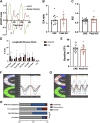
Loss of polycystin 2 (PC2) increases the incidence of dyssynchrony. A: data graphed represent the change in volume over time (dV/dt; green) and the change in velocity over time (dV/dT; red). The equivalent E and A waves are shown, representing the peak opening of the mitral valve. B and C: analysis of the E/A ratio and E/E′ ratio from n = 16–18 control (Cre) and gene polycystin 2 cardiac-specific knockout (Pkd2 KO) male mice. D: analysis of the reverse longitudinal strain from 6 segments. *P < 0.05; n = 17 Cre and 19 Pkd2 KO mice. E: baseline ejection fraction (EF) is not altered in Pkd2 KO mice; n = 20 Cre and 15 Pkd2 KO male mice. Kruskal–Wallis 1-way ANOVA statistical tests were applied. F and G: example longitudinal strain traces from a normal Cre (F) and dyskinetic Pkd2 KO (G) mouse. H: proportion of dyskinesis and dyssynchrony is increased in Pkd2 KO mice. Statistical analysis (Fisher’s exact test) was applied to the absolute number of animals that displayed either a normal echocardiogram or displayed dyskinesis/dyssynchrony; n = 17 Cre male mice and 19 Pkd2 KO male mice. *P < 0.05.
Fig. 4.
Fig. 4.
Chronic isoproterenol (ISO) application causes systolic remodeling in gene polycystin 2 cardiac-specific knockout (Pkd2 KO) mice and mismatched calcium-shortening dynamics. A and B: example intracellular calcium transients (measured with Fura 2AM) and resulting shortening activity from isolated cardiomyocytes with 0.5-Hz pacing. C: baseline (diastolic) calcium. D: rate of calcium transient decay (Tau uptake). E: amplitude of calcium transients with 0.5-Hz pacing. F: sarcomere shortening (%) with 0.5-Hz pacing. G: time required for the baseline shortening length to be returned. H: time to peak (TTP) for shortening. I: ratio between shortening and calcium transient. J and K: relationship between the individual calcium amplitude to the resultant shortening measurement under saline or ISO conditions. Linear regressions and the 95% confidence intervals (dotted lines) are shown. Data in CK taken from cells of 2–3 male mice per group; each symbol represents an individual cell. For data in CI, Kruskal–Wallis 1-way ANOVA statistical tests were applied. Cre, control mice. **P < 0.01, ***P < 0.0001, ****P < 0.00001.
Fig. 5.
Fig. 5.
Gene polycystin 2 cardiac-specific knockout (Pkd2 KO) mice pretreated with chronic isoproterenol (ISO) are desensitized to further, acute ISO application. A: evoked calcium amplitude (to 0.5-Hz pacing) is increased following acute ISO addition (0.1 μM) except in Pkd2 KO mice with chronic ISO treatment. Responses to acute ISO addition are measured in 2-min intervals and are paired recordings. B: calcium store release is unchanged between groups and is not increased following acute ISO application. C: the L-type calcium channel (CaV1.2) response is not altered between control (Cre) and Pkd2 KO mice with or without chronic ISO treatment. With the addition of acute ISO, the L-type channel response is further potentiated, but not in the chronic ISO Pkd2 KO mice. Data in AC are means and SD, n = 9–29 cells; n = 3–4 male mice/group. Kruskal–Wallis 1-way ANOVA statistical tests were applied. D and E: fraction of cells with spontaneous calcium waves. Data plotted in D and E are from n = 9–29 cells; n = 3–4 male mice/group. Statistical analysis (Fisher’s exact test) was applied to the absolute number of cells that had waves. *P < 0.05.
Fig. 6.
Fig. 6.
Chronic isoproterenol (ISO) treatment alters PKA activity and phosphorylation of ryanodine receptor (RyR) in gene polycystin 2 cardiac-specific knockout (Pkd2 KO) male mice. A: levels of β-adrenergic receptor (β-AR2) trended downward with ISO treatment in both control (Cre) and Pkd2 KO animals. The arrowhead points to the specific band quantified on the left. Pkd2 KO mice showed an overall increase in total PKA in response to saline, but not with ISO (quantification, left). Note that the total PKA phosphorylation response was varied; as an example, the arrow points to a band that shows decreased PKA phosphorylation in the ISO-treated Pkd2 KO mice. B: phosphorylation of the L-type calcium channel (CaV1.2) at S1928 was increased by ISO treatment in both genotypes. The arrowhead points to the specific band quantified. Far right graph is the quantification of total CaV1.2. C: phosphorylation at S2808 increased in the Pkd2 KO saline mice compared with the Cre saline control. Overall, there is no significant change in the RyR levels across groups (far right graph). For AC, each lane in the example Western blots represents an individual male mouse; 3–4 male mice were analyzed per group. Data are presented as means and SD. Kruskal–Wallis 1-way ANOVA statistical tests were applied. *P < 0.05, **P < 0.01.
Fig. 7.
Fig. 7.
Isoproterenol (ISO) has blunted synchronicity effects on gene polycystin 2 cardiac-specific knockout (Pkd2 KO) mice. A: left ventricle mass increases in both control (Cre) and Pkd2 KO mice following insertion of mini-osmotic pumps with isoproterenol (ISO; 30 mg·kg−1·day−1). B: ejection fraction similarly increases between groups; n = 8–12 male mice/group. Kruskal–Wallis 1-way ANOVA statistical tests were applied. C: 2 wk of chronic ISO increases dyssynchrony and dyskinesis in Cre mice but not Pkd2 KO mice; n = 7–11 male mice/group. Normalized fractions of total events are presented. Statistical analysis (Fisher’s exact test) was applied to the absolute event number. D: 2 wk of chronic ISO causes a decrease in maximal wall delay in opposing segments in both the radial and longitudinal axis for Cre mice but not Pkd2 KO mice; n = 10–20 male mice per group. Kruskal–Wallis 1-way ANOVA statistical tests were applied. *P < 0.05.

Similar articles

Cited by

References

    1. Altamirano F, Schiattarella GG, French KM, Kim SY, Engelberger F, Kyrychenko S, Villalobos E, Tong D, Schneider JW, Ramirez-Sarmiento CA, Lavandero S, Gillette TG, Hill JA. Polycystin-1 assembles with Kv channels to govern cardiomyocyte repolarization and contractility. Circulation 140: 921–936, 2019. doi:10.1161/CIRCULATIONAHA.118.034731. - DOI - PMC - PubMed
    1. Anyatonwu GI, Estrada M, Tian X, Somlo S, Ehrlich BE. Regulation of ryanodine receptor-dependent calcium signaling by polycystin-2. Proc Natl Acad Sci USA 104: 6454–6459, 2007. doi:10.1073/pnas.0610324104. - DOI - PMC - PubMed
    1. Bauer M, Cheng S, Jain M, Ngoy S, Theodoropoulos C, Trujillo A, Lin FC, Liao R. Echocardiographic speckle-tracking based strain imaging for rapid cardiovascular phenotyping in mice. Circ Res 108: 908–916, 2011. doi:10.1161/CIRCRESAHA.110.239574. - DOI - PMC - PubMed
    1. Bauer M, Cheng S, Unno K, Lin FC, Liao R. Regional cardiac dysfunction and dyssynchrony in a murine model of afterload stress. PLoS One 8: e59915, 2013. doi:10.1371/journal.pone.0059915. - DOI - PMC - PubMed
    1. Bers DM. Cardiac sarcoplasmic reticulum calcium leak: basis and roles in cardiac dysfunction. Annu Rev Physiol 76: 107–127, 2014. doi:10.1146/annurev-physiol-020911-153308. - DOI - PubMed

Publication types

Substances

LinkOut - more resources