. 2020 Sep 18;16(9):e1008739.
doi: 10.1371/journal.ppat.1008739. eCollection 2020 Sep.

Kinetics of Plasmodium midgut invasion in Anopheles mosquitoes

Affiliations

Kinetics of Plasmodium midgut invasion in Anopheles mosquitoes

Gloria Volohonsky et al. PLoS Pathog. .

Abstract

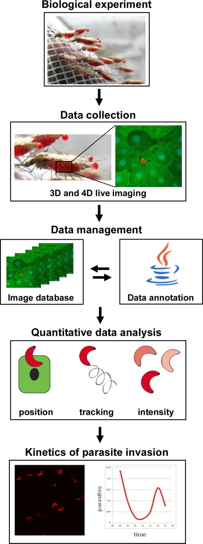
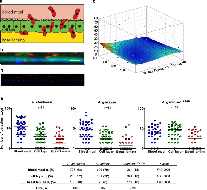
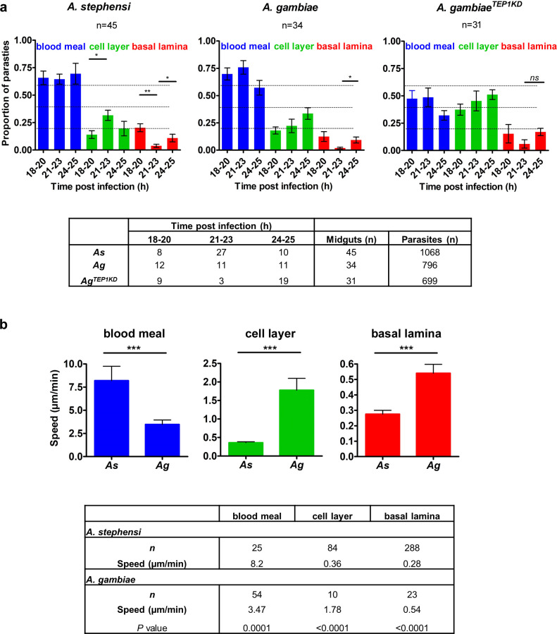
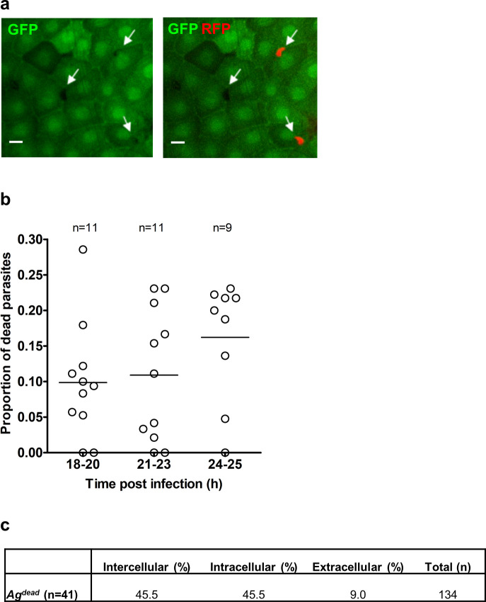
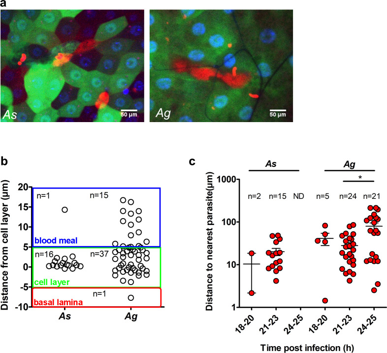
Malaria-causing Plasmodium parasites traverse the mosquito midgut cells to establish infection at the basal side of the midgut. This dynamic process is a determinant of mosquito vector competence, yet the kinetics of the parasite migration is not well understood. Here we used transgenic mosquitoes of two Anopheles species and a Plasmodium berghei fluorescence reporter line to track parasite passage through the mosquito tissues at high spatial resolution. We provide new quantitative insight into malaria parasite invasion in African and Indian Anopheles species and propose that the mosquito complement-like system contributes to the species-specific dynamics of Plasmodium invasion.


Conflict of interest statement

The authors have declared that no competing interests exist.

Figures

Fig 1
Fig 1. Workflow and experimental settings.
A. stephensi (As) and A. gambiae (Ag) mosquitoes were blood-fed on P. berghei infected mice, their midguts dissected and visualized using fast confocal microscopy. Images from all experiments collected at different time points after infection were uploaded into an image database and annotated. Quantitative data was extracted from the images in the database regarding the number, position, and intensity of visualized parasites. The results of the data analysis reveal the kinetics of parasite invasion.
Fig 2
Fig 2. Positions of the parasites relative to the midgut cells.
a. Schematic representation of the topology in the mosquito midgut. Motile ookinetes (red) traverse the mosquito midgut cells (green) and establish infection on the basal side under the basal lamina. b. A representative projection of a cross section of A. stephensi midgut, scale bar—50 μm. GFP-positive midgut cells are in green, RFP-positive P. berghei parasites are in red, nuclei are labeled by DAPI in blue. c. Schematic 3D representation of the same midgut as in (b), where the position of the cell layer is calculated relative to the nuclei. Positions of parasites are indicated as red dots, nuclei as blue dots. Deviation of the cell layer from a flat surface is color-coded from blue to red (blue no deviation, red—10 μm). Note the blood meal location of the majority of parasites (above the cell layer). d. Representation of nuclei (blue) and parasites (red) in the same midgut as (b) after segmentation. e. Pooled positions of the parasites from all records at all time points are shown for three layers relative to the midgut cells (blood meal, cell layer or basal lamina) for A. stephensi, A. gambiae and A. gambiae mosquitoes silenced for TEP1 (A. gambiaeTEP1KD). Each dot represents the number of parasites at a given position in a single midgut. The numbers of midguts analyzed (n) are indicated above the graph. Horizontal lines depict the mean number of parasites per position. The table below summarizes parasite distribution inside the mosquito midguts at 18–25 hpi. The percentage of ookinetes in the midguts of A. stephensi, A. gambiae and A. gambiae silenced for TEP1 (AgTEP1KD) at each location (blood meal, cell layer, and basal lamina) is shown in parentheses, n is the number of parasites at each position, total n is the total number of analyzed parasites. Statistical analyses were performed by Mann-Whitney t-test and the obtained P values are shown.
Fig 3
Fig 3. Kinetics of P. berghei invasion of A. stephensi and A. gambiae midguts.
a. Positions of parasites in A. stephensi (As), A. gambiae (Ag) and in A. gambiae mosquitoes silenced for TEP1 (A. gambiaeTEP1KD) between 18 and 25 h post infection (hpi). Plots show the proportion of parasites at each position (blood meal, cell layer, and basal lamina) for three time intervals (18–20, 21–23 and 24–25 hpi). Each bar represents the average proportion of parasites in midguts that contained at least 10 parasites. Parasite positions were calculated by the distance from the cell layer: blood meal for ookinetes detected more than 5 μm above the cell layer; basal lamina for parasites observed more than 5 μm below the cell layer. Statistical analyses were performed by a Mann-Whitney t-test. The table below shows the number of midguts analyzed at each time interval for each mosquito type. b. Speed of parasites as function of the parasite position in As and Ag. Speed (μm/min) was determined by tracking the parasites position over time from the time-lapse series. Four time-lapse experiments were used: guid 1615 and guid 1628 for As and guid 1622 and guid 2109 for Ag. The table below details the number of frames (n) used for speed calculations. Statistical significance of differences in the average speed at each given position between As and Ag were examined by the Mann-Whitney t-test and P ≤ 0.0001 are designated by three asterisks.
Fig 4
Fig 4. Parasite distribution in the mosquito midgut.
Parasite positions within the cellular layer calculated relative to the distance of each parasites to the nuclei of surrounding midgut cells. a. Calculations of the distance of parasites from the nuclei of the nearest neighboring midgut cell. The score (s) determine whether the parasite is intercellular (0.45 ≤ s ≤ 0.55), extracellular (s < 0.45), or intracellular (s>0.55). Example images from a z stack, scale bar = 20 μm: (I) s = 0.74, the parasite (green arrow) is intracellular; (II) s = 0.45 (red arrow) the parasite is intercellular and (III) s = 0.36, the parasite is extracellular (blue arrow). b. Schematic representation of parasite (red) and nuclei (blue) positions with distances (green lines) used to calculate distances from the nuclei. c. Positions of parasites within the cell layer in A. stephensi (As), A. gambiae (Ag) and A. gambiae mosquitoes silenced for TEP1 (A. gambiaeTEP1KD). The table indicates the percentage of parasites at each position for each mosquito. The number (n) indicates the number of midguts analyzed for each mosquito genotype. d. Comparison of the proportion of intercellular parasites between As, Ag and AgTEP1KD. Each dot represents the proportion of parasites detected between cells in a single midgut. Midguts (n) with at least six parasites within the cellular layer were used for analyses. Statistically significant differences between As and Ag and between Ag and AgTEP1KD revealed by a non-parametric Mann-Whitney t-test are indicated by asterisks (*—P = 0.03; **—P = 0.003). The table details the mean proportions of parasites in each midgut and for each position for n mosquitoes.
Fig 5
Fig 5. Quantification of dead parasites in A. gambiae.
a. Detection of dead parasites within the cellular layer. Due to uniform GFP expression with the midgut cells of the dmAct5C::dsx-eGFP line of A. gambiae, dead parasites that no longer express RFP could be distinguished in the midgut by their negative signal and a characteristic shape. Shown is a single z-section (scale bar = 20 μm) containing two live RFP-expressing parasites and one dead parasite, indicated by arrows. b. The proportion of dead parasites at different time points after Ag infection. Midguts (n) that contained at least 10 parasites were used for analyses. Each dot represents a single midgut. c. Distribution of dead parasites within the cellular layer. The table shows the percentage of parasites at each position at all time points. All images that contained dead parasites were analyzed. The number (n) is the number of midguts analyzed, total is the number of analyzed parasites.
Fig 6
Fig 6. Quantification of damaged cells.
a. Detection of dextran-positive cells in the midguts of A. stephensi (As) and A. gambiae (Ag) mosquitoes. Shown are single z-sections of GFP-expressing dissected midguts. Mosquitoes were fed on mice injected with Texas Red-conjugated dextran. Dextran-positive cells appeared red (scale bar = 50 μm). b. Positions of dextran-filled cells in the midgut layers of As and Ag. Each dot represents a single dextran-positive cell. The graph depicts positions of the dextran-positive cells within the mosquito midgut. Each layer is color coded: blood meal (blue), cell layer (green) and basal lamina (red). The number of dextran-filled cells (n) at each position is indicated. c. Distances of dextran-filled cells to the nearest parasite at different time points after infection of As and Ag. The number of dextran-positive cells analyzed (n) is shown. Statistical analysis was performed by a Mann-Whitney t-test.

Similar articles

Cited by

References

    1. WHO. WHO | World malaria report 2017. WHO. World Health Organization; 2018.
    1. Kiszewski A, Mellinger A, Spielman A, Malaney P, Sachs SE, Sachs J. A global index representing the stability of malaria transmission. Am J Trop Med Hyg. 2004;70(5):486–98. - PubMed
    1. Baton L, Ranfordcartwright L. How do malaria ookinetes cross the mosquito midgut wall? Trends Parasitol. 2005;21(1):22–8. 10.1016/j.pt.2004.11.001 - DOI - PubMed
    1. Sinden RE. Molecular interactions between Plasmodium and its insect vectors. Cell Microbiol. 2002;4(11):713–24. 10.1046/j.1462-5822.2002.00229.x - DOI - PubMed
    1. Blandin S, Levashina EA. Mosquito immune responses against malaria parasites. Curr Opin Immunol. 2004;16(1):16–20. 10.1016/j.coi.2003.11.010 - DOI - PubMed

Publication types